Abstract
Pulmonary paragonimiasis is a relatively rare cause of lung disease revealing a wide variety of radiologic findings, such as air-space consolidation, nodules, and cysts. We describe here a case of pulmonary paragonimiasis in a 27-year-old woman who presented with a 2-month history of cough and sputum. Based on chest computed tomography (CT) scans and fluorodeoxyglucose positron emission tomography (FDG-PET) findings, the patient was suspected to have a metastatic lung tumor. However, she was diagnosed as having Paragonimus westermani infection by an immunoserological examination using ELISA. Follow-up chest X-ray and CT scans after chemotherapy with praziquantel showed an obvious improvement. There have been several reported cases of pulmonary paragonimiasis mimicking lung tumors on FDG-PET. However, all of them were suspected as primary lung tumors. To our knowledge, this patient represents the first case of paragonimiasis mimicking metastatic lung disease on FDG-PET CT imaging.
-
Key words: Paragonimus westermani, paragonimiasis, positron emission tomography, tumor, metastasis
INTRODUCTION
Paragonimiasis is a zoonotic disease caused by the trematode
Paragonimus spp. About 48 species of
Paragonimus have been described; however, only 16 of these infect humans.
Paragonimus wetermani is the most common species infecting humans, causing pulmonary and pleural inflammation through ingestion of raw freshwater crabs or crayfish infected with the metacercariae, the infective stage of this parasite [
1]. However, patients diagnosed with paragonimiasis occasionally report that they had never eaten freshwater crabs or crayfish. There is a possibility that they ate food contaminated with metacercariae from the fingers or cooking utensils of people who recently handled these crustaceans [
2].
Patients with paragonimiasis reveal a wide variety of radiographic and CT findings, including pulmonary nodules or masses, and sometimes they are suspected to have a lung tumor [
3]. FDG(fluorodeoxyglucose)-PET (positron emission tomography) is used recently to differentiate benign from malignant lesions, and it has been shown to be more effective than CT [
4]. However, FDG is not a cancer-specific agent, and false positive findings in benign diseases, including paragonimiasis, have been reported [
5-
9]. Unlike those cases mimicking primary lung tumors, we report a case of pulmonary paragonimiasis with false-positive FDG-PET mimicking a metastatic lung tumor.
CASE REPORT
A 27-year-old woman was admitted to a local hospital with productive cough and blood-tinged sputum. Chest X-ray showed multiple nodules and she received anti-tuberculosis chemotherapy for 2 months. A follow-up chest X-ray taken 2 months later revealed an increased size of the nodules, and the patient was referred to our hospital. She had no history of ingestion of freshwater crabs, crayfish, or wild pig meats. On admission, she was in good general health, and the physical examination was normal. A complete blood cell count (CBC) revealed WBC levels of 11,670/mm
3, hemoglobin levels of 12.6 g/dL, platelet levels of 244,000/mm
3, absolute eosinophil count (AEC) of 6,185/mm
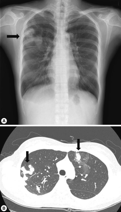
3. Serum tumor markers showed a high CA 125 level of 47.6 U/ml (normal <35 U/ml). Other laboratory tests revealed no abnormal findings. A chest X-ray showed multiple mass-like opacities in the right lung (
Fig. 1A). Chest CT revealed multiple nodules with adjacent areas of ground-glass opacity (GGO), which corresponded to hemorrhagic infarcts as in a case of hemorrhagic metastasis or other benign conditions (
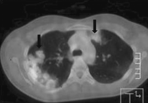
Fig. 1B). FDG-PET CT was performed to find out a possible primary site of malignancy. However, it revealed no remarkable abnormal finding except an increased FDG uptake (max SUV: 3.1) in the lung lesions (
Fig. 2). Bronchoscopic and cytological examination of the sputum showed nothing of particular significance. We could not observe parasite eggs in stool, sputum, or bronchoalvelolar lavage (BAL) fluid. However, ELISA (Genedia Ab ELISA, Green Cross MS, Yongin, Korea) was positive for antibodies against
P. westermani. She received praziquantel therapy (25 mg/kg, 3 times a day for 2 days). At a week after the medication, the patient's eosinophil count decreased from 6,185/mm
3 to 2,468/mm
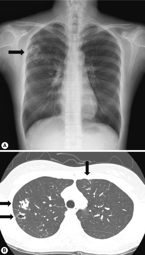
3. A follow-up chest X-ray at 4 months after the treatment showed interval decrease in the size of multiple nodules in the right lung and CT at 6 months after the treatment revealed markedly diminished nodules in size with cystic changes (
Fig. 3).
DISCUSSION
Paragonimiasis is a food-borne parasitic disease caused by the trematode
P. wertermani, which is endemic in East Asia [
1]. Adult
P. westermani worms are bean-shaped, 5-12 mm long, 3-6 mm wide, and 2-5 mm thick, a size that can be seen on radiologic images. Several studies reported that the common radiologic findings of pleuropulmonary paragonimiasis included pleural effusion, hydropneumothorax, pulmonary nodules, or air-space consolidation, and cysts [
10]. A report by Kim et al. [
11] suggested that the main CT feature of pleuropulmonary paragonimiasis in 31 patients was pulmonary nodules, including multiple nodules in 7 patients (23%) as in our patient. In the present case, areas of surrounding GGO were observed around the nodules, which suggest hemorrhage. It led us to consider the findings as hemorrhagic metastasis, though these finding are also shown in a number of benign conditions, such as angioinvasive aspergillosis and paragonimiasis [
12]. Abnormally increased FDG uptake (max SUV: 3.1) in the lung lesions on FDG-PET CT supported a possibility of malignancy further.
FDG-PFT is a highly accurate imaging in evaluating pulmonary lesions, taking advantage of the higher metabolic rate of tumor cells to show increased accumulation of FDG in malignant lesions. However, an increased metabolic rate is also observed in inflammation [
13,
14]. Many reports have demonstrated increased FDG uptake in inflammatory, granulomatous, and infectious lung diseases, including bacterial, mycobacterial, fungal, and parasitic infections.
Several cases of pulmonary paragonimiasis were reported that showed a high uptake suggestive of a malignancy on FDG-PET CT images [
5-
9]. All of them were suspected as primary lung cancers and confirmed as paragonimiasis by detecting eggs in the tissue obtained from biopsy or operation. Although the definite causes of FDG accumulation have not yet been proven in paragonimiasis, it seems likely that inflammatory cells, including eosinophilic infiltration, active inflammatory responses, and viable worms, cause a high FDG uptake [
4].
The diagnosis of paragonimiasis can be made by detecting eggs in the tissue section, or more commonly, in the stool, sputum, or BAL fluid, or by a positive anti-
Paragonimus antibody test. However, egg detection rates have been reported to be low (28-38%) and eggs are not present in the sputum until 2-3 months after an infection. Serum determination of antibodies to
Paragonimus is more accurate in early infections, and ELISA is highly sensitive (92%) and specific [
15]. In our case,
Paragonimus eggs were not found in the stool, sputum, and BAL fluid. Instead, the patient's serum strongly reacted to
P. wertermani antigen in ELISA.
Considering lung nodular lesions with GGO on chest X-ray and CT, in combination with ELISA test for paragonimiasis, FDG-PET itself was an unnecessary examination to diagnose paragonimiasis in our patient. In addition, nodular lesions were continuing to the visceral pleura, which is rather unusual for metastatic lung cancers. On the other hand,
Paragonimus invades from the pleural cavity to the lung parenchyma so that continuous lesions from the pleura to the nodular lesion in the lung parenchyma are a typical feature of radiological finding in paragonimiasis. Nevertheless, FDG-PET was performed to exclude the possibility of malignancy owing to the elevated serum level of CA 125. The serum CA 125 is often increased in patients with malignancy, such as ovarian cancer, endometrial cancer, and pancreatic cancer. However, it is also increased in a variety of benign conditions, including endometriosis, uterine fibroids, pelvic inflammatory disease, heart failure, liver and renal disease, as well as in approximately 1 percent of healthy women [
16]. There has been no report of paragonimiasis patients showing high level of CA 125.
As a possibility in the differential diagnosis of multiple pulmonary nodules or masses with FDG-PET positive finding, especially in endemic regions with a high prevalence of food-borne parasitic diseases, paragonimiasis should be considered.
References
- 1. Yokogawa M. Paragonimus and paragonimiasis. Adv Parasitol 1965;3:99-158.
- 2. Nakamura-Uchiyama F, Mukae H, Nawa Y. Paragonimiasis: a Japanese perspective. Clin Chest Med 2002;23:409-420.
- 3. Mukae H, Taniguchi H, Matsumoto N, Iiboshi H, Ashitani J, Matsukura S, Nawa Y. Clinicoradiologic features of pleuropulmonary Paragonimus westermani on Kyusyu Island, Japan. Chest 2001;120:514-520.
- 4. Chang JM, Lee HJ, Goo JM, Lee HY, Lee JJ, Chung JK, Im JG. False positive and false negative FDG-PET scans in various thoracic diseases. Korean J Radiol 2006;7:57-69.
- 5. Watanabe S, Nakamura Y, Kariatsumari K, Nagata T, Sakata R, Zinnouchi S, Date K. Pulmonary paragonimiasis mimicking lung cancer on FDG-PET imaging. Anticancer Res 2003;23:3437-3440.
- 6. Yoo IeR, Park HJ, Hyun J, Chung YA, Sohn HS, Chung SK, Kin SH. Two cases of pulmonary paragonimiasis on FDG-PET CT imaging. Ann Nucl Med 2006;20:311-315.
- 7. Osaki T, Takama T, Nakagawa M, Oyama T. Pulmonary Paragonimus westermani with false-positive fluorodeoxyglucose positron emission tomography mimicking primary lung cancer. Gen Thorac Cardiovasc Surg 2007;55:470-472.
- 8. Moon JY, Jung KH, Kim JH, Park HJ, Kim YS, Shin C. A case of pulmonary paragonimiasis presented as solitary pulmonary nodule and suspected as lung cancer on 18F-fluorodeoxyglucose positron emission tomography. Tuberc Respir Dis 2008;64:133-137.
- 9. Moon YR, Lee YD, Park SH, Cho YS, Na DJ, Cho YS, Han MS, Choi HJ, Kim DH, Yang SO, Kim KH. A case of paragonimiasis that was suspicious for a lung malignancy by PET/CT. Tuberc Respir Dis 2007;63:521-525.
- 10. Im JG, Whang HY, Kim WS, Han MC, Shim YS, Cho SY. Pleuropulmonary paragonimiasis: radiologic findings in 71 patients. AJR 1992;159:39-43.
- 11. Kim TS, Han J, Shim SS, Jeon K, Koh WJ, Lee I, Lee KS, Kwon OJ. Pleuropulmonary paragonimiasis: CT findings in 31 patients. AJR 2005;185:616-621.
- 12. Franquet T, Müller NL, Giménez A, Guembe P, de La Torre J, Bagué S. Spectrum of pulmonary aspergillosis: histologic, clinical, and radiologic findings. Radiographics 2001;21:825-837.
- 13. Hübner KF, Buonocore E, Gould HR, Thie J, Smith GT, Stephens S, Dickey J. Differentiating benign from malignant lung lesions using "quantitative" parameters of FDG PET images. Clin Nucl Med 1996;21:941-949.
- 14. Yang SN, Liang JA, Lin FJ, Kwan AS, Kao CH, Shen YY. Differentiating benign and malignant pulmonary lesions with FDG-PET. Anticancer Res 2001;21:4153-4157.
- 15. Cho SY, Hong ST, Rho YH, Choi S, Han YC. Application of micro-ELISA in serodiagnosis of human paragonimiasis. Korean J Parasitol 1981;19:151-156.
- 16. Bast RC Jr, Klug TL, St John E, Jenison E, Niloff JM, Lazarus H, Berkowitz RS, Leavitt T, Griffiths CT, Parker L, Zurawski VR Jr, Knapp RC. A radioimmunoassay using a monoclonal antibody to monitor the course of epithelial ovarian cancer. N Engl J Med 1983;309:883-887.
Fig. 1Chest X-ray and computed tomography (CT) on admission. (A) Multiple mass-like opacities are shown in the right lung (arrow). (B) Multiple nodules with adjacent areas of GGO, suggestive of hemorrhagic metastasis are observed in both lungs (arrows).

Fig. 2FDG-PET CT showed increased FDG uptake (max SUV: 3.1) (arrows).

Fig. 3Follow-up chest X-ray and CT. (A) Interval decreases in the size of multiple nodules are shown in the right lung on the chest X-ray 4 months after treatment (arrow). (B) Markedly diminished nodules in size with cystic changes are observed in both lungs on CT 6 months after treatment (arrows).

Citations
Citations to this article as recorded by

- Relationship between parasites and lung cancer: Unveiling the link
Mariam T. El Khadrawe, Nahla El Skhawy, Maha M. Eissa
Tropical Medicine & International Health.2025; 30(7): 613. CrossRef - Pulmonary paragonimiasis presenting as massive empyema requiring decortication in an adolescent: a case report
Kyo Jin Jo, Su Eun Park, Jong Myung Park, Joo-Young Na, Sungsu Jung
Journal of Medical Case Reports.2025;[Epub] CrossRef - Dynamic Computed Tomography for detecting pulmonary cysts in canine Paragonimiasis: a case report
Shinya MIZUTANI, Yuko MIZUTANI, Yoshimichi GODA, Hiroyuki SATOH, Taketoshi ASANUMA, Ayako YOSHIDA, Shidow TORISU
Journal of Veterinary Medical Science.2025; 87(9): 986. CrossRef - A clinical mimicry of malignancies in patients diagnosed with paragonimiasis
David E. Elem, Stella T. Chukwuma, Ikechukwu F. Agwu, Ukam E. Edadi, Okoi A. Ojah, Walter O. Egbara, Aje N. Ogar, Love E. Okafor, Bassey E. Ekeng
Journal of Clinical and Scientific Research.2025; 14(2): 121. CrossRef - Simple Pulmonary Eosinophilia Mimicking Lung Metastases on 18F-FDG PET/CT
Lan Yang, Ziqian Dong, Lingge Wei, Feilong Chen, Peng Xie
Clinical Nuclear Medicine.2024; 49(9): 884. CrossRef - Pulmonary, liver and cerebral paragonimiasis: An unusual clinical case in Colombia
Angel O. Donato-R., Jhon C. Donato-R.
Travel Medicine and Infectious Disease.2022; 46: 102253. CrossRef - What Findings on Chest CTs Can Delay Diagnosis of Pleuropulmonary Paragonimiasis?
Kai Ke Li, Gong Yong Jin, Keun Sang Kwon
Tomography.2022; 8(3): 1493. CrossRef - One delayed diagnosis of paragonimiasis case and literature review
Luxia Kong, Lijuan Hua, Qian Liu, Chen Bao, Jiannan Hu, Shuyun Xu
Respirology Case Reports.2021;[Epub] CrossRef - Pulmonary larval paragonimiasis mimicking lung cancer
V. V. Ermilov, A. V. Smirnov, G. L. Snigur, R. S. Dudin, S. S. Popov
Arkhiv patologii.2018; 80(2): 60. CrossRef - Electromagnetic Navigational Bronchoscopy Spares a Drunken Crab From the Surgeon’s Knife
Mihir S. Parikh, Eric Seeley, Ganesh Krishna
Journal of Bronchology & Interventional Pulmonology.2017; 24(3): 241. CrossRef - Recent Incidence of Paragonimus westermani Metacercariae in Freshwater Crayfish, Cambaroides similis, from Two Enzootic Sites in Jeollanam-do, Korea
Jin-Ho Song, Fuhong Dai, Xuelian Bai, Tae-Im Kim, Hyun-Jong Yang, Tong-Soo Kim, Shin-Hyung Cho, Sung-Jong Hong
The Korean Journal of Parasitology.2017; 55(3): 347. CrossRef - A 15-Year Old Burmese Girl With Hemoptysis: A Case Report
Charlie McLeod, Daniel Yeoh, Cameron Truarn, Christopher C Blyth, Asha C Bowen, Tom L Snelling, Ushma Wadia, Briony Hazelton, Michelle Porter
Open Forum Infectious Diseases.2017;[Epub] CrossRef - Pulmonary Paragonimiasis Misdiagnosed with Pulmonary Tuberculosis
Su Eun Park, Bokyung Song, Jae-Yeon Hwang
Pediatric Infection and Vaccine.2017; 24(3): 178. CrossRef - Imaging findings of Paragonimus westermani
Shambhu Kumar Sah, Silin Du, Yi Liu, Ping Yin, Oormila Ganganah, Manu Chiniah, Pranesh Kumar Yadav, You You Guo, Yongmei Li
Radiology of Infectious Diseases.2016; 3(2): 66. CrossRef - A Case of Intra-abdominal Paragonimiasis Mimicking Metastasis of Lung Cancer Diagnosed by Endoscopic Ultrasound-guided Fine Needle Aspiration
Cho Rong Oh, Mi-Jin Kim, Kwang Hyuck Lee
The Korean Journal of Gastroenterology.2015; 66(1): 41. CrossRef - A Case of Delayed Diagnosis of Pulmonary Paragonimiasis due to Improvement after Anti-tuberculosis Therapy
Suhyeon Lee, Yeonsil Yu, Jinyoung An, Jeongmin Lee, Jin-Sung Son, Young Kyung Lee, Sookhee Song, Hyeok Kim, Suhyun Kim
Tuberculosis and Respiratory Diseases.2014; 77(4): 178. CrossRef - Answer to February 2014 Photo Quiz
Yan Chen, Haidong Huang, Daoyin Zhou, Yanghua Qin, P. Bourbeau
Journal of Clinical Microbiology.2014; 52(2): 710. CrossRef - A Case of Ovarian Paragonimiasis Mimicking Ovarian Carcinoma
Charuwan Tantipalakorn, Surapan Khunamornpong, Theera Tongsong
Gynecologic and Obstetric Investigation.2014; 77(4): 261. CrossRef - A Case of Pulmonary Paragonimiasis Mimicking Lung Cancer Diagnosed by EBUS-TBNA
Jae June Lee, Chang Min Choi, Hwuk Hee Kwon, Min Soo Kim, Joon-Seok Kim, So Young Park, Hee Sang Hwang
Korean Journal of Medicine.2013; 84(3): 423. CrossRef - Clinical Update on Parasitic Diseases
Min Seo
Korean Journal of Medicine.2013; 85(5): 469. CrossRef - Chest CT Features of North American Paragonimiasis
Travis S. Henry, Michael A. Lane, Gary J. Weil, Thomas C. Bailey, Sanjeev Bhalla
American Journal of Roentgenology.2012; 198(5): 1076. CrossRef


