First Feline Case of Otodectosis in the Republic of Korea and Successful Treatment with Imidacloprid/Moxidectin Topical Solution
Article information
Abstract
In April 2010, pruritic symptoms were recognized in 3 privately-owned Siamese cats raised in Gwangju, Korea. Examination of ear canals revealed dark brown, ceruminous otic exudates that contain numerous live mites at various developmental stages. Based on morphological characteristics of adult mites in which caruncles were present on legs 1 and 2 in adult females and on legs 1, 2, 3, and 4 in adult males while the tarsus of leg 3 in both sexes was equipped with 2 long setae, the mite was identified as Otodectes cynotis. Ten ear mite-free domestic shorthaired cats were experimentally infected with O. cynotis to evaluate the efficacy of 10% imidacloprid/1% moxidectin spot-on. Live mites were recovered from 1 of 10 treated cats on day 9 post-treatment (PT) while no live mites were observed from the ear canals of treated cats on days 16 and 30 PT. The efficacy of 10% imidacloprid/1% moxidectin spot-on on O. cynotis in cats was, therefore, 90% on day 9 and 100% on days 16 and 30 PT. This is the first report of otodectosis in 3 cats naturally infested with O. cynotis in Gwang-ju, Korea. Both natural and experimental infestations were successfully treated with 10% imidacloprid/1% moxidectin spot-on.
INTRODUCTION
Otodectes cynotis is a mite of the family Psoroptidae, which lives predominantly in the external ear canal and occasionally on the adjacent skin of the head in dogs, cats, foxes, ferrets, and even humans [1,2]. It is a non-burrowing surface-living mite that feeds on tissue fluid and debris [1]. Infestation of the mite results in pruritic symptoms [3] and otitis externa which is characterized by ear canal erythema and the presence of dark brown, ceruminous otic exudate [1]. The prevalence of the mite in dogs ranges from 2% to 29% in London and Queensland [4,5], and that in cats was from 9% to 37% in Florida (USA), Greece, and Japan [6-9]. In the Republic of Korea, the prevalence of O. cynotis was reported to be 22.3% among dogs in an animal shelter [10], and an ear mite treatment case with selamectin in a dog has been reported [11]. However, no studies about the occurrence and treatment of O. cynotis in cats have been available in Korea.
In this study, we report 3 cats that had pruritic symptoms in Gwangju, Korea due to the infestation with O. cynotis. Using the Korean isolate of O. cynotis, we also experimentally infested domestic cats and assessed the efficacy of 10% imidacloprid/1% moxidectin spot on.
CASE RECORD
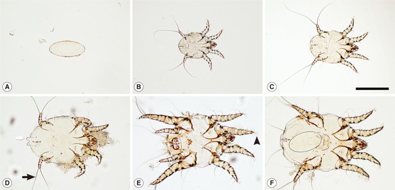
In April 2010, pruritic symptoms characterized by scratching, rubbing of the ears, and shaking of the head were recognized in 3 privately-owned Siamese cats raised in Gwangju, Korea. Two were females and 1 was male, and they were between 8 months and 2 years old. Examination of ear canals using an otoscope (Piccolight®, KaWe, Berlin, Germany) revealed dark brown, ceruminous otic exudates. Numerous live mites were collected from the ears of infested cats from which all developmental stages in life cycle (egg, larva, nymph, and adult) were observed (Fig. 1). The mites were preserved in 70% ethanol and mounted on slide glass using PVA solution [12]. The average length and width of 6 adult females were 448.3×290.0 µm, while those of 6 adult males were 360.0×277.8 µm. The mites had unsegmented and short pedicle of tarsal caruncles and striated integument. Caruncles were present on legs 1 and 2 in adult females and on legs 1, 2, 3, and 4 in the adult male (Fig. 1E, F). The tarsus of leg 3 in both sexes was equipped with 2 long setae (Fig.1D). Based on these morphological characteristics, the infesting mites were identified as O. cynotis [2,13].

Light micrographs of various developmental stages of Otodectes cynotis isolated from the external ear canal of naturally-infested cats from Korea. (A) Unembryonated egg. (B) Ventral view of a larvae. (C) Ventral view of a protonymph. (D) Ventral view of female deutonymph equipped with a couple of copulatory tubercles at the posterior end (white arrow). (E) ventral view of an adult male; (F) ventral view of an ovigerous female (adult female) containing an egg. Note all developmental stages except the egg have 2 long setae on the tarsus of leg 3 in both sexes (black arrow). Pretarsal caruncle is present on legs 1 and 2 in female and on legs 1, 2, 3, and 4 in male (arrowhead).
Efficacy evaluation of 10% imidacloprid/1% moxidectin spot-on
Ten mite-free domestic shorthaired cats (5 males and 5 females; weight range, 2.0-4.3 kg; age range, 1-2 years) were purchased from a commercial vender. Cats were thoroughly checked for the presence of any ear mite or other health problems. Experimental infestation of uninfected cats was performed twice for 2 weeks in which cerumen containing various stages of mites from the ear of the 3 naturally infested cats was collected in a petri dish and was transferred into both ears of O. cynotis-free cats using an ear swab. One month later, the presence of adult mites and eggs in both ears was confirmed in 10 cats by otoscopic and microscopic examinations. Cats did not receive any acaricidal drugs for at least 60 days prior to the treatment. Animals were housed in an appropriate accommodation that conformed to accepted guidelines for pen design/floor area, lighting, humidity, temperature, and welfare (including environmental enrichment and social interaction), as required by local and national legislations.
Ten cats were randomly allocated to the test group and 3 to the control group. Day 0 was defined as the first day of treatment for each animal. The test group was treated with 10% imidacloprid/1% moxidectin spot-on (Advocate® for Cats, Bayer Animal Health GmbH, Leverkusen, Germany, abbreviated as Im/Mox hereafter), applied at a dose of 0.1 ml/kg body weight on day 0. The Im/Mox was administered once topically to the skin in a single spot on each animal's back at the base of the neck in front of the scapulae. The ears of the cats were examined for the presence of ear mites by using the otoscope. Ear scrapes and dry cotton swabs were also used to collect debris in the ears on day 0, 9, 16, and 30 post-treatment (PT), and live ear mites were searched under a dissecting microscope (Zeiss, Stemi 2000-C, Jena, Germany). The presence of even 1 live mite in any side of ears was considered as a failure of treatment. The number of cats that had no live mites was compared to the number of all cats tested [14]. The percentage of the drug efficacy was calculated as follows [15] :
The percentage of efficacy (%)=
x=number of cats with live ear mite
Upon treatment with Im/Mox, live mites were recovered only from 1 of 10 treated cats on day 9 (Table 1). On days 16 and 30 PT, no live ear mites were observed from the ear canals of treated cats. Ear canals of 3 cats in the control group, on the other hand, showed various developmental stages of mite from day 0 to 30. The efficacy of Im/Mox on O. cynotis in cats was, therefore, 90% on day 9 and 100% on days 16 and 30 PT. The 3 cats in the control group were also treated with Im/Mox after the chemotherapy study which resulted in a successful removal of mites from the cats.
DISCUSSION
In this study, we described the first case of otodectosis in 3 cats naturally infested with O. cynotis in Gwangju, Korea, and successful treatment of experimentally infested ear mites with 10% imidacloprid/1% moxidectin spot-on. In Korea, although the prevalence of O. cynotis in dogs was 22.3% [10], that of cats has not been studied, probably because of severe bias in population of cats and dogs in Korea. According to a study in Korea, the population of dogs had been up to 97.8% of total cats and dogs in 2006 [16]. This phenomenon might have resulted by the common perception on cats by older generation of Korea who generally consider cats as unlucky and sneaky animals [17].
Treatments of the ear mite include mechanical cleaning of the ear canal followed by topical or systemic drug administration with such drugs as selamectin, ivermectin, and fipronil [18,19]. In cats, there exist many difficulties to administer the drugs via oral route which can cause esophagitis, esophageal stricture, a longer esophageal transit time, and retention in mid-cervical region of the esophagus [20,21]. Topical spot-on drugs has recently been considered as an ideal administration route because it can solve those problems of oral administration and be used without cleaning the ear before administration of the drug or treating the hair coat and living environment [22]. The topical spot-on medication can also bypass the intestinal and hepatic first-pass effects [23]. For these reasons, we have chosen the topical spot-on medication route to treat feline otocariosis in our study.
Among many treatment studies using 10% imidacloprid/1% moxidectin in cats, a single treatment with the drug applied at a dose of 0.1 ml/kg body weight resulted in a treatment success rate of 80%, as assessed 50 days after treatment in cats [15]. The efficacy of Advantage Multi™ that contains 10% imidacloprid/1% moxidectin against ear mites was 92.5% after the first treatment and 98.1% after the second treatment in client-owned cats [24]. Also, the efficacy of 10% imidacloprid/1% moxidectin against O. cynotis in another study was 100% on days 16 and 30 PT in cats [22].
In our study, the efficacy of 10% imidacloprid/1% moxidectin spot-on was 90% on day 9 and 100% on days 16 and 30 PT which was significantly higher than that of day 0 and indicates that the single spot-on medication of the drug provided more than 90% efficacy against O. cynotis-infested cats.
ACKNOWLEDGMENT
This work was supported in part by the Ministry of Education and Human Resources Development through the Brain Korea 21 Project in Korea.
