Molecular Diagnosis of an Ocular Toxocariasis Patient in Vietnam
Article information
Abstract
An ocular Toxocara canis infection is reported for the first time in Vietnam. A 34-year-old man residing in a village of Son La Province, North Vietnam, visited the National Eye Hospital (NEH) in August 2011. He felt a bulge-sticking pain in his left eye and loss of vision occurred over 3 months before visiting the hospital. The eye examination in the hospital showed damage of the left eye, red eye, retinal fibrosis, retinal detachment, inflammation of the eye tissues, retinal granulomas, and a parasitic cyst inside. A larva of Toxocara was collected with the cyst by a medical doctor by surgery. Comparison of 264 nucleotides of internal transcribed spacer 2 (ITS2) of ribosomal DNA was done between our Vietnamese Toxocara canis and other Toxocara geographical isolates, including Chinese T. canis, Japanese T. canis, Sri Lankan T. canis, and Iranian T. canis. The nucleotide homology was 97-99%, when our T. canis was compared with geographical isolates. Identification of a T. canis infection in the eye by a molecular method was performed for the first time in Vietnam.
INTRODUCTION
Humans are an accidental host for Toxocara, yet human toxocariasis is seen throughout the world. Human toxocariasis is the parasitic disease caused by the larvae of 2 Toxocara species, namely, T. canis from dogs and, less commonly, T. cati from cats [1]. People become infected by accidental swallowing of dirt that has been contaminated with dog or cat feces containing Toxocara eggs, and although it is rare, people can become infected through eating undercooked meat containing Toxocara larvae of the paratenic hosts such as cats, foxes, cows, monkeys, pigs, lambs, mice, rats, chickens, pigeons, and ostriches [2-4]. After entering the body, the eggs hatch into larvae that penetrate through walls of the digestive tract, and the larvae then migrate through the organs and tissues of the accidental host (including humans), most commonly the lungs, liver, eyes, brain, and elsewhere.
Toxocariasis is a zoonotic infection that can cause serious illness, including organ damage and eye diseases in humans. There were 4% ocular toxocariasis among lesions simulating retinoblastoma (pseudoretinoblastoma) patients [5]. Vision loss was reported in 85% of 54 patients with ocular toxocariasis [6]. Kwon et al. [7] reported that Toxocara granuloma was found in 52% of ocular toxocariasis patients in Korea. Morais et al. [8] analyzed ocular toxocariasis in which 63.6% occurred as posterior pole granuloma; 9.1% as chronic endophthalmitis; 18.2% as peripheral granuloma, and 9.1% as posterior pole granuloma associated with chronic endophthalmitis. Serology by ELISA was 100% positive among these cases. Symptoms include pallor, fatigue, weight loss, anorexia, fever, headache, rash, cough, asthma, chest tightness, increased irritability, abdominal pain, nausea, and vomiting [9]. Ocular infection is rare compared with visceral larva migrans. In Asian countries including Korea [7] and China [10], ocular larva migrans have been frequently reported, but in Vietnam there was no report of ocular larva migrans patients.
Recently, a Toxocara larva was collected from the eye of a Vietnamese man, which was identified by a molecular method as T. canis. This is the first report of ocular toxocariasis in Vietnam.
CASE RECORD
The patient was a 34-year old male residing in Phieng Ngua Village, Chieng Xom Commune, Son La City, Son La Province of mountainous North Vietnam. In August 2011, he felt a bulge-sticking pain in his left eye and loss of vision occurred over 3 months before visiting the national hospital. A funduscopic examination showed damage of the left eye as red eye, retinal fibrosis, retinal detachment, inflammation of the eye tissues, retinal granulomas, and a parasitic cyst inside and under retina, while his right eye was normal. The rate of eosinophils was 30.2% and the patient was seropositive against T. canis antigen in ELISA. A larva of Toxocara (about 350 µm in length and 20 µm in width) embbeded in the surrounding cyst was collected from his left eye by a medical doctor with surgery.
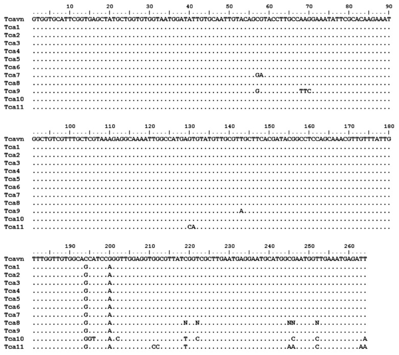
The larva from this patient was identified by the molecular method using the internal transcribed spacer 2 (ITS-2) of rDNA in comparison with standard strains in GenBank. Comparison of 264 nucleotides in ITS-2 of rDNA between Vietnamese T. canis (Tcavn) and other strains of T. canis, including Chinese T. canis (Tca1, Tca2, and Tca3; GenBank no. JF837170.1, JF837169.1, and JN617989.1, respectively), Japanese T. canis (Tca5 and Tca8; GenBank no. AB110032.1 and AB110034.1, relatively), Sri Lankan T. canis (Tca4; GenBank no. FJ418788.1) and Iranian T. canis (Tca7, Tca9, Tca10, and Tca11; GeneBank no. AB743615.1, AB743614.1, AB743617.1, and AB743616.1, relatively), and GenBank (Tca6; GenBank no. Y09489.1) [11-16] (Tables 1, 2; Fig. 1).

Sequencing of internal transcribed spacer (ITS-2) of rDNA of different Toxocara species from GenBank compared with the present case

Percentage of identity of nucleotide of ITS-2 sequences of Vietnamese Toxocara canis and others Toxocara in GenBank

Comparison of 264 nucleotides of internal transcribed spacer 2 (ITS-2) of rDNA between Vietnamese Toxocara canis and other geographical isolates including Chinese T. canis (Tca1, Tca2, and Tca3), Japanese T. canis (Tca5 and Tca8), Sri Lankan T. canis (Tca4), Iranian T. canis (Tca7, Tca9, Tca10, and Tca11), and an unknown origin Toxocara (Tca6). Note the difference between Vietnamese T. canis (Tcavn) and other isolates as shown by their sign nucleotides. Mark (.) is similar to each other in the nucleotide.
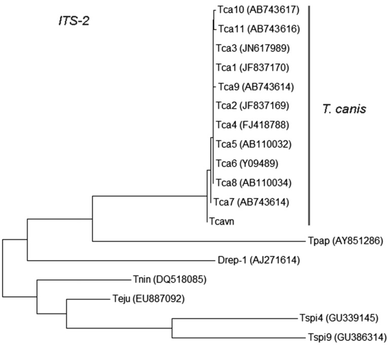
The results showed that the nucleotide homologies between the Vietnamese Toxocara and T. canis from China, Sri Lanka, Japan, Iran, and 1 from GenBank were 97-99%. Phylogenetic tree of Vietnamese T. canis and other isoaltes based on ITS-2 nucleotide sequence estimated by neighbor-joining (NJ) method using the MEGA5.1 [17]; Vietnamese T. canis was a group together with other T. canis isolates in the world (Fig. 2). The Toxocara larva collected from the eye of this patient (Vietnam) was identified as T. canis by the molecular methods.

Phylogenetic tree of Vietnamese Toxocara canis and other isolates based on ITS-2 nucleotide sequence as estimated by neighbor-joining (NJ) using MEGA5 (Tamura et al., 2004) [13]. Tcavn=Vietnamese T. canis; Tca1, Tca2, and Tca3=Chinese T. canis; Tca4=Sri Lankan T. canis; Tca5 and Tca8=Japanese T. canis; Tca6=unknown Toxocara; Tca7, Tca9, Tca10, and Tca11=Iranian T. canis; Tpap=Trichinella papuae (GenBank no. AY851286); Teju=Troglosiro cf. juberthiei (GenBank no. EU887092); Tnin=Troglosiro ninqua (GenBank no. DQ518085); Tspi4=Chinese Trichinella spiralis (GenBank number GU339145); Tspi9=GenBank Trichinella (GenBank no. GU386314); Drep-1=Italian Dirofilaria repens (GenBank no. AJ271614).
DISCUSSION
In Vietnam, in recent years, toxocariasis are increasingly detected by serodiagnosis and there are hundreds of toxocariasis patients diagnosed each year. Based on ELISA test on suspected patients who visited hospitals, a Toxocara infection rate of 32.5% was found in the south, 30.2% in the middle, and 33.3% in the northern part of this country (unpublished). The clinical symptoms in these patients were similar to those found in visceral larva migrans. However, ocular larva migrans due to Toxocara larvae was never reported in Vietnam, and the present patient is the first ocular case in Vietnam.
The patient's family fed the dog and the patient was always in contact with his dogs. We regret that we could not perform fecal examination of the dogs. Feeding dogs is very common in Vietnam, especially in rural areas; however, human toxocariasis is widespread in the whole country. Hundreds of toxocariasis patients are detected every year in Vietnam with many complicated symptoms, most of them are visceral larva migrans and some of them might have been ocular larva migrans. In our report, a single larva was detected in the eye of a patient. This case was similar to the report of John and Petri [9] and Review of Optometry Online, Handbook of Ocular Disease Management. The time onset of our patient was longer (over 3 months) compared with the report of Holland and Smith [1], in which loss of vision occurred over days or weeks [1]. The signs and symptoms were damage of the left eye including red eye, retinal fibrosis, retinal detachment, inflammation of the eye tissues, retinal granulomas, and a parasitic cyst inside and under retina, while his right eye was normal. These signs were similar with the report of Stewart et al. [18]. Toxocara damage in the eye is permanent and can result in blindness [5,19].
In our study, the larva from an eye of a human was identified as T. canis using the molecular method. This is the first time when T. canis extracted from the eye of human infections was identified by the molecular method in Vietnam.
ACKNOWLEDGMENTS
We acknowledge the funds supported from the National Foundation for Science and Technology Development (NAFOSTED) of Vietnam (No. 106.12-2011.13 to Nguyen Van De), Ministry of Health Project 2012-2014, and National Eye Hospital (NEH), Hanoi, Vietnam.
Notes
We have no conflict of interest related to this study.