Abstract
Holostephanus metorchis(Digenea: Cyathocotylidae) is a parasite of birds, transmitted by freshwater fishes. H. metorchis adults were recovered from chicks experimentally infected with metacercariae collected from freshwater fishes, Pseudorasbora parva. The metacercariae were oval, surrounded with thick fibrous capsules. In adult flukes, the holdfast organ occupied the ventral concavity, and the anterior testis did not reach the level of the ventral sucker. Based on these morphological characteristics, these flukes were identified as H. metorchis.
-
Key words: Holostephanus metorchis, trematode, Pseudorasbora parva, holdfast organ
INTRODUCTION
Holostephanus Szidat, 1936, belongs to the family Cyathocotylidae. Its ventral pouch exhibits a distinctive morphology and encloses the holdfast organ [
1]. Other unique features of this species include the gonad position (testes are diagonally arranged and well-developed) and the location of vitelline follicles which extend to the level of the ventral sucker or pharynx [
2]. Twelve species belong to the genus
Holostephanus; H.
luehei (type),
H. anhinga,
H. calvusi,
H. corvi,
H. curonensis,
H. dubius,
H. ibisi,
H. ictaluri,
H. lutzi,
H. metorchis,
H. nipponicus, and
H. phalacrocoraxus [
1].
Holostephanus species were originally described as arasites of birds, and adult flukes of
H. nipponicus and
H. metorchis were first recovered from the black kite,
Milvus migrans lineatus, in Japan [
3]. The first intermediate hosts are snails of the
Parafossarulus spp., including
P. spiridonovi, and the second hosts were proven to be freshwater fish,
Pseudorasbora parva [
3]. Adult worms of
H. ictaluri were discovered from the intestine of a catfish [
5],and Besprozvannykh et al. [
4] succeeded in propagating
H. nipponicus in chickens.
Infection status of freshwater fishes with digenetic trematode metacercariae has been extensively studied in the Republic of Korea. However, trematode fauna of wild birds in Korea has not been a subject of extensive investigation. As
Gymnophalloides seoi was isolated from a pancreatitis patient [
6], parasites of birds were recognized as a possible source of human infections. Hence, it is necessary to explore on digeneans infecting birds.
To our knowledge, there are 3 reports which studied on
Holostephanus spp. in Korea. The presence of
H. nipponicus was confirmed by the removal of 21 metacercariae from
P. parva [
7]. Nam et al. [
8] isolated 8 metacercariae from the pond smelt
Hypomesus olidus.
H. nipponicus adults were identified and reported in 2007 [
9]. In the present study, we collected metacercariae of
H. metorchis from
P. parva, and succeeded in rearing them into adult flukes in chicks, and identified to
H. metorchis.
MATERIALS AND METHODS
A total of 200 P. parva were purchased from a local fish market near Nakdong-river, Kyongsangbuk-do, in May 2006. They were brought to the laboratory and digested using an artificial pepsin-HCl solution. The digested material was filtered through a sieve and washed several times with 0.85% physiological saline. Metacercariae of H. metorchis were collected from particulate sediment under stereomicroscope. The collected metacercariae were measured using a light microscope. Chicks, 7-day-old, free from intestinal helminth infections by fecal examination, were orally fed with about 100 metacercariae, and sacrificed on day 7 post-infection (PI). The small intestine was resected, opened along the mesenteric border, and washed several times with 0.85% saline. Adult flukes were recovered from the intestinal contents using Baermann's apparatus, and counted under stereomicroscope. They were washed several times with saline, fixed in 10% neutral formalin, stained with Semichon's acetocarmine, mounted in permount (Fischer), and observed under a light microscope.
RESULTS
Metacercariae of H. metorchis
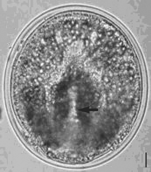
The metacercaria was oval, 164.7 µm long and 140.3 µm wide, similar in size and shape with that of
H. nipponicus. It was enclosed in a thick fibrous capsule, consisting of 2 layers (
Fig. 1). Cyst wall was 5 µm thick, which was thinner than that of
H. nipponicus, 9.5 µm. The larvae were underneath the inner cyst wall, and a holdfast organ was observed (
Fig. 1). The oral sucker was discernible but not other internal organs.
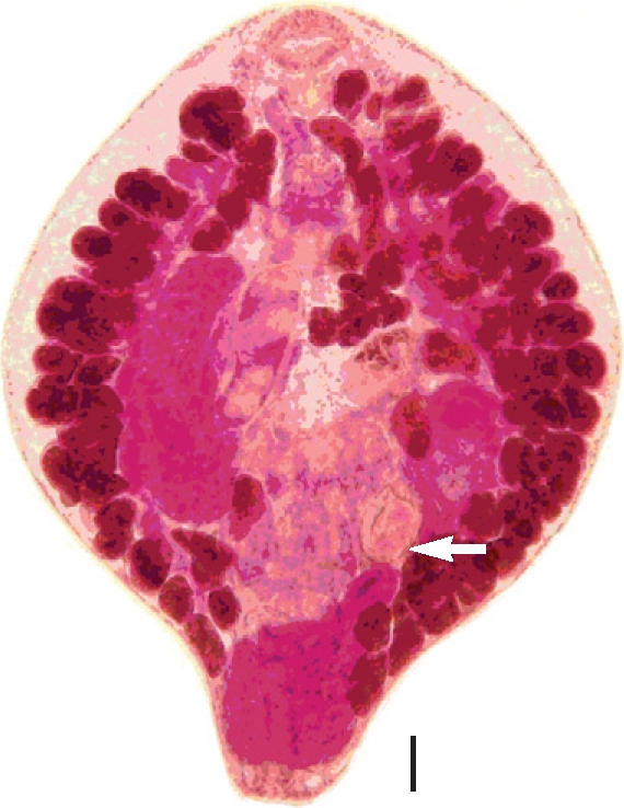
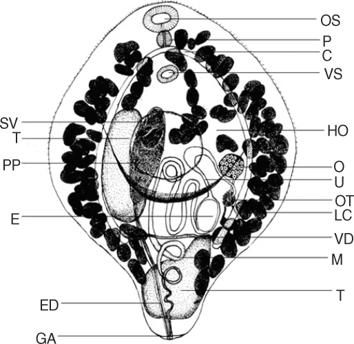
From 5 experimental chicks, 7 flukes were recovered (recovery rate; 1.4%). Morphological characteristics observed were as follows (units in µm). Body ovoid with narrow posterior part, measuring 1,020-1,450 × 740-920 (1,289 × 806, in average). Oral sucker, 87.5-125.0 × 115.0-142.5 (110.8 × 131.1 in average). Pharynx, 62.5-120.5 (89.3) in length. Esophagus unrecognized. Ventral sucker, 52.5-72.5 × 75-107.5 (63.0 × 89.0), situated immediately posterior to cecal arch. Ceca long, extending to level of the posterior testis. Holdfast organ large and well developed, occupying nearly whole ventral concavity. Testes irregular in shape, and apart each other. Anterior testis, ellipsoid, lied adjacent to the left cecum and extended parallel with cirrus pouch, from midline to posterior one-third of the body. Posterior testis occupied posterior junction and the narrow projection. The anterior testis 355.0-460.0 × 135.0-215.0 (396.1 × 130.4), and the posterior testis 270.0-495.0 × 120.0-212.0 (361.4 × 173.7). Cirrus pouch elongate, 210.0-350.0 × 65.0-120.0 (298.3 × 100.7), and close to the anterior testis. Seminal vesicle bipartite. Pars prostatica well discernible, surrounded by prostate cell. Ejaculatory duct slender, extending to posterior extremity, and had a common opening with the metraterm forming the genital atrium. Ovary round to oval, 115.0-147.5 × 87.5-130.0 (126.8 × 103.9), inside the left cecal arch on the level of midline. Below the ovary located the ootype, and short Laurer's canal could be seen. Uterus rolled up dorsally to the holdfast organ, and an intrauterine egg observed. The majority of worms had an egg in the uterus, but one worm had two intrauterine eggs. The eggs were 100.0-102.5 × 62.5-67.5 (102.2 × 64.1). Metraterm spirally extended into the posterior extremity, joining the ejaculatory duct. Numerous vitelline follicles distributed along the ceca, from level of pharynx to posterior extremity, beyond the posterior testis. A high magnification view revealed the vitelline duct located transversely in front of the posterior testis (
Figs. 2,
3). From these results, this species was identified to be
H. metorchis.
DISCUSSION
The adult worms propagated in this study were identified as
H. metorchis. Morphologic differences are evident upon examination of
H. nipponicus and
H. metorchis adults [
3]. The anterior testis of
H. metorchis does not extend beyond the level of the ventral sucker and is adjacent to the cirrus pouch, whereas the testis of
H. nipponicus passes the ventral sucker and reaches the level of the pharynx. The distribution of vitellaria illustrates another difference between the 2 species. The vitelline follicles do not extend below the level of posterior testis in
H. nipponicus, but extend over the posterior testis to the posterior extremity in
H. metorchis [
3]. The worms of present study were 1,289 µm by 806 µm, fitting better to the measurements of
H. metorchis (960-1,400 × 650-900) than those of
H. nipponicus (1,000-1,100 × 650-800) [
3].
The characteristic organ of the genus
Holostephanus is the holdfast organ which occupies a large portion of the ventral concavity. The trematode,
Alaria mustelae, a member of the family Diplostomatidae, has a holdfast organ which is involved in extracorporeal digestion and absorption, not for attaching to the host mucosa [
10].
Phrixocephalus cincinnatus, a blood-feeding parasitic copepod, has a holdfast organ that functions for digestion of host erythrocyte as well as detoxification and storage of iron liberated from the catabolism of hemoglobin [
11]. The tribocytic organ of
Neodiplostomum seoulense plays a role in self-protection and host tissue lysis [
12]. Hence, the holdfast organ of
H. metorchis might play a role in digestion rather than adhesion, but its precise function remains to be elucidated.
Though metacercariae of this genus were sometimes observed in the Republic of Korea [
7,
8], there has been only one report on
Holostephanus adult flukes [
9]. It should result from the fact that
P. parva is the second intermediate host for
H. metorchis, which is also an important fish for
Clonorchis sinensis, the Chinese liver fluke. The members of
Holostephanus sp. generally have a small number of intrauterine eggs, and the adult
H. metorchis obtained in the present study had 1-2 eggs in the uterus. It is of note that the incidence of
H. ictaluri, a member of the family Cyathocotylidae, was extremely low in the host in comparison with other parasites [
5]. Since
H. metorchis are bird parasites, chicks were used as experimental hosts in the present study. However, the worm recovery rate was only 1.4%, lower than that of
H. nipponicus (9.6%) [
9]. Further experiments using more suitable experimental hosts are required.
Human infections with
H. metorchis have never been documented. This is not surprising since
P. parva is typically not consumed raw in the Republic of Korea. The adult fluke of
Mesostephanus indicum, a member of the Cyathocotylidae, prefers highly specific hosts and adults were recovered only from
Milvus migrans govinda, and other birds were refractory to the infection [
13]. However, the majority of cyathocotylid trematodes lack host specificity and
Mesostephanus longisaccus, was isolated from a naturally infected dog [
14]. Therefore, occurrence of human infections is likely if
H. metorchis metacercariae encyst in freshwater fish other than
P. parva. In fact, metacercariae of
H. nipponicus were detected from pond smelts,
Hypomesus olidus [
8], which are consumed raw by some Korean people. Screening for human trematode infections should be regularly performed in villages near river basins or ponds. Rigorous identification of intermediate hosts will also provide valuable information regarding the natural history of trematode infections in the Republic of Korea.
References
Fig. 1A metacercaria of Holostephanus metorchis from Pseudorasbora parva. The holdfast organ is indicated (arrow). Bar = 2.35 µm.

Fig. 2An adult of Holostephanus metorchis recovered from an experimental chick. An intrauterine egg is indicated (arrow). Bar = 100 µm.

Fig. 3
A line drawing of Holostephanus metorchis adult.
OS, oral sucker; P, pharynx; VS, ventral sucker; HO, holdfast organ; O, ovary; U, uterus; OT, ootype; LC, Laurer's canal; VD, vitelline duct; M, metraterm; T, testis; GA, genital atrium; ED, ejaculatory duct; E, egg; PP, pars prostatica; SV, seminal vesicle.


