A Case of Ectopic Peritoneal Paragonimiasis Mimicking Diverticulitis or Abdominal Abscess
Article information
Abstract
Paragonimiasis is a parasitic disease caused by Paragnonimus species. The primary site of infection is the lung, and extrapulmonary involvement is also reported. When infected with Paragonimus westermani, which is the dominant species in Korea, the central nervous system is frequently involved along with the liver, intestine, peritoneal cavity, retroperitoneum, and abdominal wall. Ectopic paragonimiasis raises diagnostic challenge since it is uncommon and may be confused with malignancy or other inflammatory diseases. Here, we report an ectopic paragonimiasis case initially presented with recurrent abdominal pain. The patient developed abdominal pain 3 times for the previous 3 years and the computed tomography (CT) of the abdomen revealed fluid collection with wall enhancement. Recurrent diverticulitis was initially suspected and part of the ascending colon was resected. However, the specimen showed intact colon wall without evidence of diverticulitis and multiple parasite eggs and granulomas were found instead. The size of about 70 μm, the presence of an operculum and relatively thick egg shell suggested eggs of Paragonimus species. With appropriate exposure history and a positive antibody test, the definitive diagnosis was made as peritoneal paragonimiasis.
INTRODUCTION
Paragonimiasis is a parasitic disease caused by Paragonimus species or lung flukes. Most common site of infection is the lung, and the infected person usually presents with cough, hemoptysis, and pleural effusion. However, extrapulmonary involvement is also reported. In cases infected with Paragonimus westermani, which is the dominant species in Korea, the central nervous system involvement comprised 45% of the total extrapulmonary paragonimiasis [1]. We present here a case of peritoneal paragonimiasis, which was initially considered as recurrent diverticulitis and intra-abdominal abscess.
CASE RECORD
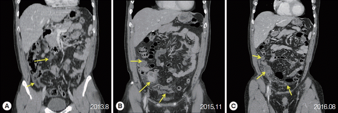
A 45-year-old male was admitted to our hospital for recurrent abdominal pain. The abdominal pain first developed suddenly 3 years before this admission. The patient remembered that it was localized in the right lower quadrant area, and its intensity was about 4 to 5 on a scale of 0 to 10, with 10 indicating the most severe pain. He denied febrile sense or chills. On the next day, computed tomography (CT) of the abdomen and pelvis was performed and revealed fluid dispersion and fat infiltration around the cecum and small bowel mesentery. Although there was no colonic wall thickening, a presumptive diagnosis of acute diverticulitis was made. The abdominal pain was improved 4 days after intravenous antibiotics treatment and fasting. Later, he underwent colonoscopy and esop-hagogastroduodenoscopy, and no abnormal finding was found.
At 27 months after the initial episode (10 months before this admission), the abdominal pain developed again at the same area. The patient also denied any systemic symptoms. The CT of the abdomen and pelvis was repeated and revealed several thick-walled cavitary lesions, which are suggestive of intra-abdominal abscesses with adjacent peritoneal fat infiltration. Recurred diverticulitis and peritoneal abscess was suspected, and surgical resection of the cecum was discussed with the patient. Since he refused the surgery, medical treatment with intravenous antibiotics started and resulted in the improvement of symptoms in 1 week.
At 2 weeks before admission, the patient reported abdominal pain for the third time. The pain started over the whole abdomen, and later it was localized at the right lower quadrant area. The patient complained of continuous pain and rated its intensity as 6 to 7 on a scale of 0 to 10. This time he felt febrile sense and chills. Other gastrointestinal symptoms, such as anorexia, nausea, vomiting, and diarrhea were absent. On examination, localized tenderness and rebound tenderness was found at the right lower quadrant area. A complete blood count showed white-cell count 5,800×103 per mm3 with 56.7% of neutrophils and 3.3% of eosinophils. Total bilirubin was slightly increased as 1.5 mg/dl, and the results of other liver function tests, such as aspartate aminotransferase, alanine aminotransferase, alkaline phosphatase, and glutamyl transferase were within normal range. C-reactive protein was elevated to 4.66 mg/dl. The chest radiography showed no active lung lesions. The repeated CT of the abdomen and pelvis showed that the majority of the previous peritoneal lesions remained although the overall extent of intra-abdominal abscesses and peritoneal fat infiltration was slightly decreased. Colonic wall thickening or enhancement was not definite (Fig. 1).

Serial CT findings of peritoneal paragonimiasis. (A) A coronal CT scan performed on first episode, 2013, showing fluid dispersion and fat infiltration around the cecum and small bowel mesentery (arrows). (B) CT images performed on second episode, 2015, demonstrating several thick-walled cavitary lesions (arrows) suggestive of intra-abdominal abscess with adjacent peritoneal fat infiltration. (C) A CT performed on recent episode showing that the overall extent of intra-abdominal abscesses (arrows) and peritoneal fat infiltration was slightly decreased, but the majority of lesions remained.
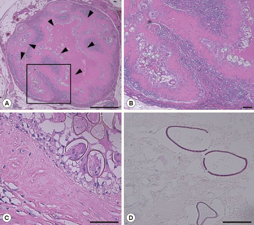
Intravenous antibiotics infusion started with piperacillin and tazobactam. In consideration of his recurrent episodes, the patient underwent laparoscopic right hemicolectomy after 2 weeks of antibiotics treatment. Multiple abscess-like lesions were found adjacent to the proximal colon and also numerous peritoneal nodules were identified. The terminal ileum, cecum, and proximal ascending colon were removed along with adjacent lesions. Multiple biopsies also were performed on peritoneal nodules. When the specimen was examined, colonic wall revealed no abnormality. The appendix showed reactive follicles and acute serositis, and lymph nodes showed reactive hyperplasia. The abscess was shown as necrotizing granulomas with eosinophilic infiltrations and many parasite eggs. Peritoneal nodules were identified as a granuloma, and parasite eggs were identified inside.
When questioned about the consumption of raw freshwater fish or crab, the patient disclosed that he frequently ate the soybean sauce marinated crabs and boiled snails. He remembered that he visited Thailand, the Philippines, China, and Turkey during the previous 5 years. However, he could not recall any consumption of raw food and freshwater exposure abroad except swimming in swimming pools in hotels. Antibodies for Clonorchis sinensis, P. westermani, Schistosoma haematobium, cysteicercus, and Anisakis species were checked with ELISA method. Only antibodies for P. westermani were positive. When the surgical specimen was carefully examined, the size of the eggs was about 60 to70 μm in the longest diameter. The thick egg shell and the operculum suggested that the eggs were those of Paragonimus species (Fig. 2). Collectively, the diagnosis of peritoneal paragonimiasis was made, and the patient was treated with praziquantel 25 mg/kg 3 times a day for 2 days.
DISCUSSION
Here, we presented a case of peritoneal paragonimiasis who suffered from recurrent abdominal pain. Since the thick-walled cavitary lesions were adjacent to the cecum on abdominal CT, recurrent diverticulitis and intra-abdominal abscess were initially considered. Only after the surgery, paragonimiasis was diagnosed based on the pathologic findings and subsequent serology.
When the abdominal pain is localized to the right lower quadrant, usual differential diagnosis includes acute appendicitis, diverticulitis, colitis, and inflammatory bowel diseases. Some of these diseases can cause recurrent episodes. For example, underlying colonic diverticulosis could result in repeated diverticulitis and unresolved vascular problem can cause recurrent ischemic colitis. In our case, the initially identified lesions on CT were peritoneal fluid dispersion with fat infiltration. They developed into thick-walled cavitary lesions containing dirty fluid or debris over 27 months and remained in the similar position. The chronic nature of the lesions and an absence of diverticulosis in colonoscopy suggested some kinds of chronic peritoneal inflammatory diseases, including parasite infections. The definitive diagnosis was made as peritoneal paragonimiasis after a surgery.
Paragonimiasis is a parasitic disease caused by Paragonimus species. Humans are usually infected when they consume inadequately cooked crabs or crayfish which contain the encysted metacercariae of Paragonimus species. In Korea, P. westermani is the most common etiology, and soybean sauce marinated freshwater crabs are well-known as the source of human paragonimiasis. When a metacercaria contained in the crab is swallowed, it excysts in the small intestine, penetrates the intestinal wall to the peritoneal cavity, and moves through the diaphragm and pleura into the lung. The larva makes a cyst-like capsule in the lung and matures inside the cavity. Clinical symptoms of pulmonary paragonimiasis occur when the cyst capsule ruptures into the bronchioles. The parasite eggs are discharged through the airway when the infected patients cough.
Although the lung is the most common site of infection, other organs such as the central nervous system, liver, intestine, peritoneal cavity, retroperitoneum, and abdominal wall are also known to be involved [2–7]. In many cases, mass-like lesions or abscess were found in the involved organs. Malignancy is a concern, so surgical resection is usually performed to treat the abscess and to exclude the malignancy. On surgical removal, worms or eggs of Paragonimus species could be found. When “PubMed” and “KoreaMed” were searched with terms of “ectopic paragonimiasis”, 10 articles of either English or Korean language were found after the year 2010 [6,8–16]. These cases included 5 cases of subcutaneous infection, 5 of intra-abdominal infection, 2 of central nervous system infection, and 1 case of mediastinal cavity infection. All the cases underwent either radiological tissue biopsy or surgery, and the final diagnoses were made from pathological examinations revealing the parasite or eggs.
The definitive diagnosis of ectopic paragonimiasis can be made when worms or eggs of characteristic shape are found on a surgical specimen. When the specimen could not reveal the typical characteristics of Paragonimus worm or eggs, molecular approaches, such as PCR can be helpful [17]. Some cases are diagnosed with exposure history, radiological findings, and immunological test. Commonly, ELISA is used to measure antibodies to Paragonimus species. However, a positive ELISA test does not always imply that active infection is present, since it took 4 to 18 months for the antibody level to decrease to a normal level [18]. Praziquantel 25 mg/kg 3 times for 2–3 days are usually used to treat ectopic paragonimiasis, including cerebral paragonimiasis [19]. Surgery is usually performed when the lesions are relatively superficial and easy to remove or when the dead worm accompanied complications [17].
In addition to Paragonimus species, the metacercariae of Fasciola hepatica migrate to the peritoneal cavity to reach the liver and bile duct, the usual site of infection. Occasionally, the metacercaria is entrapped in the peritoneum, grows to be an adult, and produces abscess [20,21]. Larvae of Anisakis species and the plerocercoid of Spirometra species (=sparganum) can also penetrate the wall of the gastrointestinal tract and could be found in the peritoneal cavity [22,23]. These parasitic infections are known to produce varying degrees of mesenteric infiltrations, peritoneal nodules, and peritoneal thickening in CT of the abdomen [24]. In our case, the underlying problem for the multiple peritoneal abscesses was considered to be diverticulitis, but later found to be the granulomatous inflammation caused by parasite eggs.
The exact prevalence of human paragonimiasis in a defined community is hard to estimate since no single satisfactory diagnostic method exists. Regular national intestinal parasite surveys examine the stools of the systematically sampled population. Since the passage of parasite eggs in the stool only occurs in a limited number of patients, the eggs of Paragonimus species are hardly detected even in national surveys since 1981. Only several seroprevalence data suggests that the prevalence of paragonimiasis is decreasing [25]. Our case clearly shows that the human paragonimiasis is still endemic in Korea, and ectopic paragonimiasis should be included in the differential diagnosis of peritoneal inflammatory lesions.
Notes
CONFLICT OF INTEREST
The authors declare that they have no conflict of interest related to this study.
