Gastrointestinal Parasite Infection in Cats in Daegu, Republic of Korea, and Efficacy of Treatment Using Topical Emodepside/Praziquantel Formulation
Article information
Abstract
The purpose of this study was 2-fold: 1) to investigate the prevalence of gastrointestinal parasite infection in cats reared in Daegu, Republic of Korea and 2) to assess the efficacy and safety of a topical emodepside/praziquantel formulation for cats with parasitic infections. The gastrointestinal parasite infections were examined microscopically using the flotation method. Of 407 cats, 162 (39.8%) were infected by at least one gastrointestinal parasite, including Toxocara cati (63.0%), Toxascaris leonina (31.5%), Taenia taeniaeformis (3.7%), and Cystoisospora felis (1.9%). None of the infected animals had multiple infections. When the data were analyzed according to sex, age, and type of cat, stray cats showed statistically higher prevalence than companion cats (P<0.05). On the 5th day after treatment, no parasitic eggs were detected using microscopic examination. In addition, no adverse effects, such as abnormal behaviors and clinical symptoms, were observed in the cats treated with the drug. These results quantify the prevalence of gastrointestinal parasites in cats in Daegu, Republic of Korea, and show that topical emodepside/praziquantel is a safe and effective choice for treating the parasitic infections in cats.
INTRODUCTION
Cats are susceptible to various gastrointestinal parasites, including Nematoda (Toxocara spp., Aelurostrongylus abstrusus, Strongyloides spp., and Ancylostoma spp.), Trematoda (Clonorchis sinensis and Paragonimus spp.), Cestoda (Taenia taeniaeformis, Spirometra mansoni, Diphyllobothrium spp., and Echinococcus multilocularis), Coccidia (Cystoisospora spp., Toxoplasma gondii, Cryptosporidium spp., and Sarcocystis spp.), and Trepomonadea (Giardia duodenalis). Some of these can infect humans as well as animals [1–5]. Of these parasites, infections by helminths are the most common in cats and dogs, despite the availability of numerous effective anthelmintic products [6]. However, less data exist for parasitism on cats than for dogs [7]. In the Republic of Korea, several studies of gastrointestinal parasites in cats are available, however, the studies were done in restricted regions, several decades ago [2,8,9]. Up-to-date information on gastrointestinal parasite infection in cats is scarce.
Concerning administration routes, topical treatment with anthelmintics is preferred because it is more convenient and less stressful than oral administration for both cats and their owners [10]. Emodepside 2.1% (w/v) plus praziquantel 8.6% (w/v) (Profender® spot-on, Bayer, Kiel, Germany) is a topically applied anthelmintic drug with broad-spectrum activity. The product is intended for treatment of gastrointestinal parasites in cats, including nematodes (Toxocara cati, Toxascaris leonina, and Ancylostoma tubaeforme) and cestodes (Dipylidium caninum, T. taeniaeformis, and E. multilocularis). Previous studies showed that the product has high efficacy (>98.5%) against susceptible gastrointestinal parasites [4,6]. Emodepside belongs to a new class of anthelmintic compounds called cyclooctadepsipeptides. It is a derivative of PF1022A, and shows various anthelmintic activities in animals [11]. Praziquantel is a pyrazinoisoquinoline derivative that has shown effectiveness against cestodes in large and small animals [10,12].
The purpose of the present study was to investigate the prevalence of gastrointestinal parasites in cats from Daegu, Republic of Korea, based on microscopic examination. In addition, this study evaluated the efficacy and safety of topical emodepside/praziquantel in naturally parasite-infected cats.
MATERIALS AND METHODS
Sample collection
Before the study was conducted, written consent was obtained from the owners. Daegu, Republic of Korea, is located at longitude 128°36′ East and latitude 35°52′ North, with a mean annual temperature of 14.1°C and a mean annual precipitation of 1,064.4 mm [13]. A total of 407 cat fecal samples were collected from a local veterinary clinic and animal shelter in Daegu, Republic of Korea, from June 2012 to August 2015. The average weight of the cats was 2.2 kg, with a standard deviation of 1.5 kg. The cats selected were classified according to sex (male or female), age (young, <1-year-old; adult, ≥1-year-old), and type of cat (companion or stray cat). The fecal samples were collected from the cages where the cats were kept. The samples were fresh, not dried. After collection, the fecal samples were transported to the College of Veterinary Medicine, Kyungpook National University, Daegu, Republic of Korea, and were stored at 4°C until examined microscopically.
Identification of gastrointestinal parasite eggs
Identification of gastrointestinal parasite eggs from cat fecal samples was based on morphology, using a flotation method with a saturated sodium chloride solution [3]. The severity of infection was judged by the number of parasite eggs, using a modified McMaster method [3]. Eggs per gram of feces (EPG) were calculated and then categorized into three groups: mild, EPG<1,000; moderate, 1,000≤EPG<10,000; severe, EPG≥10,000.
Treatment of anthelmintics
After the fecal examination, cats that were positive for gastrointestinal parasites (n=162) received a single topical application of emodepside/praziquantel (Profender® spot-on), according to the manufacturers’ instructions. Cystoisospora felis-infected cats were excluded, because the drug is not considered effective for this species. The treatment was applied to the skin of the cat’s neck at the base of the skull while hairs were put aside without shaving.
Evaluation of efficacy and safety
The efficacy of the emodepside/praziquantel treatment was determined through microscopic examination of gastrointestinal parasite eggs from cat fecal samples, as previously described, five days after treatment. Safety was determined by identification of any behavior abnormalities or clinical symptoms by the veterinarians or owners.
Statistical analysis
To evaluate statistically significant differences in prevalence as a function of sex, age, or companion/stray status, the Chi-square test was applied using SPSS V.21.0 (IBM Corporation, Armonk, New York, USA). A P-value less than 0.05 was regarded as statistically significant. Also, 95% confidence intervals were calculated.
RESULTS
Prevalence of gastrointestinal parasites
Of the 407 cat fecal samples tested, 162 (39.8%) were infected by at least one of the gastrointestinal parasites (Table 1). Concerning sex, 76 of 195 male cats (39.0%) and 86 of 212 female cats (40.6%) showed positive fecal samples; the difference in prevalence was not statistically significant (P=0.743). Concerning age, 79 of 218 young cats (36.2%) and 83 of 189 old cats (43.9%) showed positive samples; the difference was not statistically significant (P=0.115). Concerning the type of cat, stray cats showed a higher prevalence (97 of 183, 39.8%) than cats with owners (65 of 224, 29.0%); this difference was statistically significant (P<0.001).

Prevalence of gastrointestinal parasite infection in cats in Daegu, Republic of Korea, according to sex, age, and type of cat (companion/stray)
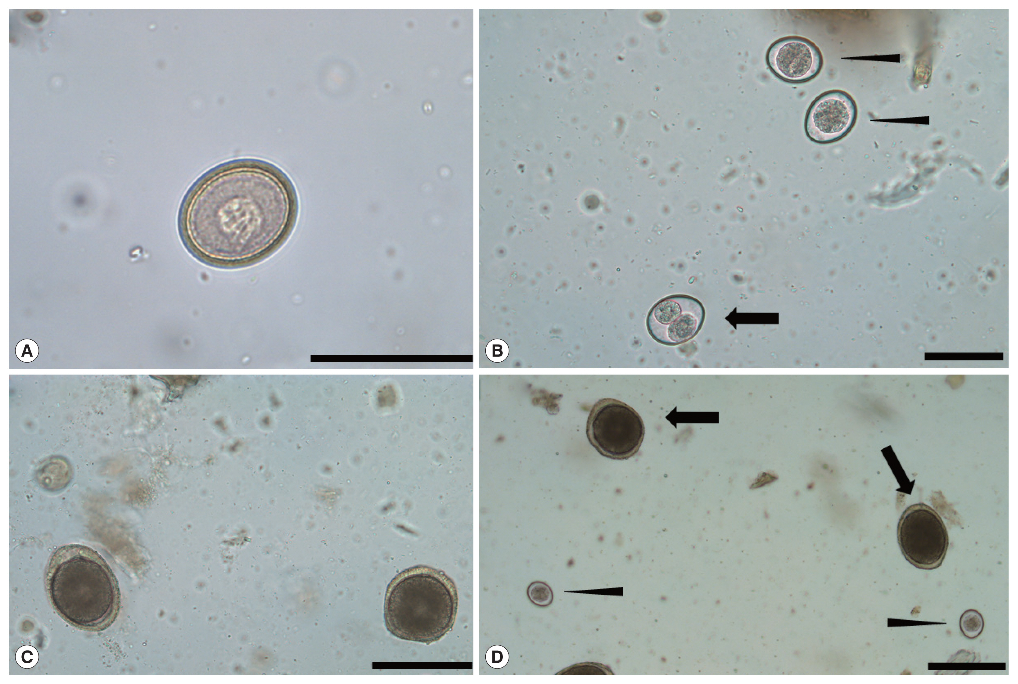
Based on morphology, T. cati, T. leonina, T. taeniaeformis, and C. felis were identified in 102 (63.0%), 51 (31.5%), 6 (3.7%), and 3 (1.9%) cat fecal samples, respectively (Fig. 1; Tables 2, 3). When the data were analyzed according to the parasite species, there were no statistically significant differences as a function of sex or age (P>0.05); however, T. cati and T. leonina both showed significantly higher prevalence in stray cats than in companion cats, with P-values of 0.003 and 0.001, respectively. Most of the cats were mildly infected (140/162; 86.4%); no severe infections were observed (Table 3). No concurrent infections with two or more parasites were recorded.

Eggsand cysts of gastrointestinal parasites in the cats. (A) Taenia taeniaeformis egg. (B) Cystoisospora felis sporulated (black arrow) and unsporulated (black arrow heads) oocysts. (C) Toxocara cati eggs and (D) mixed infection of C. felis (black arrow heads) and T. cati (black arrows). Scale bars=50 μm (A and B) and 100 μm (C and D).

Prevalence of gastrointestinal parasite infection in cats in Daegu, Republic of Korea, for the four observed parasite species
Efficacy and safety of the treatment
At the day 5 after treatment, all the fecal samples were reexamined but no parasite eggs were detected under microscopy. In addition, there were no reports of adverse drug effects, including abnormal behavior or clinical symptoms, from the veterinarians or owners, following administration of the topical treatment.
DISCUSSION
Feline gastrointestinal parasites have been reported worldwide, but with greatly different prevalence, for example, 39.6% (71/179) in the USA [14], 58.3% (271/465) in Argentina [15], 34.3% (142/414) in Romania [1], 22.8% (1,952/8,560) in Germany [16], 88.5% (46/52) in Iran [17], 35.1% (533/1,519) in Europe [7], and 86.3% (44/51) in Iran [5]. The variability in prevalence seen in these studies might be attributed to differences in sampling region, season, habitat, and the number of samples. In the Republic of Korea, a few decades ago, there were several studies regarding gastrointestinal parasites in cats. In most cases, the studies were restricted to specific regions, and the prevalence varied accordingly, from 75.6% to 85.6% [2,8,9].
The overall prevalence of gastrointestinal parasite infections in cats in this study was 39.8% (162/407). The results differed according to age, with adults showing higher prevalence than young cats, but this was not statistically significant (P=0.115). There have been debates regarding the relationship between gastrointestinal infection and age [5,7,16,17], however, each study was based on a different age classification, study region, and sample size; therefore, better-controlled studies are required. Regardless of the debate, the prevalence of gastrointestinal parasites in both young and adult cats indicates a susceptibility to infection in all life stages [5]. There were no significant differences between male and female cats in prevalence. Many previous studies have reported similar observations for different regions [1,5,17,18]. Stray cats showed significantly higher prevalence than companion cats, for infections in general as well as for the specific parasite species T. cati and T. leonina. Previous studies targeted at stray cats showed a very high prevalence (>85%) of gastrointestinal parasites [2,5,17–19], compared to house cats (<35%) [1,7,16]. The prevalence in stray cats in the present study are not as high as in the previous studies; however, it was significantly higher than in the companion cats. Because the stray cats roamed freely before they were taken to the animal shelter, they had many more chances to be exposed to parasites. It is worth noting that the companion cats, although generally reared inside houses, also showed a high enough prevalence to require regular anthelmintic treatment.
The following parasites were detected in this study: T. cati (63.0%), T. leonina (31.5%), T. taeniaformis (3.7%), and C. felis (1.9%). The study found that T. cati was the most prevalent parasite, which is consistent with reports of 53.3% in England [20], 55.2% in Spain [19], 61.2% in Argentina [15], 20.3% in Romania [1], and 28.8% in Iran [17]. In addition, previous studies in different regions of the Republic of Korea also showed T. cati forming the largest fraction of gastrointestinal parasite cases [2,8,9]. Two factors may explain the high prevalence of T. cati in cats. First, cats often roam freely and discharge eggs of the parasite in the environment, which then contaminate soils [21]. Second, the parasite can be transmitted by breastfeeding, so T. cati-infected maternal cats can pass the infection to their non-infected kittens [22]. In contrast to our results showing T. leonina as the second most prevalent helminth in cats, at 30.6%, previous studies reported variable prevalences according to countries (0.1–12.9%) [16,18,23]. To evaluate accurate prevalence and distribution of T. leonina, more samples from different regions and countries are needed because there were not many studies on T. leonina in cats.
In terms of the efficacy and safety of the topical emodepside/praziquantel formulation (Profender® spot-on), the results of this study showed that the drug is safe and effective against gastrointestinal parasites of cats. Five days after applying the treatment, all the gastrointestinal parasites (n=159) yielded negative results in fecal examination, which means that the treatment had high efficacy. In a study conducted using the same treatment, 100% efficacy was recorded against mature and immature A. tubaeforme in domestic cats [24]. According to Reinemeyer et al. [25], this emodepside/praziquantel topical solution is effective against all stages of T. cati, yielding 100% efficacy against both mature and immature adults. Compared to oral adminstration of anthelmintics, topical application is more convenient, especially for cats [26].
In conclusion, the results in this study showed that gastrointestinal parasites in cats in Daegu, Republic of Korea, are prevalent and T. cati forming the largest portion of cases. The high prevalence of gastrointestinal parasites in cats suggests a possibility that cats are easily exposed not only to gastrointestinal parasites but also other pathogens, emphasizing the need for regular medical check-ups. In addition, treatment of gastrointestinal parasites in cats using emodepside/praziquantel formulation showed high safety and efficacy.
Notes
CONFLICT OF INTEREST
The authors report no conflict of interest related to this study.
