Concurrent Capillaria and Heterakis Infections in Zoo Rock Partridges, Alectoris graeca
Article information
Abstract
Two adult rock partridges raised in a city zoo were examined parasitologically and pathologically. Two distinctive eggs resembling those of Capillaria and Heterakis were detected in the feces. At necropsy, a markedly-dilated duodenum with severe catarrhal exudates, containing adult worms of Capillaria sp. and Heterakis sp. in the cecum, was observed. Male Capillaria had the cloacal aperture extended almost terminally with a small bursal lobe and an unsheathed spicule with transverse folds without spines. Female Capillaria had a vulva that was slightly prominent and slightly posterior to the union of the esophagus and intestine. The esophagus of the adult Capillaria was more than a half as long as the body in the male, but was much shorter in the female. Based on these morphological features, the capillarid nematode was identified as Capillaria obsignata. The male adult worms of Heterakis was identifiable by 2 dissimilar spicules, a unique morphological feature where the right spicule was considerably longer than the left, which is also a characteristic feature of Heterakis gallinarum. This is the first report of concurrent infections with C. obsignata and H. gallinarium in rock partridges.
INTRODUCTION
Capillarid nematodes of birds, also known as hairworms because of their extreme thinness in size, are divided into 2 groups: those that burrow into the epithelium of the upper digestive tract (the esophagus and the crop) and those that burrow into the epithelium of the lower digestive tract (the small intestine and rarely the ceca) [1,2]. Species of the genus Capillaria commonly found in the crop and in the esophagus are C. annulata (Molin, 1958) and C. contorta (Creplin, 1839). C. caudinflata (Molin, 1858), C. bursata (Freitas and Almeida, 1934) and C. obsignata (Madsen, 1945) parasitizes the small intestine, whereas C. anatis (Schrank, 1790) (syn. A. brevicollis, C. retusa, C. collaris, C. anseris, and C. mergi) is found in the ceca [3]. All 6 species have been reported to occur in domesticated and wild birds, and are cosmopolitan in their distribution. However, there has been no report on avian Capillaria infection in Korea.
Adult worms of the genus Heterakis live principally in the lumen of the ceca of birds. Three species are known to be prominent in poultry: H. gallinarum, H. dispar, and H. isolonche. Birds affected by Heterakis sp. include chickens, turkeys, guinea fowls, quails, ducks, pheasants, and geese [1,2]. The parasite usually has a direct cycle but may spread to birds that ingest earthworms carrying the ova of the parasite [1,2,4].
The veterinary literature does not contain detailed parasitological and histological descriptions of Capillaria and Heterakis infections in rock partridges [2,5-8]. Here, we describe parasitological and histological characteristics of concurrent Capillaria and Heterakis infections in rock partridges.
CASE DESCRIPTION
Two 2-year-old rock partridges (Alectoris graeca, 1 male and 1 female) were submitted to the College of Veterinary Medicine, Chonnam National University, for necropsy by a local zoo veterinarian in Gwangju Metropolitan City. The birds were found dead due to severe head injuries that presumably had resulted from either mating rituals or mating fights.
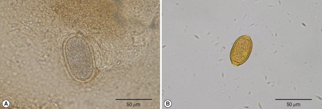
At necropsy, the duodenums of both birds were markedly dilated with severe catarrhal exudates in the lumen. The ceca contained fibrinopurulent exudates and numerous nematode parasites in the lumen. The subcutaneous tissues of the head of both birds had multiple hemorrhages with edema due to picking during mating. Using the modified Sheather's sugar flotation technique, 2 distinctive eggs were detected in the feces of both birds (Fig. 1). The heterakid eggs measured 66.7-75.2 by 35.3-40.8 µm (average 70.7 × 37.9 µm), were thick, smooth shelled, ellipsoid in shape, and difficult to differentiate from Ascaridia galli eggs (Fig. 1A). The capillarid eggs measured 46.1-53.9 by 28.3-33.1 µm (average 49.6 × 30.5 µm), were barrel-shaped with clear plugs on each pole, and had a reticulate pattern on the shell surface (Fig. 1B).

(A) An egg of Heterakis gallinarum is thick, smooth shelled, ellipsoidal, and difficult to differentiate from eggs of Ascaridia galli. (B) The barrel-shaped egg of Capillaria obsignata with clear plugs on each pole.
Hair-like worms of Capillaria spp. were found by careful examination of catarrhal exudates of the duodenums of both birds under a dissecting microscope (Fig. 2). Male worms were 7.8-10.6 mm (average 8.93 mm) long and 36.9-53.9 µm (average 47.4 µm) wide. The cloacal aperture extended almost terminally of the body and had a small bursal lobe on either side, where the 2 lobes were connected dorsally by a delicate bursal membrane. The spicule measured 1.4-1.5 µm (average 1.4 µm) long and had a sheath with transverse folds without spines (Fig. 2A). Females were 10.1-12.6 mm (average 11.4 mm) long and 73.1-85.4 µm (average 80.6 µm) wide. The esophagus was more than a half as long as the length of the body in the male (range 4.2-5.2, average 4.6 µm), which was found to be slightly shorter in the female (5.0-5.4, average 5.2 µm) (Fig. 2B). Characteristically, the vulva was slightly prominent and slightly posterior to the union of the esophagus and the intestine (Fig. 2C).

(A) Male Capillaria obsignata having no spines on the spicule, and the esophagus more than half as long as the body. (B) Female C. obsignata having shorter esophagus as long as the body. (C) Female C. obsignata characterized by the vulva which is slightly prominent (black arrow), and posterior to the union of the esophagus and the intestine (white arrow).
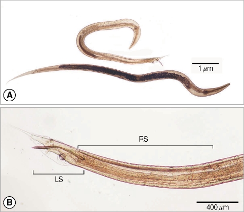
Adult Heterakis worms found in the ceca of both birds were small and white (Fig. 3). The males were 4.6-7.4 mm (average 5.8 mm) long and 0.26-0.31 mm (average 0.29 mm) wide in size, and the females were 8.6-9.1 mm (average 8.8 mm) long and 0.28-0.32 mm (average 0.31 mm) wide. Male worms had 2 characteristic dissimilar spicules in length, the right one being 0.9-2.8 (generally 2.0) mm long and the left one being 0.4-1.1 mm long with a curved tip (Fig. 3). The mouth was surrounded by 3 equal-sized lips. Narrow lateral alae extended almost to the end of the body on both sides, and the esophageus ended in a well-developed bulb containing a valvular apparatus. The tail was straight, ending in a subulate point in the male and being long, narrow and pointed in the female. The vulva of the female was not prominent and was positioned slightly posterior to the middle of the body. Two large lateral bursal wings were present in the male. The preanal sucker of the male was well-developed with strongly chitinized walls. A small semicircular incision was recognized in the posterior margin of the sucker wall. There were 12 pairs of caudal papillae on the abdominal surface of the male, the 2 most posterior pairs being stout and superimposed.

(A) Adult male and female Heterakis gallinarum isolated from a rock partridge. The bottom female exhibits the esophagus ending in a well-developed bulb containing a valvular apparatus. (B) The adult male H. gallinarum shows unique spicules, the right one being very long (RS) and the left one being short (LS).
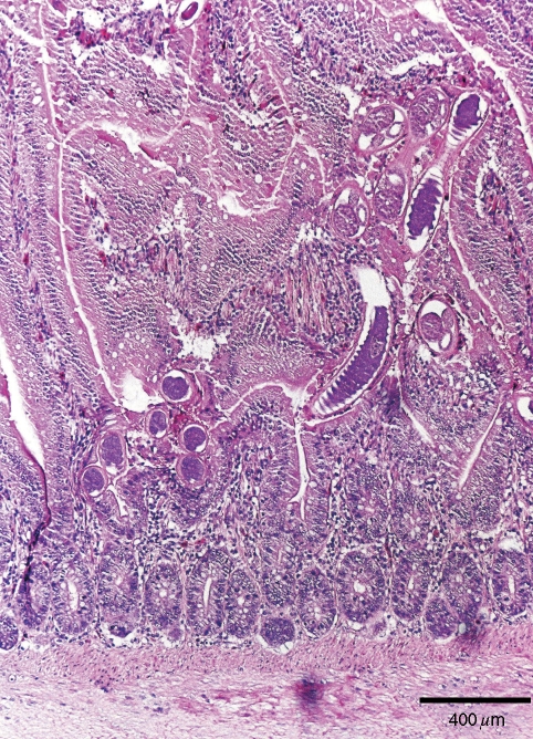
Histopathologically, most lesions caused by capillarid parasites were observed in the duodenum in both birds where most adult Capillaria were found (Fig. 4). The thin anterior ends of many adult Capillaria worms invaded into the mucosa between the villi and burrowing into the lamina propria, while the posterior ends of many of the parasites extended into the lumen of the intestine (Fig. 4). Lamina propria showed usually moderate-to-severe infiltration of lymphoid cells with some heterophils. The number of goblet cells was increased in the villi of the small intestine. Occasional distension of the glands of Lieberkühn with mucus and/or heterophils was also observed.

Adult worms of C. obsignata penetrating the lamina propria of the duodenum. Hematoxylin and eosin stain. Bar = 80 µm.
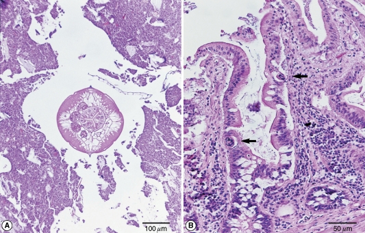
Adult and larval Heterakis worms were found only in the cecum of the 2 birds. Adult worms remained free in the cecal lumen (Fig. 5A), while larvae were present in the lamina propria, glandular epithelium, or in the crypt lumen (Fig. 5B). The crypt lumen was dilated and contained mucus and/or heterophils. Mononuclear cells with some heterophils infiltrated into the lamina propria. The ulcer was covered with eosinophilic necrotic debris in which heterophils were mainly observed. Goblet cells were increased in number.
DISCUSSION
Infections with Capillaria spp. can be highly pathogenic for birds in deep-litter systems or in free-range systems where big numbers of infective eggs may build up in the litter or in the soil [3]. C. obsignata infections are highly pathogenic in pigeons and may cause high mortality rates [3]. Capillarid parasites can be identified by the characteristic morphology of adult male and female worms. The morphological characteristics of male and female worms observed in the rock partridges were identical to those of C. obsignata documented previously [3,8]. For instance, spines were not found on the surface of the sheath of the male spicule with the cloacal aperture almost terminal; presence of a small bursal lobe on either side in the male worm; and the adult female having vulva slightly prominent and slightly posterior to the union of the esophagus and intestine [1,2]. The length and the width of the eggs from female worms; however, were slightly bigger in size than those of C. obsignata documented previously. The egg size was not in accordance with either of C. anatis, C. bursata, or C. caudinflata. This may indicate that the Capillaria sp. observed in the rock partridges may have to be classified to a new species. However, key features of the adult worm led authors to identify the worm as C. obsignata. Most species of Capillaria from birds have been described under a variety of names, leading to confusing reports in the literature [1].
Although the morphology of adult H. gallinarum, H. dispar, and H. isolonche is very similar, they can be easily differentiated based on the shape of the spicules in the male [3]. The morphological characteristic of spicules of male Heterakis from rock partridges, with the right one being extensively longer than the left one, agreed with those of H. gallinarum. Other morphological features of male and female heterakid worms and eggs also coincided with those of H. gallinarum. This is the first report of a concurrent Capillaria and Heterakis infections in rock partridges.
Although the ceca of infected birds with H. gallinarum may show marked inflammation and thickening of the mucosa with petechial hemorrhages, clinical signs may not be seen except in H. isolonche infections in which nodular thyplitis, diarrhea, emaciation, and death may be seen [3]. The most important role of H. gallinarum is its capability of transferring the protozoon Histomonas meleagridis to fowls [9]. A previous report indicated that an outbreak of histomoniasis due to H. meleagridis developed in a chuchar partridge farm in Korea [10]. However, pathologic changes of the liver and the cecum due to H. meleagridis were not observed in the rock partridges reported in this paper.
ACKNOWLEDGMENTS
The author thanks Drs. Mun-Il Kang and Kyoung-Oh Cho for helpful discussions.
