Warning: mkdir(): Permission denied in /home/virtual/lib/view_data.php on line 81

Warning: fopen(upload/ip_log/ip_log_2024-07.txt): failed to open stream: No such file or directory in /home/virtual/lib/view_data.php on line 83

Warning: fwrite() expects parameter 1 to be resource, boolean given in /home/virtual/lib/view_data.php on line 84
Han, Goo, Park, Hwang, Kim, Yang, Ahn, and Ryu: Proinflammatory Cytokine and Nitric Oxide Production by Human Macrophages Stimulated with Trichomonas vaginalis
Cited By
Citations to this article as recorded by
Leukotriene B4 receptors BLT1 and BLT2 are involved in interleukin-8 production in human neutrophils induced by Trichomonas vaginalis-derived secretory products
Young Hee Nam, Arim Min, Seong Hoon Kim, Young Ah Lee, Kyeong Ah Kim, Kyoung-Ju Song, Myeong Heon Shin
Inflammation Research.2012; 61(2): 97.     CrossRef
Trichomonas vaginalis triggers the release of THP-1 extracellular traps
Li Fei, Wei Zhengkai, Jiang Weina, Cao Lili, Gao Yuhang, Yang Zhengtao, Li Jianhua, Yu Biao, Zhang Xichen, Gong Pengtao
Parasitology Research.2019; 118(1): 267.     CrossRef
Involvement of purinergic signaling on nitric oxide production by neutrophils stimulated with Trichomonas vaginalis
Amanda Piccoli Frasson, Geraldo Attilio De Carli, Carla Denise Bonan, Tiana Tasca
Purinergic Signalling.2012; 8(1): 1.     CrossRef
Adenosine reduces reactive oxygen species and interleukin-8 production by Trichomonas vaginalis-stimulated neutrophils
Amanda Piccoli Frasson, Camila Braz Menezes, Gustavo Krumel Goelzer, Simone Cristina Baggio Gnoatto, Solange Cristina Garcia, Tiana Tasca
Purinergic Signalling.2017; 13(4): 569.     CrossRef
Impact of microparticles released during murine systemic inflammation on macrophage activity and reactive nitrogen species regulation
Weronika Ortmann, Anna Such, Elzbieta Kolaczkowska
Immunologic Research.2024; 72(2): 299.     CrossRef
Trichomoniasis immunity and the involvement of the purinergic signaling
Camila Braz Menezes, Tiana Tasca
Biomedical Journal.2016; 39(4): 234.     CrossRef
Local cytokine/chemokine profiles in BALB/c and C57BL/6 mice in response to T. vaginalis infection
Sonal Yadav, Vivek Verma, Rakesh Singh Dhanda, Sumeeta Khurana, Manisha Yadav
Experimental Parasitology.2022; 239: 108287.     CrossRef
Leukotriene B4 receptor BLT-mediated phosphorylation of NF-κB and CREB is involved in IL-8 production in human mast cells induced by Trichomonas vaginalis-derived secretory products
Young Hee Nam, Deulle Min, Hyoung-Pyo Kim, Kyoung-Ju Song, Kyeong Ah Kim, Young Ah Lee, Seong Hoon Kim, Myeong Heon Shin
Microbes and Infection.2011; 13(14-15): 1211.     CrossRef
BLT1-mediated O-GlcNAcylation is required for NOX2-dependent migration, exocytotic degranulation and IL-8 release of human mast cell induced by Trichomonas vaginalis-secreted LTB4
Arim Min, Young Ah Lee, Kyeong Ah Kim, Myeong Heon Shin
Microbes and Infection.2018; 20(6): 376.     CrossRef
Effect of iNOS inhibitor LNMMA along with antibiotics Chloramphenicol or Ofloxacin in murine peritoneal macrophages regulates S.aureus infection as well as inflammation: An in vitro study
Somrita Dey, Biswadev Bishayi
Microbial Pathogenesis.2017; 105: 307.     CrossRef
OsmC and incomplete glycine decarboxylase complex mediate reductive detoxification of peroxides in hydrogenosomes of Trichomonas vaginalis
Eva Nývltová, Tamara Smutná, Jan Tachezy, Ivan Hrdý
Molecular and Biochemical Parasitology.2016; 206(1-2): 29.     CrossRef
ReducedTrichomonas vaginalisviability in mice pretreated with parasite DNA
Marco Antonio Barajas-Mendiola, Laura E. Castellano, Miriam Barrios-Rodiles, Martha A. Deveze-Alvarez, Eva E. Avila, Patricia Cuéllar-Mata
Parasitology.2019; 146(13): 1636.     CrossRef
Immune cell arrival kinetics to peritoneum and role during murine-experimental trichomoniasis
F. J. Rangel-Mata, E. E. Ávila-Muro, J. E. Reyes-Martínez, L. M. Olmos-Ortiz, M. E. Brunck, L. A. Arriaga-Pizano, P. Cuéllar-Mata
Parasitology.2021; 148(13): 1624.     CrossRef
In Vitro Activation of Macrophages by an MHC Class II-restricted Trichomonas Vaginalis TvZIP8-derived Synthetic Peptide
Victor Ermilo Arana-Argáez, Emanuel Ceballos-Góngora, María Elizbeth Alvarez-Sánchez, Antonio Euan-Canto, Julio Lara-Riegos, Julio César Torres-Romero
Immunological Investigations.2022; 51(1): 88.     CrossRef
IL‐22 Levels are Associated with Trichomonas vaginalis Infection in the Lower Genital Tract
Hadijat Moradeke Makinde, Reza Zariffard, Paria Mirmonsef, Richard M Novak, Olamide Jarrett, Alan L Landay, Gregory T Spear
American Journal of Reproductive Immunology.2013; 70(1): 38.     CrossRef
The anti‐Trichomonas vaginalis phloroglucinol derivative isoaustrobrasilol B modulates extracellular nucleotide hydrolysis
Camila Braz Menezes, Graziela Vargas Rigo, Henrique Bridi, Danielle da Silva Trentin, Alexandre José Macedo, Gilsane Lino von Poser, Tiana Tasca
Chemical Biology & Drug Design.2017; 90(5): 811.     CrossRef
Involvement of mast cells in inflammation induced by Trichomonas vaginalis via crosstalk with vaginal epithelial cells
I. H. HAN, S. J. PARK, M. H. AHN, J. S. RYU
Parasite Immunology.2012; 34(1): 8.     CrossRef
Immunity in urogenital protozoa
N. Malla, K. Goyal, R. S. Dhanda, M. Yadav
Parasite Immunology.2014; 36(9): 400.     CrossRef
Trichomonas vaginalis exosome‐like vesicles modify the cytokine profile and reduce inflammation in parasite‐infected mice
L. M. Olmos‐Ortiz, M. A. Barajas‐Mendiola, M. Barrios‐Rodiles, L. E. Castellano, S. Arias‐Negrete, E. E. Avila, P. Cuéllar‐Mata
Parasite Immunology.2017;[Epub]     CrossRef
Humoral and T cell–mediated immune response against trichomoniasis
M. Nemati, N. Malla, M. Yadav, H. Khorramdelazad, A. Jafarzadeh
Parasite Immunology.2018;[Epub]     CrossRef
Inflammatory responses during trichomoniasis: The role of Toll‐like receptors and inflammasomes
Abdollah Jafarzadeh, Maryam Nemati, Ehsan Salarkia, Sonal Yadav, Najmeh Aminizadeh, Sara Jafarzadeh, Manisha Yadav
Parasite Immunology.2023;[Epub]     CrossRef
Activation of murine macrophages by membrane proteins from Tritrichomonas foetus grown on iron‐ and calcium‐rich conditions
Antonio Euan‐Canto, Julio César Torres‐Romero, María Elizbeth Alvarez‐Sánchez, Victor Ermilo Arana‐Argáez, Karla Acosta‐Viana, Emanuel Ceballos‐Góngora, Laura Vázquez‐Carrillo, Leidi Alvarez‐Sánchez
Parasite Immunology.2024;[Epub]     CrossRef
Heterogeneous macrophages: Supersensors of exogenous inducing factors
Caiyun Qian, Zehui Yun, Yudi Yao, Minghua Cao, Qiang Liu, Song Hu, Shuhua Zhang, Daya Luo
Scandinavian Journal of Immunology.2019;[Epub]     CrossRef
SNAP23-Dependent Surface Translocation of Leukotriene B 4 (LTB 4 ) Receptor 1 Is Essential for NOX2-Mediated Exocytotic Degranulation in Human Mast Cells Induced by Trichomonas vaginalis-Secrete
Arim Min, Young Ah Lee, Kyeong Ah Kim, Jamel El-Benna, Myeong Heon Shin, Judith A. Appleton
Infection and Immunity.2017;[Epub]     CrossRef
Differential Regulation of Proinflammatory Cytokine Expression by Mitogen-Activated Protein Kinases in Macrophages in Response to Intestinal Parasite Infection
Mei Xing Lim, Chin Wen Png, Crispina Yan Bing Tay, Joshua Ding Wei Teo, Huipeng Jiao, Norbert Lehming, Kevin Shyong Wei Tan, Yongliang Zhang, J. A. Appleton
Infection and Immunity.2014; 82(11): 4789.     CrossRef
Analysis of the Oxidative Stress Status in Nonspecific Vaginitis and Its Role in Vaginal Epithelial Cells Apoptosis
Zhaojie Chen, Zhen Zhang, Haiyan Zhang, Beibei Xie
BioMed Research International.2015; 2015: 1.     CrossRef
Trichomonas vaginalis Induces NLRP3 Inflammasome Activation and Pyroptotic Cell Death in Human Macrophages
Angelica Montenegro Riestra, J. Andrés Valderrama, Kathryn A. Patras, Sharon D. Booth, Xing Yen Quek, Chih-Ming Tsai, Victor Nizet
Journal of Innate Immunity.2019; 11(1): 86.     CrossRef
T. vaginalis Infection Is Associated with Increased IL-8 and TNFr1 Levels but with the Absence of CD38 and HLADR Activation in the Cervix of ESN
Olamide D. Jarrett, Kirsten E. Brady, Sharada P. Modur, Jill Plants, Alan L. Landay, Mahmood Ghassemi, Elizabeth T. Golub, Greg T. Spear, Richard M. Novak, Clive M. Gray
PLOS ONE.2015; 10(6): e0130146.     CrossRef
Prostatic Disease Associated withTrichomonas vaginalis
Jae-Sook Ryu
The Korean Journal of Urogenital Tract Infection and Inflammation.2014; 9(2): 61.     CrossRef
IL-10 release by bovine epithelial cells cultured with Trichomonas vaginalis and Tritrichomonas foetus
Ricardo Chaves Vilela, Marlene Benchimol
Memórias do Instituto Oswaldo Cruz.2013; 108(1): 110.     CrossRef
NF-κB and CREB Are Involved in IL-8 Production of Human Neutrophils Induced byTrichomonas vaginalis-Derived Secretory Products
Young Hee Nam, Deulle Min, Soon-Jung Park, Kyeong Ah Kim, Young Ah Lee, Myeong Heon Shin
The Korean Journal of Parasitology.2011; 49(3): 291.     CrossRef
Involvement of PI3K/AKT and MAPK Pathways for TNF-α Production in SiHa Cervical Mucosal Epithelial Cells Infected with <i>Trichomonas vaginalis</i>
Jung-Bo Yang, Juan-Hua Quan, Ye-Eun Kim, Yun-Ee Rhee, Byung-Hyun Kang, In-Wook Choi, Guang-Ho Cha, Jae-Min Yuk, Young-Ha Lee
The Korean Journal of Parasitology.2015; 53(4): 371.     CrossRef
Trichomonas vaginalis α-Actinin 2 Modulates Host Immune Responses by Inducing Tolerogenic Dendritic Cells via IL-10 Production from Regulatory T Cells
Hye-Yeon Lee, Juri Kim, Jae-Sook Ryu, Soon-Jung Park
The Korean Journal of Parasitology.2017; 55(4): 375.     CrossRef
Immune Response of BALB/c Mice toward Putative Calcium Transporter Recombinant Protein of Trichomonas vaginalis
Tahali Mendoza-Oliveros, Victor Arana-Argáez, Leidi C. Alvaréz-Sánchez, Julio Lara-Riegos, María Elizbeth Alvaréz-Sánchez, Julio C. Torres-Romero
The Korean Journal of Parasitology.2019; 57(1): 33.     CrossRef
Immunopathology of Recurrent Vulvovaginal Infections: New Aspects and Research Directions
Namarta Kalia, Jatinder Singh, Manpreet Kaur
Frontiers in Immunology.2019;[Epub]     CrossRef
Symbiotic Association with Mycoplasma hominis Can Influence Growth Rate, ATP Production, Cytolysis and Inflammatory Response of Trichomonas vaginalis
Valentina Margarita, Paola Rappelli, Daniele Dessì, Gianfranco Pintus, Robert P. Hirt, Pier L. Fiori
Frontiers in Microbiology.2016;[Epub]     CrossRef
Trichomonas vaginalis Induces Production of Proinflammatory Cytokines in Mouse Macrophages Through Activation of MAPK and NF-κB Pathways Partially Mediated by TLR2
Ling Li, Xin Li, Pengtao Gong, Xichen Zhang, Zhengtao Yang, Ju Yang, Jianhua Li
Frontiers in Microbiology.2018;[Epub]     CrossRef
Modulation of dendritic cell function by Trichomonas vaginalis-derived secretory products
Min-Ji Song, Jong-Joo Lee, Young Hee Nam, Tae-Gyun Kim, Youn Wook Chung, Mikyoung Kim, Ye-Eun Choi, Myeong Heon Shin, Hyoung-Pyo Kim
BMB Reports.2015; 48(2): 103.     CrossRef
Trichomoniasis
Jae-Sook Ryu
Hanyang Medical Reviews.2010; 30(3): 213.     CrossRef

Abstract

Trichomonas vaginalis commonly causes vaginitis and perhaps cervicitis in women and urethritis in men and women. Macrophages are important immune cells in response to T. vaginalis infection. In this study, we investigated whether human macrophages could be involved in inflammation induced by T. vaginalis. Human monocyte-derived macrophages (HMDM) were co-cultured with T. vaginalis. Live, opsonized-live trichomonads, and T. vaginalis lysates increased proinflammatory cytokines, such as TNF-α, IL-1β, and IL-6 by HMDM. The involvement of nuclear factor (NF)-κB signaling pathway in cytokine production induced by T. vaginalis was confirmed by phosphorylation and nuclear translocation of p65 NF-κB. In addition, stimulation with live T. vaginalis induced marked augmentation of nitric oxide (NO) production and expression of inducible NO synthase (iNOS) levels in HMDM. However, trichomonad-induced NF-κB activation and TNF-α production in macrophages were significantly inhibited by inhibition of iNOS levels with L-NMMA (NO synthase inhibitor). Moreover, pretreatment with NF-κB inhibitors (PDTC or Bay11-7082) caused human macrophages to produce less TNF-α. These results suggest that T. vaginalis stimulates human macrophages to produce proinflammatory cytokines, such as IL-1, IL-6, and TNF-α, and NO. In particular, we showed that T. vaginalis induced TNF-α production in macrophages through NO-dependent activation of NF-κB, which might be closely involved in inflammation caused by T. vaginalis.

INTRODUCTION

Trichomonas vaginalis is a protozoan parasite that infects the urogenital tract of humans. It is one of the most common causes of non-viral sexually transmitted diseases in the world [1]. T. vaginalis infection typically elicits aggressive local cellular immune responses with inflammation of the vaginal epithelium and exocervix in women and the urethra of men [2]. This inflammatory response induces the recruitment of leukocytes, including HIV target cells, such as CD4+-bearing lymphocytes and macrophages to which HIV can bind and gain access [3-5].
However, few studies have reported how macrophages showed immune responses against T. vaginalis infection. Our previous experiments showed that natural cell-mediated cytotoxicity against T. vaginalis is mediated by macrophages in vitro, cytotoxicity by lymphokine-activated peritoneal macrophages increased more than non-activated peritoneal macrophages, and their nitric oxide (NO) plays an important role in the host defense against T. vaginalis [6-9].
Macrophages are expected to come to infection loci later than the first attacker, neutrophils, against extracellular parasites, including T. vaginalis. During the infection progress, a limited number of immune cells could not defend against proliferating trichomonads. Macrophages have been presumed to attack trichomonads remaining in the infection loci and to be involved in inflammatory reactions. However, it was not previously known whether macrophages have an influence on vaginal inflammation.
We suggest that human macrophages may be involved in vaginal inflammation by T. vaginalis via proinflammatory cytokine and NO production. In addition, trichomonad-triggered proinflammatory TNF-α production might be regulated by NO. In this study, we examined proinflammatory cytokine and NO production by human macrophages stimulated with T. vaginalis.

MATERIALS AND METHODS

Reagents

Histopaque 1077 and PDTC were purchased from Sigma (St. Louis, Missouri, USA). Dextran T500 was supplied by Pharmacia (Uppsala, Sweden). Fetal calf serum (FCS), horse serum, and Iscove's DMEM (IMDM) were bought from Gibco BRL (Gaithersburg, Maryland, USA). LPS was from List Biological Laboratories (Campbell, California, USA). Goat anti-mouse IgG and Cy3-conjugated anti-mouse antibodies (Ab) were from Jackson ImmunoResearch (Baltimore, Maryland, USA). M-CSF (recombinant human macrophage colony stimulating factor) (BioSource, Camarillo, California, USA), L-NMMA (NG-Monomethyl-L-arginine monoacetate salt) (Calbiochem, San Diego, California, USA), Bay11-7082 (BioMol, Plymouth Meeting, Pennsylvania, USA), rabbit anti-phospho NF-κB p65 Ab (Cell Signaling, Danvers, Massachusetts, USA), mouse anti-phospho NF-κB p65 Ab, mouse anti-inducible NO synthase (iNOS) Ab (Santa Cruz, California, USA), and Vectashield mounting medium with DAPI (Vecta Labs, Burlingame, California, USA) were used.

T. vaginalis culture and trichomonal lysate preparation

T. vaginalis isolate (T016) was grown in a complex trypticase-yeast extract-maltose (TYM) medium supplemented with 10% heat-inactivated horse serum. T. vaginalis lysates were prepared by harvesting during the log phase of T. vaginalis growth, sonicating in 0.1 M PBS, and centrifuging at 10,000 g for 1 hr at 4℃. The resulting supernatants were passed through a 0.22 µm filter [10]. To make opsonized trichomonads, live T. vaginalis was treated with 5% human plasma for 1 hr.

Human monocytes-derived macrophages (HMDM)

Human polymorphonuclear leucocytes (PMNs) and monocytes were isolated from peripheral venous blood drawn from healthy volunteers as previously described [10]. Briefly, mononuclear cells were separated by centrifugation on a Histopaque 1077 density gradient and plated at 5 × 10⁶ cells/ml IMDM supplemented with 10% human and M-CSF (10 ng/ml). Non-adherent cells were removed after 2 hr, and macrophages were cultured for 3 days in IMDM containing 10% human plasma and M-CSF (10 ng/ml). The medium was changed every 3 days. For all experiments, cells were cultured for 6-8 days, and then used as HMDM. Cell viability was determined using the trypan blue exclusion test (> 99%). Monocytes were detached by scraping with a rubber policeman, and their purity (~95%) was examined by phenotypic analysis using flow cytometry (FACSCalibur, Becton Dickinson, San Jose, California, USA) after staining with fluorescein isothiocyanate (FITC)-conjugated anti-human CD14 Ab.

Cytokine production by macrophages stimulated with T. vaginalis

Human macrophages were reacted with T. vaginalis (live trchomonads, lysate, opsonized trichomonads) for 6 hr or 24 hr. The culture supernatants were centrifuged to remove particle debris and stored in aliquots at -70℃. Cytokine concentrations of TNF-α, IL-6, and IL-1β in the culture supernatants were determined by ELISA using a BD TNF-α, IL-6, and IL-1β OptEIA set (BD Biosciences, San Diego, California, USA) according to the instructions provided by the manufacturers.

Western blot analysis for pNF-κB p65 and iNOS

After human macrophages were coincubated with live trichomonads (macrophages : trichomonads = 5 : 1) for 1 hr (iNOS) or 2 hr (NF-κB), cells (2 × 10⁶) were lysed with ice-cold lysis buffer, i.e., 20 mM Tris-HCl (pH 7.5), 60 mM β-glycerophosphate, 10 mM EDTA, 10 mM MgCl2, 10 mM NaF, 2 mM DTT, 1 mM Na3VO4, 1 mM APMSF, 1% NP-40, leupeptin (5 µg/ml), 2 mM levamisol, pepstatin A (10 µg/ml), 0.5 mM benzamidine, and 1 tablet of complete Mini® (protease inhibitor cocktail) (Roche, Indianapolis, Illinois, USA). Lysed samples were mixed with SDS-PAGE sample buffer and boiled at 100℃ for 5 min. The total cellular proteins were subjected to 15% SDS polyacrylamide gel and electrophoretically transferred to a PVDF membrane. The blots were incubated with rabbit anti-phospho NF-κB p65 monoclonal Ab (1 : 100) or mouse anti-iNOS monoclonal Ab (1 : 100) as a primary Ab, and anti-rabbit horseradish peroxidase-conjugated IgG or anti-mouse horseradish peroxidase-conjugated IgG (Amersham Pharmacia, Uppsala, Sweden) as a secondary Ab. The protein band detection was performed with ECL plus kits as described by the manufacturer (Amersham Pharmacia).

Immunofluorescent staining of NF-κB p65

The localization and translocation of NF-κB p65 in HMDM grown on coverslips was determined by immunofluorescence assay. After coincubation of HMDM and live trichomonads for 30 min, macrophages were fixed with 4% paraformaldehyde for 15 min, permeabilized with 0.5% Triton X-100 for 10 min. Coverslips were then incubated overnight at room temperature in mouse anti-phospho NF-κB p65 monoclonal Ab (1 : 50). Cells were then washed and incubated in Cy3-conjugated anti-mouse Ab for 90 min. The cover slips were mounted in anti-fade mounting medium with DAPI to visualize the nucleus. Fluorescence was measured using the fluorescence microscope (Nikon, Tokyo, Japan).

Transient transfection and luciferase reporter gene assays

THP-1 cells cultured in RPMI medium in the presence of 10% FBS were induced to become an adherent, matured macrophage-like phenotype by the addition of 10 ng/ml phorbol 12-myristate 13-acetate (PMA) to the culture for 72 hr. PMA-treated adherent THP-1 cells were washed 3 times and cultured in fresh medium.
The PCH110 and PGL3 NF-κB promoter constructs were transfected into THP-1 cells with LipofectAmine 2000 (Invitrogen, Carlsbad, California, USA). Luciferase activity was assayed with a luciferase assay kit (Promega, Madison, Wisconsin, USA). Cell extracts obtained from THP-1 stimulated with live trichomonads for 1 hr were prepared in 500 µl of 1 × reporter lysis buffer. Lysates were centrifuged at 13,000 g for 5 min and supernatants were used to measure luciferase activity with a Luminometer (Turner Biosystems, Sunnyvale, California, USA).

Nitrite production

The concentration of NO in culture supernatants was determined as nitrite using Griess reagent as described previously [9].

Detection of TNF-α mRNA by RT-PCR

After HMDM coincubated with live T. vaginalis for 30 min, mRNA expressions of TNF-α was measured by RT-PCR [11]. Total RNA was extracted from cells using Trizol reagent (Invitrogen) according to the method described previously [11]. Primer sequences and PCR conditions used for amplification of β-actin and TNF-α were as follows: β-actin (sense) 5'-CCAGAGCAAGAGAGGTATCC-3', (antisense) 5'-CTGTGGTGGTGAAGCTGTAG-3'; PCR conditions of initial DNA denaturation at 95℃ for 10 min and 30 rounds of denaturation (95℃ for 45 sec), annealing (58℃ for 2.5 min), and extension (72℃ for 1 min). TNF-α (sense) 5'-ACT CTT CTG CCT GCT GCA CTT TGG-3', (antisense) 5'-GTT GAC CTT TGT CTG GTA GGA GAC GG-3'; PCR conditions of initial DNA denaturation at 95℃ for 2 min and 31 rounds of denaturation (94℃ for 30 sec), annealing (55℃ for 30 sec), and extension (72℃ for 1 min). The PCR products were electrophoresed in 2% agarose gel containing 0.5 µg/ml ethidium bromide and photographed under ultraviolet light. The band intensity was quantified using the Quantity One program (BioRad, Hercules, California, USA) [12].

Pretreatment of macrophages with pharmacological inhibitors

The HMDM and THP-1 cells were pretreated with NO synthase inhibitor (L-NMMA) or NF-κB inhibitors (PDTC, Bay11-7082) for 30 min or 60 min, and then reacted with live trichomonads. Expressions of TNF-α of the both macrophages were measured by ELISA and/or RT-PCR.

Statistical analysis

Data are expressed as means±SEM of 3-4 independent experiments and were analyzed with Mann-Whitney U-test. Significant difference was accepted at a P-value of < 0.05.

RESULTS

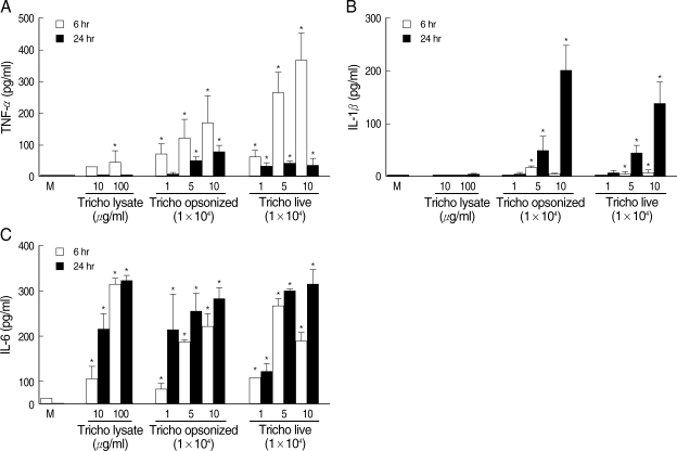
When T. vaginalis (T. vaginalis lysate, opsonized trichomonads, or live trichomonads) was coincubated with human monocyte-derived macrophage monolayer (HMDM) for 6 hr or 24 hr, TNF-α, 1L-1β, and IL-6 showed increased levels compared with untreated macrophages. The cytokine amount was generally increased with each trichomonads in dose- and time-dependent manner. The TNF-α concentration at 6 hr of coincubation showed increased levels than those of 24 hr. The live and opsonized trichomonads strongly stimulated HMDM to produce higher level of cytokines than T. vaginalis lysate. IL-6 showed increased levels regardless of the incubation time (Fig. 1).
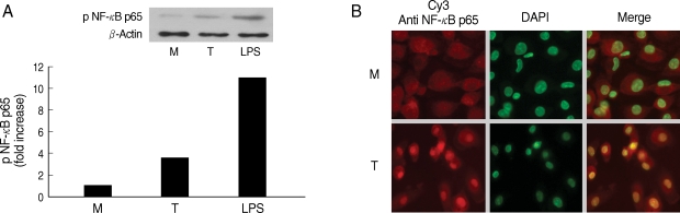
Next, we examined involvement of NF-κB in the cytokine production by HMDM. When human macrophages were stimulated with live T. vaginalis, increased phospho p65 NF-κB activity, and translocation of NF-κB p65 into nucleus were observed by western blot and fluorescent microscopy, respectively (Fig. 2).
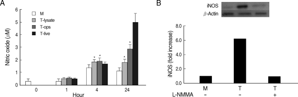
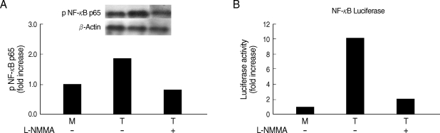
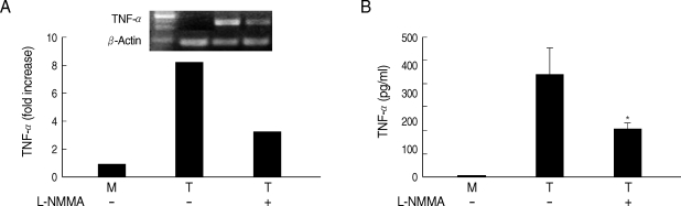
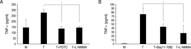
Whether NO is involved in cytokine production by HMDM stimulated with trichomonads, NO levels were measured by the Griess reagent. After 24 hr of coincubation, T. vaginalis, particularly live trichomonads, caused macrophages to produce significantly increased levels of NO. The iNOS expression of HMDM activated with live trichomonads was also increased 6 times more than untreated macrophages, and showed decreased levels by pretreatment with NO synthase inhibitor, L-NMMA by western blot (Fig. 3). Then, we investigated whether NO could influence NF-κB activity. When L-NMMA, NO synthase inhibitor-pretreated HMDM was coincubated with live trichomonads, decreased phospho NF-κB p65 activities, compared with untreated macrophages, were observed in HMDM or THP-1 by western blot and NF-κB luciferase assay, respectively (Fig. 4). Also, NO synthase inhibitor was expected to induce decreased production of cytokines by macrophages. TNF-α mRNA and protein expressions were significantly decreased by pretreatment with L-NMMA compared to the untreated group, examined by RT-PCR and ELISA, respectively (Fig. 5). Therefore, TNF-α production might be regulated by NO. Human macrophages (THP-1 and HMDM) showed decreased production of TNF-α on stimulation of live T. vaginalis by pretreatment with NF-κB inhibitor, such as PDTC or Bay11-7082, or NO synthase inhibitor, L-NMMA (Fig. 6).

DISCUSSION

In this study, human macrophages were shown to be involved in vaginal inflammation by T. vaginalis via proinflammatory cytokine and NO production. In addition, TNF-α induced by live trichomonads in macrophages might be regulated through NO-dependent activation of NF-κB. Our results are consistent with previous reports in which macrophages infected with various protozoan parasites, such as Trypanosoma congolense, Eimeria bovis, Leishmania chagasi, and Leishmania donovani, showed increased expressions of many inflammatory mediators, such as cytokines, chemokines, and NO [13-16].
NO is gaseous water and lipid-soluble molecule that is one of the most widespread signaling molecules in mammals. NO is implicated in modulating a variety of physiological reactions, including vasodilation and smooth muscle relaxation associated with the circulation, functioning as a neurotransmitter in various neural processes, and regulation of immunological defense mechanisms [17]. The role of NO in inflammation and inflammatory responses remains unclear [18]. However, it is known that NO production in human macrophages differs from the rodent macrophages and the regulation of macrophage NO in humans appears to be a more selective and variable process than that seen in the rodent macrophages [19].
Human macrophage in the study showed increased levels of NO by stimulation with T. vaginalis after 24 hr of coincubation. The iNOS expression in HMDM by T. vaginalis was also about 6 times higher than the control group, however, trichomonad-induced iNOS levels were inhibited by pretreatment with NO synthase inhibitor, L-NMMA, suggesting that NO plays a significant role in immune responses by T. vaginalis.
It is widely known that NF-κB is normally present in the cytosol and exists as an inactive complex with a class of inhibitory proteins, known as inhibitor κB (IκB) proteins. Following an inflammatory stimulus, the phosphorylation of IκB triggers its degradation and the translocation of NF-κB to the nucleus, where it binds to promoter regions and induces the expression of a wide variety of genes involved in inflammation, including those encoding cytokines (such as IL-1, IL-6, and TNF-α), enzymes (including NO synthase), adhesion molecules, and acute-phase proteins [20]. Expectedly, in this study, we have demonstrated similar results that involvement of NF-κB activation in the production of cytokines, such as TNF-α, induced by T. vaginalis stimulation was identified by nuclear translocation and phosphorylation of NF-κB p65 in human macrophages (Fig. 2).
NO production is generally upregulated by some stimuli (including LPS) known to induce iNOS gene expression via a NF-κB-mediated pathway [21]. We found that human macrophages pretreated with an iNOS inhibitor, L-NMMA, showed decreased NF-κB activity and TNF-α production triggered by T. vaginalis (Figs. 2-5), although the inhibitory effects of NF-κB inhibitor on iNOS protein expression or NO production induced by T. vaginalis was not checked in this study. Therefore, it is speculated that decreased iNOS levels with L-NMMA may occur through suppressed NF-κB activities.
On the other hand, few reports have been shown about the cytokine production regulated by NO; NO production activates the release of cytokines, cytokine receptors, and adhesion molecules [22-24]. The effect of NO on expression of TNF-α is reported to be likely via activation of a TNF-α-converting enzyme (TACE), which is responsible for membrane-bound TNF and thereby activating release of this cytokine [22]. TNF-α production caused by T. vaginalis in the study was significantly decreased by pretreatment with L-NMMA in human macrophages, suggesting an important role of NO in T. vaginalis-induced TNF-α production. Our result is in line with a previous report by Huang et al. [25] in that NO inhibitors decreased TNF-α synthesis by an LPS-activated J774 macrophage cell line. In this study, we also found that L-NMMA-pretreated macrophages reduced NF-κB activity. Therefore, it is strongly suggested that T. vaginalis stimulates human macrophages to produce TNF-α through NO-dependent activation of NF-κB.
Neutrophil migration is a complex process, which results mainly from the release of neutrophil chemotactic factors by resident cells. Apart from its importance in host defense, the migration of neutrophils to the inflammatory site is, at least in part, responsible for tissue damage observed in several inflammatory diseases, such as rheumatoid arthritis, glomerulonephritis, and inflammatory bowel disease [26,27]. In trichomoniasis, neutrophils are known to be the predominant inflammatory cells found in the vaginal discharge of the patients. We previously indicated that T. vaginalis-induced recruitment of neutrophils may be mediated via CXC chemokine IL-8 [28]. IL-1β, TNF-α, C5a, and lipid mediators, such as platelet-activating factor 4 and leukotriene (LTB4), are known the main chemotactic mediators involved in neutrophil recruitment to the sites of inflammation [29]. Therefore, IL-1β and TNF-α produced by macrophages stimulated with T. vaginalis may play a crucial role in vivo as mediators of neutrophilic tissue recruitment at the infected sites in vaginal trichomoniasis in women.
Our results showed that T. vaginalis caused human monocyte-derived macrophage (HMDM) to produce proinflammatory cytokines, such as IL-1, IL-6, TNF-α, and NO, via NF-κB. It is suggested that human macrophages may be involved in inflammation caused by T. vaginalis via the cytokine and NO production.

ACKNOWLEDGEMENTS

This work was supported by the Korea Research Foundation Grant funded by the Korean Government (MOEHRD) (KRF-2006-E00019) and Anti-Communicable Diseases Control Program of the National Institute of Health (NIH-348-6111-215), Ministry of Health and Welfare, Republic of Korea.

REFERENCES

1. World Health Organization. Global prevalence and incidence of selected curable sexually transmitted infections: overview and estimates. 2001, Geneva, Switzerland. WHO.

2. Shafir SC, Sorvillo FJ, Smith L. Current issues and considerations regarding trichomoniasis and human immunodeficiency virus in African-Americans. Clin Microbiol Rev 2009;22:37-45. PMID: 19136432.
crossref pmid pmc
3. Kiviat NB, Paavonen JA, Brockway J, Critchlow CW, Brunham RC, Stevens CE, Stamm WE, Kuo CC, DeRouen T, Holmes KK. Cytological manifestations of cervical and vaginal infections. I. Epithelial and inflammatory cellular changes. JAMA 1985;253:989-996. PMID: 3968836.
crossref pmid
4. Levine WC, Pope V, Bhoomkar A, Tambe P, Lewis JS, Zaidi AA, Farshy CE, Mitchell S, Talkington DF. Increase in endocervical CD4 lymphocytes among women with nonulcerative sexually transmitted diseases. J Infect Dis 1998;177:167-174. PMID: 9419184.
crossref pmid
5. Sardana S, Sodhani P, Agarwal SS, Sehgal A, Roy M, Singh V, Bhatnagar P, Murthy NS. Epidemiologic analysis of Trichomonas vaginalis infection in inflammatory smears. Acta Cytol 1994;38:693-697. PMID: 8091899.

6. Landolfo S, Martinotti MG, Martinetto P, Forni G. Natural cell-mediated cytotoxicity against Trichomonas vaginalis in the mouse. I. Tissue, strain, age distribution, and some characteristics of the effector cells. J Immunol 1980;124:508-514. PMID: 6965381.
pmid
7. Ryu JS, Ahn MH, Min DY. Cytotoxicity of resident and lymphokine-activated mouse peritoneal macrophage against Trichomonas vagianalis. Korean J Parasitol 1990;28:85-89.
crossref pmid
8. Yoon K, Ryu JS, Min DY. Cytotoxicity of lymphokine activated peritoneal macrophages against Trichomonas vaginalis. Korean J Parasitol 1991;29:381-388.
crossref pmid
9. Park GC, Ryu JS, Min DY. The role of nitric oxide as an effector of macrophage-mediated cytotoxicity against Trichomonas vaginalis. Korean J Parasitol 1997;35:189-195. PMID: 9335184.
crossref pmid
10. Ahn MH, Song HO, Ryu JS. Trichomonas vaginalis-induced neutrophil apoptosis causes anti-inflammatory cytokine production by human monocyte-derived macrophages. Parasite Immunol 2008;30:410-416.
crossref pmid
11. Lee EJ, Heo YM, Choi JH, Song HO, Ryu JS, Ahn MH. Suppressed production of pro-inflammatory cytokines by LPS-activated macrophages after treatment with Toxoplasma gondii lysate. Korean J Parasitol 2008;46:145-151. PMID: 18830053.
crossref pmid pmc
12. Kim YS, Song HO, Choi IH, Park SJ, Ryu JS. Hydrogenosomal activity of Trichomonas vaginalis cultivated under different iron conditions. Korean J Parasitol 2006;44:373-378. PMID: 17170580.

13. Magez S, Radwanska M, Drennan M, Fick L, Baral TN, Allie N, Jacobs M, Nedospasov S, Brombacher F, Ryffel B, De Baetselier P. Tumor necrosis factor (TNF) receptor-1 (TNFp55) signal transduction and macrophage-derived soluble TNF are crucial for nitric oxide-mediated Trypanosoma congolense parasite killing. J Infect Dis 2007;196:954-962. PMID: 17703428.
crossref pmid
14. Taubert A, Behrendt JH, Sühwold A, Zahner H, Hermosilla C. Monocyte-and macrophage-mediated immune reactions against Eimeria bovis. Vet Parasitol 2009;doi:10.1016/j.vetpar.2009.06.003.

15. Gantt KR, Goldman TL, McCormick ML, Miller MA, Jeronimo SMB, Nascimento ET, Britigan BE, Wilson ME. Oxidative responses of human and murine macrophages during phagocytosis of Leishmania chagasi. J Immunology 2001;167:893-901. PMID: 11441096.
crossref pmid
16. Dasgupta B, Roychoudhury K, Ganguly S, Akbar MA, Das P, Roy S. Infection of human mononuclear phagocytes and macrophage-like THP1 cells with Leishmania donovani results in modulation of expression of a subset of chemokines and a chemokine receptor. Scand J Immunol 2003;57:366-374. PMID: 12662300.
crossref pmid
17. Wink DA, Mitchell JB. Chemical biology of nitric oxide: insights into regulatory, cytotoxic, and cytoprotective mechanisms of nitric oxide. Free Radic Biol Med 1998;25:434-456. PMID: 9741580.
crossref pmid
18. Rogers JA, Fuseler JW. Regulation of NF-κB activation and nuclear translocation by exogenous nitric oxide (NO) donors in TNF-α activated vascular endothelial cells. Nitric Oxide 2007;16:379-391. PMID: 17374495.
crossref
19. Thomassen MJ, Kavuru MS. Human alveolar macrophages and monocytes as a source and target for nitric oxide. Int Immunopharmacol 2001;1:1479-1490. PMID: 11515813.
crossref pmid
20. Jung WK, Choi IH, Lee DY, Yea SS, Choi YH, Kim MM, Park SG, Seo SK, Lee SW, Lee CM, Park YM, Choi IW. Caffeic acid phenethyl ester protects mice from lethal endotoxin shock and inhibits lipopolysaccharide-induced cyclooxygenase-2 and inducible nitric oxide synthase expression in RAW 264.7 macrophages via the p38/ERK and NF-κB pathways. Int J Biochem Cell Biol 2008;40:2572-2582. PMID: 18571461.

21. Xie QW, Kashiwabara Y, Nathan C. Role of transcription factor NF-κB/Rel in induction of nitric oxide synthase. J Biol Chem 1994;269:4705-4708. PMID: 7508926.

22. Zhang Z, Kolls JK, Oliver P, Good D, Schwarzenberger PO, Joshi MS, Ponthier JL, Lancaster JRJr. Activation of tumor necrosis factor-α-converting enzyme-mediated ectodomain shedding by nitric oxide. J Biol Chem 2000;275:15839-15844. PMID: 10747938.
crossref
23. Stevanin TM, Laver JR, Poole RK, Moir JWB, Read RC. Metabolism of nitric oxide by Neisseria meningitidis modifies release of NO-regulated cytokines and chemokines by human macrophages. Microbes Infect 2007;9:981-987. PMID: 17544805.
crossref pmid
24. Yan L, Wang S, Rafferty SP, Wesley RA, Danner RL. Endogenously produced nitric oxide increases tumor necrosis factor-α production in transfected human U937 cells. Blood 1997;90:1160-1167. PMID: 9242548.
pmid
25. Huang FP, Niedbala W, Wei XQ, Xu D, Feng GJ, Robinson JH, Lam C, Liew FY. Nitric oxide regulates Th1 cell development through the inhibition of IL-12 synthesis by macrophages. Eur J Immunol 1998;28:4062-4070. PMID: 9862342.
crossref pmid
26. Guo RF, Ward PA. Mediators and regulation of neutrophil accumulation in inflammatory responses in lung: insights from the IgG immune complex model. Free Radic Biol Med 2002;33:303-310. PMID: 12126752.
crossref pmid
27. Hanai H, Watanabe F, Yamada M, Sato Y, Takeuchi K, Iida T, Tozawa K, Tanaka F, Maruyama Y, Matsushita I, Iwaoka Y, Saniabadi A. Correlation of serum soluble TNF-α receptors I and II levels with disease activity in patients with ulcerative colitis. Am J Gastroenterol 2004;99:1532-1538. PMID: 15307873.
crossref pmid
28. Ryu JS, Kang JH, Jung SY, Shin MH, Kim JM, Park H, Min DY. Production of Interleukin-8 by human neutrophils stimulated with Trichomonas vaginalis. Infect Immun 2004;72:1326-1332. PMID: 14977935.
crossref pmid pmc
29. Schröder JM. Chemoattractants as mediators of neutrophilic tissue recruitment. Clin Dermatol 2000;18:245-263. PMID: 10856658.
crossref
Fig. 1
Trichomonas vaginalis stimulates human monocyte-derived macrophages (HMDM) to produce proinflammatory cytokines. Human macrophages (1.25 × 10⁶/well of 96-well culture plate) were incubated with live trichomonads (1, 5, and 10 × 104), opsonized trichomonads (1, 5, and 10 × 104) and T. vaginalis lysate (10 and 100 µg/ml). Cell culture supernatant was harvested after 6 hr and 24 hr of incubation, and analyzed for levels of TNF-α, IL-1β, and IL-6 using cytokine-specific ELISA. *P < 0.05 versus untreated macrophages.
kjp-47-205-g001
Fig. 2
NF-κB activation induced by Trichomonas vaginalis (T) in human macrophages (M). (A) Phosphorylation of NF-κB p65 expression in human macrophages reacted with T. vaginalis was determined by western blot. (B) Nuclear translocation of NF-κB p65 in human macrophages stimulated with T. vaginalis (low) compared with medium alone (up). Human macrophages (5 × 10⁶/cover glass) were incubated with live T. vaginalis (1 × 10⁶) for 30 min. Cells were then fixed and subjected to immunofluorescent microscopy after staining with anti-NF-κB p65 Ab (Red) and DAPI (Green) to visualize nuclei.
kjp-47-205-g002
Fig. 3
NO production (A) and iNOS expression (B) by HMDM stimulated with T. vaginalis (T). Human macrophages (1.25 × 10⁶/well of 96-well culture plate) were incubated with live trichomonads (1.25 × 10⁵), opsonized trichomonads (1.25 × 10⁵), or T. vaginalis lysate (100 µg/ml). NO was determined by the Griess reagent after 1 hr, 4 hr, and 24 hr of incubation (A). For identification of iNOS expression, human macrophages (1.25 × 10⁶/well) were incubated with live trichomonads (1.25 × 10⁵) for 1 hr. The iNOS expression was examined by western blot with anti-iNOS Ab (B). *P < 0.05 versus untreated macrophages.
kjp-47-205-g003
Fig. 4
Effects of L-NMMA (an iNOS inhibitor) on NF-κB activity of human macrophages induced by T. vaginalis. L-NMMA-preteated HMDM or THP-1 cells was stimulated with live T. vaginalis. NF-κB activities were determined by western blot (A) or luciferase assay (B). Human macrophages were coincubated with live trichomonads (macrophage : trichomonads = 5 : 1) for 2 hr, and then western blot was done with rabbit anti-phospho NF-κB p65 monoclonal Ab (A). The PCH110 and PGL3 NF-κB promoter constructs were transfected into THP-1 cells with LipofectAmine 2000. Luciferase activity was assayed with a luciferase assay kit. Cell extracts obtained from THP-1 cells stimulated with live trichomonads for 1 hr were prepared in 500 µl of 1 × reporter lysis buffer. The luciferase activity was measured with a Luminometer (B).
kjp-47-205-g004
Fig. 5
Effects of L-NMMA on TNF-α production in HMDM cells induced by live T. vaginalis. L-NMMA-pretreated macrophage suppressed T. vaginalis-triggered TNF-α expression, measured by RT-PCR (A) and ELISA (B), respectively. *P < 0.05 versus macrophages stimulated with T. vaginalis.
kjp-47-205-g005
Fig. 6
Effects of iNOS inhibitor or NF-κB inhibitor on TNF-α production in macrophages induced by T. vaginalis. Pretreatment of human macrophages (THP-1 or HMDM) with L-NMMA or NF-κB inhibitor (PDTC or Bay11-7082) showed significant reduction of trichomonad-induced TNF-α production in THP-1 (A) and HMDM (B). Protein levels of TNF-α were measured by ELISA. *P < 0.05 versus untreated macrophages. P < 0.05 versus T. vaginalis-stimulated macrophages.
kjp-47-205-g006