Warning: mkdir(): Permission denied in /home/virtual/lib/view_data.php on line 81

Warning: fopen(upload/ip_log/ip_log_2024-07.txt): failed to open stream: No such file or directory in /home/virtual/lib/view_data.php on line 83

Warning: fwrite() expects parameter 1 to be resource, boolean given in /home/virtual/lib/view_data.php on line 84
Pandey, Madan, Asthana, Das, Kumar, and Jain: External Ophthalmomyiasis Caused by Oestrus ovis: A Rare Case Report from India
Cited By
Citations to this article as recorded by
La myiase conjonctivale humaine à Oestrus ovis dans le sud tunisien
S. Anane, L. Ben Hssine
Bulletin de la Société de pathologie exotique.2010; 103(5): 299.     CrossRef
Human myiasis cases originating and reported in africa for the last two decades (1998–2018): A review
Simon K. Kuria, Adebola O. Oyedeji
Acta Tropica.2020; 210: 105590.     CrossRef
External ophthalmomyiasis. A case report from Spain
C. Berrozpe-Villabona, N. Avalos-Franco, S. Aguilar-Munoa, P. Bañeros-Rojas, J. Castellar-Cerpa, D. Diaz-Valle
Journal Français d'Ophtalmologie.2015; 38(9): e219.     CrossRef
External ophthalmomyiasis: A case report
Mohammad Al-Amry, Fahad I. Al-Saikhan, Saad Al-Dahmash
Saudi Journal of Ophthalmology.2014; 28(4): 322.     CrossRef
Spatial Distribution of Necrophagous Flies of Infraorder Muscomorpha in Iran Using Geographical Information System
Kamran Akbarzadeh, Abedin saghafipour, Nahid Jesri, Moharram Karami-Jooshin, Koroush Arzamani, Teymour Hazratian, Razieh Shabani Kordshouli, Abbas Aghaei Afshar
Journal of Medical Entomology.2018;[Epub]     CrossRef
Unusual presentation of cutaneous myiasis in the knee: case report
Omar S Dahduli, Sarah A Aldeghaither, Abdullah M Alhossan
Journal of Surgical Case Reports.2024;[Epub]     CrossRef
Ophthalmomyiasis externa from Hakkari, the south east border of Turkey
Şeref Istek
BMJ Case Reports.2014; : bcr2013201226.     CrossRef
Second Report of Accidental Intestinal Myiasis due toEristalis tenax(Diptera: Syrphidae) in Iran, 2015
Ramezani Awal Riabi Hamed, Ramezani Awal Riabi Hamid, Naghizade Hamid
Case Reports in Emergency Medicine.2017; 2017: 1.     CrossRef
Ophthalmomyiasis externa: three cases caused by Oestrus ovis larvae in Turkey
Sinan Çalışkan, Sılay Cantürk Ugurbaş, Murat Sağdık
Tropical Doctor.2014; 44(4): 230.     CrossRef
External ophthalmomyiasis caused by Oestrus ovis in east China
Aihui Zhang, Qiaoli Nie, Jingjing Song
Tropical Doctor.2018; 48(2): 169.     CrossRef
Ophthalmomyiasis externa due to sheep nasal botfly in rural Jamaica
Valence Jordan, Lizette Mowatt
Tropical Doctor.2019; 49(1): 48.     CrossRef
External Ophthalmomyiasis: Case Reports of Two Cases Associated With Agrarian Practices
Meenakshi Sharma
Journal of Bacteriology & Mycology: Open Access.2017;[Epub]     CrossRef
Unusual Case: Ophthalmomyiasis
Fatma Kesmez Can, Handan Alay, Emine Çinici
Revista da Sociedade Brasileira de Medicina Tropical.2021;[Epub]     CrossRef
Ophthalmomyiasis secondary to infected scleral buckle
Magna Mary Kuruvila, Sunil G Biradar
IP International Journal of Ocular Oncology and Oculoplasty.2021; 6(4): 266.     CrossRef
Opthalmomyiasis: A rare case report
Shushruta Mohanty, Sujata Panda, Leesa Mohanty, Bijay Kumar Pattnayak
IP Journal of Diagnostic Pathology and Oncology.2022; 7(2): 130.     CrossRef
Eyelid myiasis caused byCordylobia anthropophaga
Paolo Sivelli, Riccardo Vinciguerra, Luigi Tondini, Elena Cavalli, Andrea Galli, Paolo Chelazzi, Simone Donati, Luigi Bartalena, Paolo Grossi, Claudio Azzolini
Ocular Immunology and Inflammation.2015; 23(3): 259.     CrossRef
A Case of Nosocomial Nasal Myiasis in Comatose Patient
Sung Jae Heo, Mi Jin Lee, Chang Mook Park, Jung Soo Kim
Korean Journal of Otorhinolaryngology-Head and Neck Surgery.2013; 56(10): 664.     CrossRef
A Nasal Myiasis in a 76-Year-Old Female in Korea
Jae-Soo Kim, Pil-Won Seo, Jong-Wan Kim, Jai-Hyang Go, Soon-Cheol Jang, Hye-Jung Lee, Min Seo
The Korean Journal of Parasitology.2009; 47(4): 405.     CrossRef
Ophthalmomyiasis Externa and Importance of Risk Factors, Clinical Manifestations, and Diagnosis: Review of the Medical Literature
Hugo Martinez-Rojano, Herón Huerta, Reyna Sámano, Gabriela Chico-Barba, Jennifer Mier-Cabrera, Estibeyesbo Said Plascencia-Nieto
Diseases.2023; 11(4): 180.     CrossRef
A Case of Aural Myiasis in Chronic Alcoholism Patient
Sung Hoon Jung, Jae Hwan Jung, Seok Hyun Kim, Yang Jae Kim
Journal of Clinical Otolaryngology Head and Neck Surgery.2016; 27(2): 332.     CrossRef
Oestrus ovis (Diptera: Oestridae) un importante ectoparásito en ovinos de cuatro cantones del municipio de Sorata provincia Larecaja, departamento de La Paz
Graciela Cristina Choque-Fernández, Manuel Gregorio Loza-Murguia, Nicolasa Lourdes Vino-Nina, Luis Alfredo Coria-Conde
Journal of the Selva Andina Animal Science.2017; 4(1): 3.     CrossRef
External ophthalmomyiasis by Oestrus ovis larvae: A Rare Case Report
B. M. Shanker VENKATESH, P. Ganga BHAVANI
Journal of Microbiology and Infectious Diseases.2021; : 159.     CrossRef
Miasis intestinal humana por Eristalis tenax en un niño de la zona urbana del municipio de Policarpa, Nariño, Colombia
Álvaro Francisco Dulce-Villarreal, Angélica María Rojas-Bárcenas, José Danilo Jojoa-Ríos, José Fernando Gómez-Urrego
Biomédica.2020; 40(4): 599.     CrossRef
Bilateral Ophthalmomyiasis Externa of Lid by Musca domestica: A Rare Presentation
Manjiri P Sune, Mona P Sune, Shital M Mahajan, Pradeep Sune
Cureus.2024;[Epub]     CrossRef

Abstract

Myiasis of different organs has been reported off and on from various regions in the world. We report a human case of external ophthalmomyiasis caused by the larvae of a sheep nasal botfly, Oestrus ovis, for the first time from Meerut city in Western Uttar Pradesh, India. A 25-year-old farmer presented with severe symptoms of conjunctivitis. The larvae, 3 in number, were observed in the bulbar conjunctiva, and following removal the symptoms of eye inflammation improved within a few hours.

INTRODUCTION

Myiasis is infection of tissues or organs of animals or man by larvae of a fly. The common sites are skin wounds. Eyes, nose, nasal sinuses, throat, and urogenital tract are less common sites [1-3]. Ocular involvement occurs in about < 5% of all cases of human myiasis [4]. Ophthalmomyiasis is classified as ophthalmomyiasis externa if the larvae are present on the conjunctiva, and ophthalmomyiasis interna when there is intraocular penetration of larvae [5]. Cases of ophthalmomyiasis externa have been reported from various parts of the world [6-8]. However, this condition is rare in India and there are only a few reported cases [5,9-12]. To the best of our knowledge, no case of ophthalmomyiasis externa caused by Oestrus ovis (the sheep nasal botfly) has been reported from this part of India. The present case therefore highlights the awareness among the ophthalmologists regarding larval conjunctivitis in spring and summer season, with timely diagnosis and treatment of this rather rare infestation.

CASE REPORT

A 25-year-old male farmer came to our outpatient department in April 2008 with a 2-day history of foreign body sensation, burning, and excessive watering from his right eye. He gave a history of something falling in his eye while he was resting below a tree after lunch in the field. He was absolutely fine before that, and there was no significant history of ocular or medical problems preceding this.
On examination it was found that his visual acuity was 20/20 in both eyes. Eyelids of the affected right eye were absolutely normal. The conjunctiva was mildly congested with profuse lacrimation. Extraocular movements were full. On slit lamp examination, the most remarkable finding was the presence of tiny translucent worms, 1-2 mm in size with dark heads, crawling over the bulbar conjunctiva and cornea. The larvae actively avoided the bright light of the slit lamp and tried to burrow deep into the conjunctival fornices. Pupillary reaction was normal. Slit lamp examination did not reveal any anomaly. Using a topical anesthesia with 4% xylocaine drops these larvae were allowed to crawl on the sterile cotton swab sticks placed over the conjunctiva. The larvae (3 in number) were mounted on a glass slide and sent to department of microbiology for identification.
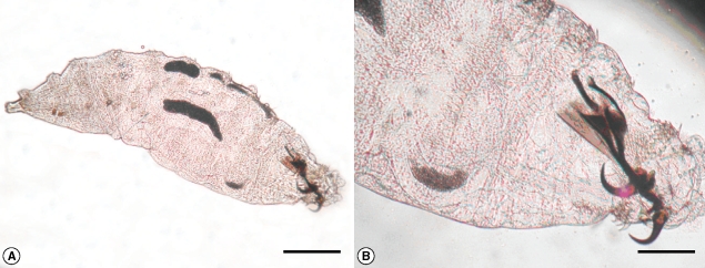
Direct and indirect ophthalmoscopy showed no evidence of intraocular organisms or inflammation. Examination of his left eye was normal. Topical antihistaminic and antibiotic drops were prescribed. When the patient came for a follow-up 2 days later, he was completely relieved of his symptoms of foreign body sensation and excessive watering. A repeat slit lamp examination of the anterior segment and fundus was normal. The larvae mounted on a microscopic slide were examined carefully and photographed under a microscope. It was identified as the first stage larvae of O. ovis, which is a larviparous dipteran on the basis of their spindle shaped skeleton (Fig. 1). The larvae also showed a pair of sharp dark brown oral hooks and tufts of numerous brown hooks on the anterior margin of each body segment (Fig. 1). The posterior spiracles were found, in the eighth segment [12].

DISCUSSION

The sheep nasal botfly, O. ovis, are large dark gray flies with dark spots on the dorsum of the thorax and abdomen, and are covered by a moderate amount of light brown hair [13]. Although myiasis in man is generally rare, members of the Oestridae (Diptera) may produce human myiasis in countries where the standard of hygiene is low and there is abundance of flies around the locality [8]. The female sheep nasal botfly dashes to deposit their freshly hatched larvae in the naris, on the conjunctiva, on the lip, and in the mouth of the usual hosts like sheep, cattle, and horse. Man serves as an accidental host [13].
Opthalmomyiasis due to O. ovis was described for the first time in 1947 by James [8]. External ophthalmomyiasis manifests as acute catarrhal conjunctivitis with symptoms similar to those presented in this case [1]. Therefore, ophthalmomyiasis externa caused by O. ovis should not be regarded as a benign condition and should be treated promptly to prevent serious complication such as corneal ulcer, decreased vision, and invasion into eye globe causing endophthalmitis, iridocyclitis, and even blindness which has been reported in the past [14]. However, none of these complications were encountered in our patient. It may have been due to a small number of larvae, i.e., only 3 in our case, and a short history of 2 day duration.
Our patient came in April (the spring season). A previous paper presenting clinical manifestations and seasonal variations of 8 ophthalmomyiasis cases have reported that most of them occurred in the spring and summer seasons [1]. The present case highlights 2 things first, it creates awareness among the ophthalmologists regarding larval conjunctivitis as 1 of the causes of conjunctivitis during the spring and summer seasons especially in developing countries like India, where the general standard of hygiene is low and there are a large number of flies around. Second, irrigation of the conjunctiva with normal saline is unsuccessful in washing out the larvae because the larvae grab the conjunctiva firmly with the help of a pair of oral hooks and numerous hooks on each segment. Therefore, after anesthetizing the conjunctiva, a thorough examination of the eye under magnification and prompt removal of the larvae manually with sterile cotton swab sticks should be done to avoid disastrous complications of internal ophthalmomyiasis.

REFERENCES

1. Mohsen M, Keramatalab H. External ophthalmomyiasis caused by sheep botfly (Oestrus ovis) larvae: a report of 8 cases. Arch Iran Med. 2004. 7:136-139.

2. Al-Dabagh M, Al-Mufti N, Shafiq M, Al-Rawas AY, Al-Saffar S. A second record from Iraq of human myiasis caused by larvae of the sheep botfly Oestrus ovis L. Ann Trop Med Parasitol. 1980. 74:73-77. PMID: 7377876.
pmid
3. Amr ZS, Amr BA, Abo-Shehada MN. Ophthalmomyiasis externa caused by Oestrus ovis L. in the Ajloun area of northen Jordan. Ann Trop Med Parasitol. 1993. 87:259-262. PMID: 8257236.
pmid
4. Ziemianski MC, Lee KY, Sabates FN. Ophthalmomyiasis interna. Arch Ophthalmol. 1980. 98:1588-1589. PMID: 7191701.
crossref pmid
5. Gupta VP, Baveja UK. Ophthalmomyiasis externa caused by the sheep nasal botfly Oestrus ovis. Indian J Ophthalmol. 1988. 36:41-42. PMID: 3253202.

6. McDermott S, Schafer MD. Ophthalmomyiasis due to Oestrus ovis in South Australia. Med J Aust. 1983. 1:129-130. PMID: 6571540.
pmid
7. Victor R, Bhargva K. Ophthalmomyiasis in Oman: a case report and comments. Wilderness Environ Med. 1998. 9:32-35. PMID: 11990179.
crossref pmid
8. Patel SJ. Extra-ocular myiasis due to the larva of Oestrus ovis. East Afr Med J. 1975. 52:167-169. PMID: 1140145.
pmid
9. Narayanan S, Jayaprakash K. Incidence of ocular myiasis due to infection with the larva of Oestrus ovis (Oestridae: Diptera). Indian J Ophthalmol. 1991. 39:176-178. PMID: 1810881.

10. Misra S, Misra N, Reddy B. External ophthalmomyiasis by Oestrus ovis: an unknown endemic eye disease in rural parts of central India. Trop Doct. 2008. 38:120-122. PMID: 18453513.
crossref pmid
11. Chandra DB, Agrawal TN. Ocular myiasis caused by Oestrus ovis - a case report. Indian J Ophthalmol. 1981. 29:199-200. PMID: 7346425.

12. Mathai E, George JA, Lalitha MK, Raju R. Ophthalmomyiasis externa caused by Oestrus ovis-a case report from South India. Indian J Pathol Microbiol. 1994. 37:205-207. PMID: 7959990.
pmid
13. Beaver PC, Jung RC, Cupp EW. Clinical Parasitology. 1984. 9th ed. Philadelphia, USA: Lea & Febiger; 680-695.

14. Hunt EW Jr. Unusual case of ophthalmomyiasis interna posterior. Am J Ophthalmol. 1970. 70:978-980. PMID: 5490626.
crossref pmid
Fig. 1
A wet mount showing the spindle shaped skeleton (A) and dark brown oral hooks (B) of an Oestrus ovis larva extracted from the patient. Scale bars = 0.5 mm (A) and 0.2 mm (B).
kjp-47-57-g001