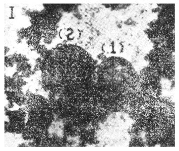
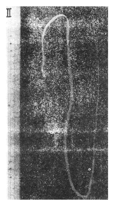
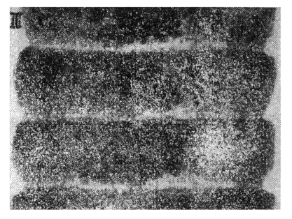
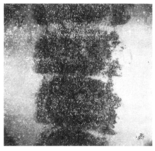
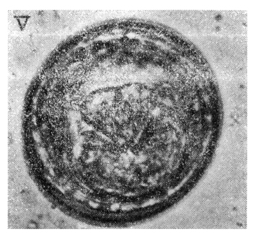
There has been no report on human infection of Hymenolepis diminuta in Korea until the first 3 cases were reported by our members after the identification of those eggs in stool in 1964. However, the distinct differentiation between H. diminuta and H. nana would often be difficult by the shape of eggs without adult worm. In 1965, authors found the additional case revealed the eggs in stool and succeeded to obtain three adult worms of H. diminuta from 10 years old boy in Pusan. The characteristic morphology of egg and adult worm were discussed to compare to those of H. nana. Conclusively, the first human infection of H. diminuta in Korea was reported after the identification both the eggs and adults worms. |