Wakid: A Laboratory-Based Study for First Documented Case of Urinary Myiasis Caused by Larvae of Megaselia scalaris (Diptera: Phoridae) in Saudi Arabia
Cited By
Citations to this article as recorded by
Morphology of immature stages of Megaselia spiracularis Schmitz (Diptera: Phoridae)
Feng Dian‐Xing, Liu Guang‐Chun
Microscopy Research and Technique.2012; 75(9): 1297.     CrossRef
Interaction effects of temperature and food on the development of forensically important fly, Megaselia scalaris (Loew) (Diptera: Phoridae)
Raja Muhammad Zuha, Tasnim Abdul Razak, Nazni Wasi Ahmad, Baharudin Omar
Parasitology Research.2012; 111(5): 2179.     CrossRef
Neglected Tropical Diseases (NTDs) in Saudi Arabia: Systematic Review and Meta-analysis
Najm Z. Alshahrani, Abdullah M. Alarifi, Abdullah M. Assiri
Journal of Epidemiology and Global Health.2025;[Epub]     CrossRef
Human urogenital myiasis: A systematic review of reported cases from 1975 to 2017
Roghiyeh Faridnia, Masoud Soosaraei, Hamed Kalani, Mahdi Fakhar, Pikka Jokelainen, Reza Zolfaghari Emameh, Elham Sadat Banimostafavi, Hajar Ziaei Hezarjaribi
European Journal of Obstetrics & Gynecology and Reproductive Biology.2019; 235: 57.     CrossRef
Pupal age estimation of forensically important Megaselia scalaris (Loew) (Diptera: Phoridae)
Dian-Xing Feng, Guang-Chun Liu
Forensic Science International.2014; 236: 133.     CrossRef
A complete morphological characterization of all life stages of the phorid fly Megaselia scalaris
Jayakumar Pallavi, Harshita Snehal, Rakshita Sukruth Kolipakala, Daniela Salazar, Mrunal Hanbar, Larina Bejoy Chiramel, Khushi Alok Jha, Sai Bhumica Lakshmi Venkatesh, Tanishka Dayanand Shetty, Navya Madhusudan, Amrutha Mohan, Amulia John, Naomi Deep D’so
Scientific Reports.2023;[Epub]     CrossRef
Morphological and Molecular Characteristic of Megaselia scalaris (Diptera: Phoridae) Larvae as the Cause of Urinary Myiasis
R. Solgi, N. Dinparast Djadid, A. Eslamifar, A. Raz, S. Zakeri
Journal of Medical Entomology.2017; 54(3): 781.     CrossRef
Occurrence of human urogenital myiasis due to neglected personal hygiene: a review
Amandeep Singh, Jasneet Kaur
Transactions of The Royal Society of Tropical Medicine and Hygiene.2019; 113(1): 4.     CrossRef
Megaselia scalaris reared on Rhipicephalus (Boophilus) microplus laboratory cultures
E. MIRANDA-MIRANDA, R. COSSIO-BAYUGAR, F. MARTINEZ-IBAÑEZ, C. R. BAUTISTA-GARFIAS
Medical and Veterinary Entomology.2011; 25(3): 344.     CrossRef
Laboratory Identification of Arthropod Ectoparasites
Blaine A. Mathison, Bobbi S. Pritt
Clinical Microbiology Reviews.2014; 27(1): 48.     CrossRef
Myiasis
Fabio Francesconi, Omar Lupi
Clinical Microbiology Reviews.2012; 25(1): 79.     CrossRef
A case of uncomplicated urinary myiasis in a healthy female
Olugbemisola Oredein McCoy, Andrew Rabley, Michaella M Prasad, Eric S Rovner
BMJ Case Reports.2016; : bcr2016214783.     CrossRef
Prevalence and Salient Morphological Features of Myiasis Causing Dipteran Flies in Jeddah, Saudi Arabia
Masroor Alikhan, K. Al Ghamdi, Fahad S Al Zahrani, Emad I Khater, Ahmad M Allam
Biosciences, Biotechnology Research Asia.2018; 15(1): 101.     CrossRef
The scuttle flies (Diptera: Phoridae) of Iran with the description of Mahabadphora aesthesphora as a new genus and species
Roya Namaki-Khameneh, Samad Khaghaninia, R. Henry L. Disney, Naseh Maleki-Ravasan, Feng ZHANG
PLOS ONE.2021; 16(10): e0257899.     CrossRef
Utilizing Ecological Modeling to Follow the Potential Spread of Honey Bee Pest (Megaselia scalaris) from Nearby Countries towards Saudi Arabia under Climate Change Conditions
Areej A. Alkhalaf
Diversity.2022; 14(4): 261.     CrossRef
The Price of Hospital Reshaping: Nasal Myiasis Caused by Flesh Fly (Diptera: Sarcophagidae) in Reallocated COVID-19 Intensive Care Unit
Vladimir Dolinaj, Jasmina Grujić, Davor Križanović, Aleksandar Potkonjak, Thomas Pape, Pavle Banović
Healthcare.2023; 11(11): 1533.     CrossRef
Human urinary myiasis due to larvae of Clogmia (Telmatoscopus) albipunctata Williston (Diptera: Psychodidae) first report in Egypt
AymanA El-Badry, HosniKhairy Salem, YusufAbd El-Aziz Edmardash
Journal of Vector Borne Diseases.2014; 51(3): 247.     CrossRef
Native parasitoids of the fall webworm, Hyphantria cunea (Drury, 1773) (Lepidoptera, Erebidae), an invasive alien pest in northern Iran
Amene Karami, Ali Asghar Talebi, Ebrahim Gilasian, Yaghoub Fathipour, Mohammad Mehrabadi
Journal of Insect Biodiversity and Systematics.2023; 9(1): 81.     CrossRef

Abstract

Megaselia scalaris (Diptera: Phoridae) is one of the medically important insects. Maggots from a urine sample of a 5-year-old Saudi girl were examined microscopically for identification. These maggots were cultured to become adult flies. Larvae and adults were identified using standard keys. Protozoan flagellates were obtained from the gut of the larvae. This is the first report of M. scalaris as a causative agent of urinary human myiasis in Saudi Arabia.

INTRODUCTION

Myiasis is a term used to describe the invasion of tissues or organs of vertebrates with the larvae (maggots) of dipterous flies. It could be external or internal, and the invasion for the maggot could be obligatory, facultative, and sometimes accidental [1]. This is the first report to describe a laboratory-based diagnosis of accidental human urinary myiasis due to invasion of Megaselia scalaris (Diptera: Phoridae) in Saudi Arabia. In addition, we report the detection of protozoan motile flagellates in the larvae.

CASE RECORD

The urine sample containing 5 larvae was collected in a clean container after washing the urogenital area, from a 5-yr-old female living in Jeddah, Saudi Arabia. She complained of difficulty in urination and discomfort for several days. All symptoms disappeared on the next day of specimen collection. Two larvae were placed in 70% alcohol for identification, and then were examined under a stereo dissection binocular microscope. Another 2 larvae were cultured on a stool sample to allow development into adult flies. The fifth larva was crushed and examined microscopically as a wet mount with saline, and then certain parts were transferred onto another slide, allowed to air dry, fixed with methanol, and finally stained with 10% (v / v) Giemsa stain. Identification of larvae and adult flies was done according to keys described by previous workers [2-5].
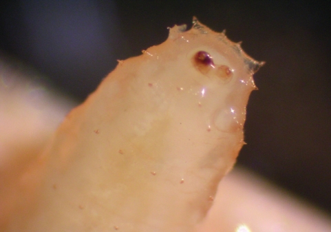
Macroscopic examination showed that all the 5 larvae were alive, whitish in color with dark internal contents in the third terminal part. The length of larvae ranged 2.5-3.5 mm. Microscopic examination of the preserved 2 larvae under a dissection binocular microscope revealed the presence of rows of many tiny short spines on the body (Figs. 1-3). Dense, tooth-like sharp pointed spines were on the anterior part (Fig. 2). A pair of posterior spiracles is situated dorsally (Fig. 3). According to the description in previous workers, the larvae were assumed as the first and second-instar larvae of M. scalaris [2-5].
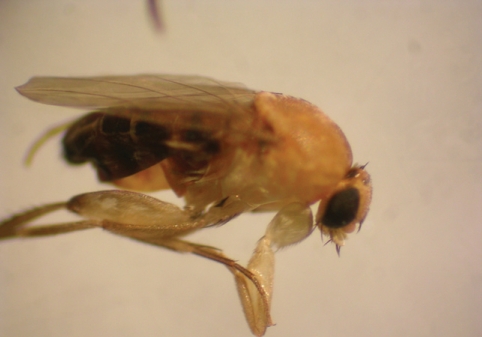
For confirmation of the identity of the fly, 2 larvae were cultured on a stool sample. On the second day, one of these larvae was clearly lager than the other. On the fourth day, the bigger larvae buried itself inside the stool except the posterior end. The color of this larva was light brown. Some soil was added to maintain humid condition and to include the possibility of sand burying phenomenon if applicable. The second larva started burying on the next day. On day 12, the first adult fly immerged (Fig. 4). This tiny fly was 1.8 mm long, with a hump-backed appearance, light brown in color with striated back and a characteristic quick jerky motion. Two days later (Day 14), the second fly immerged. According to general appearance of adult flies and the characteristic morphology of the larvae, the identity was confirmed as M. scalaris.
The fifth larva was crushed on a microscope slide, and examined as a wet preparation after adding a drop of saline and covered with a cover glass. Using light microscopy, it was possible to observe 4 motile flagellates in the ruptured gut. This part of the gut was carefully smeared onto another microscope slide, allowed to air dry, fixed with methanol, and finally stained with 10% (v / v) Giemsa stain. The size of the flagellates ranged between 30-35 µm and each flagellate was clearly consisting of a body with a nucleus and a single flagellum. The motility and morphology of the flagellates were similar to the protozoan flagellated form belonging to the family Trypanosomatidae.

DISCUSSION

Megaselia scalaris is a small hump-backed fly with characteristic rapid jerky movements on surfaces, hence the common name is the scuttle fly [3,6]. The females are highly attracted by strong odors and lay their eggs on deferent decomposing materials including feces, flesh, vegetables, and fruits.
Several previous workers reported myiasis due to M. scalaris in man, animals, and plants. Several workers reported cases of intestinal myiasis due to M. scalaris [7-9]. Hira et al. [10] described in 2004 a case of nosocomial myiasis in Kuwait caused by M. scalaris. The first report of human urogenital myiasis due to a species of Megaselia was represented in 1978 by Disney and Kurahashi [11], and then more cases were reported [12-14]. Other workers studied invasion of ticks [15], bananas [16], and snacks [17] by the larvae of M. scalaris. The use of M. scalaris specimens in forensic investigations was also documented 18,19].
Phorids can live on many types of decomposing organic matters and in the present study the larvae were reared in stool cultures; however, the stool or other materials were used by previous workers [3,7,20]. Bacterial culture plates were also found to be suitable for maintaining the larvae of Megaselia [14].
Many flagellates of the family Trypanosomatidae have been isolated from a wide range of plants, animals, and insects [21]. Flagellates of the genus Herpetomonas are usually found colonizing in the digestive tract of M. scalaris [22], so the observed flagellates in the present study could belong to that genus. We plan for a further investigation using electron microscopy and molecular techniques to confirm the identity of those flagellates. Monoxenous trypanosomatids are not believed to infect vertebrate cells. However, recent in vitro work suggested that monoxenous trypanosomatids could be potentially pathogenic to vertebrate hosts [23].
People in Jeddah, Saudi Arabia, used to see scuttle flies in different places, such as vegetable and fruit markets, toilets, and around decomposing materials. The present myiasis case is probably accidental infestation related to a lack of personal hygiene and it is likely that Megaselia adults deposited their eggs on the underwear cloths of the patient or directly in the urogenital area after being attracted by urine odor. To follow up the case, for several days later, urine specimens were collected and examined but no larvae were observed. It is possible that the occurrence of many myiasis cases much more than are reported.
To the best of our knowledge, only one paper documented the distribution of M. scalaris in Saudi Arabia [24], so we hope that in the coming future other specialized workers will study the common species of the phorid flies in Saudi Arabia.

ACKNOWLEDGEMENTS

The author would like to thank the Faculty of Applied Medical Sciences at King Abdulaziz University in Jeddah, Saudi Arabia, for providing the space and facilities for achievement of this work. Thanks extend to Mr. A. Salwati for his assistance in photography.

REFERENCES

1. Service MW. Medical Entomology for Students. 2000. 2nd ed. Cambridge, UK: University Press.

2. Boonchu N, Sukontason K, Sukontason KL, Chaiwong T, Piangjai S, Vogtsberger RC. Observations on first and second-instar larvae of Megaselia scalaris (Loew) (Diptera: Phoridae). J Vector Ecol. 2004. 29:79-83. PMID: 15266744.
pmid
3. Harrison DA, Cooper RL. Characterization of development, behavior and neuromuscular physiology in the phorid fly, Megaselia scalaris. Comp Biochem Physiol A Mol Integr Physiol. 2003. 136:427-439. PMID: 14511761.
crossref pmid
4. Sukontason KL, Sukontason K, Letthamnongtham S, Boonchu N. Surface ultrastructure of third-instar Megaselia scalaris (Di-ptera: Phoridae). Mem Inst Oswaldo Cruz. 2002. 97:663-665. PMID: 12219132.
crossref pmid
5. Disney RHL. Scuttle Flies: the Phoridae. 1994. 2nd ed. London, UK: Chapman and Hall.

6. Miller PL. A possible sensory function for the stop-go pattern of running in phorid flies. Physiol Entomol. 1979. 4:361-370.
crossref
7. Mazayad SA, Rifaat MM. Megaselia scalaris causing human intestinal myiasis in Egypt. J Egypt Soc Parasitol. 2005. 35:331-340. PMID: 15881016.
pmid
8. Singh NB, Singh TK, Singh YI, Razaque MA. Intestinal myiasis caused by Megaselia scalaris (Diptera: Phoridae): a case report. J Commun Dis. 1988. 20:163. PMID: 3241077.
pmid
9. Trape JF, Vattier_Bernard G, Trouillet J. A case of intestinal myiasis caused by larvae of Megaselia scalaris (Diptera, Phoridae) in the Congo. Bull Soc Pathol Exot Filiales. 1982. 75:443-446. PMID: 7172362.

10. Hira PR, Assad RM, Okasha G, Al-Ali FM, Iqbal J, Mutawali KE, Disney RH, Hall MJ. Myiasis in Kuwait: nosocomial infections caused by Lucilia sericata and Megaselia scalaris. Am J Trop Med Hyg. 2004. 70:386-389. PMID: 15100451.
pmid
11. Disney RH, Kurahashi H. A case of urogenital myiasis caused by species of Megaselia (Diptera: Phoridae). J Med Entomol. 1978. 14:717. PMID: 691012.

12. Singh TS, Rana D. Urogenital myiasis caused by Megaselia scalaris (Diptera: Phoridae): a case report. J Med Entomol. 1989. 26:228-229. PMID: 2724321.
crossref pmid
13. Meinhardt W, Disney RH. Urogenital myiasis caused by scuttle fly larvae (Diptera: Phoridae). Br J Urol. 1989. 64:547-548. PMID: 2611630.
crossref pmid
14. Biery TL, Clegern RW, Hart WW. Two cases of phorid (Diptera: Phoridae) myiasis in Texas. J Med Entomol. 1979. 15:122-123. PMID: 448690.
crossref pmid
15. Andreotti R, Koller WW, Tadei WJ, Do Prado AP, Barros JC, Santos FD, Gomes A. Occurrence of the Megaselia scalaris (Loew, 1866) (Diptera, Phoridae) as a parasitoid of Boophilus microplus in Campo Grande, MS, Brazil. Rev Bras Parasitol Vet. 2003. 12:46-47.

16. Karunaweera ND, Ihalamulla RL, Kumarasinghe SP. Megaselia scalaris (Diptera: Phoridae) can live on ripe bananas-a potential health hazard? Ceylon Med J. 2002. 47:9-10. PMID: 12001616.
pmid
17. da Silva RJ, do Prado AP, Rodrigues RR, Lopes CA, Godoy WA. Megaselia scalaris (Diptera: Phoridae) causing myiasis in Crotalus durissus terrificus (Serpentes: Viperidae) in Brazil. J Med Entomol. 1999. 36:630. PMID: 10534959.
crossref pmid
18. Sukontason K, Sukontason K, Vichairat K, Piangjai S, Lertthamnongtham S, Vogtsberger RC, Olson JK. The first documented forensic entomology case in Thailand. J Med Entomol. 2001. 38:746-748. PMID: 11580050.
crossref pmid
19. Greenberg B, Wells JD. Forensic use of Megaselia abdita and Megaselia scalaris (Phoridae: Diptera): case studies, development rates, and egg structure. J Med Entomol. 1998. 35:205-209. PMID: 9615535.
crossref pmid
20. Disney RH. Duration of development of two species of carrion-breeding scuttle flies and forensic implications. Med Vet Entomol. 2005. 19:229-235. PMID: 15958029.
crossref pmid
21. Vickerman K, Lee JJ, Leedale GF, Bradbury P. Order Kinetoplastea Honigberg, 1963. An Illustrated Guide to the Protozoa. 2000. Lawrence, Kansas, USA: American Society of Protozoologists; 1159-1184.

22. Podlipaev SA, Strum NR, Fiala I, Fernandes O, Westenberger SJ, Dollet M, Campbell DA, Lukes J. Diversity of insect trypanosomatids assessed from the spliced leader RNA and 5S rRNA genes and intergenic regions. J Eukaryot Microbiol. 2004. 51:283-290. PMID: 15218696.
crossref
23. Santos DO, Bourguignon SC, Castro CC, Silva JS, Franco LS, Hespanhol R, Soares MJ, Corte-Real S. Infection of mouse dermal fibroblasts by monoxenous trypanosomatid protozoa Critrhidia deanei and Herpetomonas roitmani. J Eukaryot Microbiol. 2004. 51:570-574. PMID: 15537092.
crossref
24. Amoudi MD, Diab FM, Abu-Fannah SS. The occurrence of Megaselia scalaris (Loew) (Diptera: Phoridae) in Saudi Arabia with some aspects on the life history and distribution in Ryiadh province. J K Saud Univ Sci. 1989. 1-2:43-51.

Fig. 1
A whole larva of Megaselia scalaris showing anterior and posterior ends (×10).
kjp-46-33-g001
Fig. 2
The anterior end of Megaselia scalaris larva showing the tooth-like sharp-pointed end spines. Also rows of the short spines are very clear (×50).
kjp-46-33-g002
Fig. 3
The posterior end of Megaselia scalaris larva showing the 2 spiracles. Also rows of the short spines are very clear (×50).
kjp-46-33-g003
Fig. 4
An adult fly of Megaselia scalaris showing the typical hump-backed appearance and striated back (×4).
kjp-46-33-g004
1