Warning: mkdir(): Permission denied in /home/virtual/lib/view_data.php on line 81

Warning: fopen(upload/ip_log/ip_log_2024-07.txt): failed to open stream: No such file or directory in /home/virtual/lib/view_data.php on line 83

Warning: fwrite() expects parameter 1 to be resource, boolean given in /home/virtual/lib/view_data.php on line 84
Kim, Huh, and Chung: Seroprevalence of Toxocariasis among Healthy People with Eosinophilia
Cited By
Citations to this article as recorded by
In vitro production of Toxocara canis excretory-secretory (TES) antigen
Divyamol Thomas, N. Jeyathilakan, S. Abdul Basith, T. M. A. Senthilkumar
Journal of Parasitic Diseases.2016; 40(3): 1038.     CrossRef
Seroprevalence of toxocariasis in hypereosinophilic individuals in Ahwaz, south-western Iran
S. Maraghi, A. Rafiei, R. Hajihossein, S. M. Sadjjadi
Journal of Helminthology.2012; 86(2): 241.     CrossRef
Keys to Unlock the Enigma of Ocular Toxocariasis: A Systematic Review and Meta-analysis
Milad Badri, Aida Vafae Eslahi, Meysam Olfatifar, Sahar Dalvand, Elham Houshmand, Amir Abdoli, Hamidreza Majidiani, Ali Eslami, Mohammad Zibaei, Morteza Ghanbari Johkool, Ali Taghipour, Sima Hashemipour
Ocular Immunology and Inflammation.2021; 29(7-8): 1265.     CrossRef
Toxocara Optic Neuropathy after Ingestion of Raw Meat Products
Hee Kyung Yang, Se Joon Woo, Jeong-Min Hwang
Optometry and Vision Science.2014; 91(11): e267.     CrossRef
Latent and AsymptomaticToxocaraInfection among Young Population in Northwest Iran: The Necessity of Informing People as a Potential Health Risk
Tina Momeni, Mahmoud Mahami-Oskouei, Esmaeil Fallah, Abdolrasoul Safaiyan, Leyla Mahami-Oskouei
Scientifica.2016; 2016: 1.     CrossRef
Seroprevalence of fascioliasis, toxocariasis, strongyloidiasis and cysticercosis in blood samples diagnosed in Medic Medical Center Laboratory, Ho Chi Minh City, Vietnam in 2012
Toan Nguyen, Fei Wen Cheong, Jonathan Wee Kent Liew, Yee Ling Lau
Parasites & Vectors.2016;[Epub]     CrossRef
Urticaria and silent parasitism by Ascaridoidea: Component-resolved diagnosis reinforces the significance of this association
Marta Viñas, Idoia Postigo, Ester Suñén, Jorge Martínez, Maria Victoria Periago
PLOS Neglected Tropical Diseases.2020; 14(4): e0008177.     CrossRef
Frequency of eosinophilia and risk factors and their association with Toxocara infection in schoolchildren during a health survey in the north of Lima, Peru
William H. Roldán, Yrma A. Espinoza, Arturo Atúncar, Emperatriz Ortega, América Martinez, Melissa Saravia
Revista do Instituto de Medicina Tropical de São Paulo.2008; 50(5): 273.     CrossRef
SEROPOSITIVITY FOR ASCARIOSIS AND TOXOCARIOSIS AND CYTOKINE EXPRESSION AMONG THE INDIGENOUS PEOPLE IN THE VENEZUELAN DELTA REGION
Zaida Araujo, Sietze Brandes, Elena Pinelli, María A. Bochichio, Andrea Palacios, Albina Wide, Bruno Rivas-Santiago, Juan Carlos Jiménez
Revista do Instituto de Medicina Tropical de São Paulo.2015; 57(1): 47.     CrossRef
A Systematic Review and Meta-analysis of Toxocariasis in Iran: Is it Time to Take it Seriously?
Mohammad Javad Abbaszadeh Afshar, Farzaneh Zahabiun, Peyman Heydarian, Hossein Mozafar Saadati, Sina Mohtasebi, Farzad Khodamoradi, Vahid Raissi
Acta Parasitologica.2020; 65(3): 569.     CrossRef
The Clinical Characteristics of Ocular Toxocariasis in Jeju Island Using Ultra-wide-field Fundus Photography
Jong Young Lee, Jin Young Kim, Sang-Yoon Lee, Hye Jin Lee, Jin Ho Jeong, Eun Kyoung Lee
Journal of the Korean Ophthalmological Society.2017; 58(5): 554.     CrossRef
Significance of Serum Antibody Test for Toxocariasis in Healthy Healthcare Examinees with Eosinophilia in Seoul and Gyeongsangnam-do, Korea
Hong Seok Kim, Yan Jin, Min-Ho Choi, Jae-Hwan Kim, Young Ha Lee, Cheong Ha Yoon, Eui-Hyuk Hwang, Hun Kang, Sang-Yong Ahn, Gi Jin Kim, Sung-Tae Hong
Journal of Korean Medical Science.2014; 29(12): 1618.     CrossRef
Ultrastructural Localization of Toxocara canis Larval Antigen Reacted with a Seropositive Human Serum
Soo-Ung Lee, Jae-Ran Yu, Sun Huh
The Korean Journal of Parasitology.2009; 47(1): 65.     CrossRef
A Seroepidemiological Survey of Toxocariasis among Eosinophilia Patients in Chungcheongnam-do
Min Seo, Sung Chul Yoon
The Korean Journal of Parasitology.2012; 50(3): 249.     CrossRef
A Case of Ocular Toxocariasis Successfully Treated with Albendazole and Triamcinolon
San Seong, Daruchi Moon, Dong Kyu Lee, Hyung Eun Kim, Hyun Sup Oh, Soon Hyun Kim, Oh Woong Kwon, Yong Sung You
The Korean Journal of Parasitology.2014; 52(5): 537.     CrossRef
Draft Genome of Toxocara canis, a Pathogen Responsible for Visceral Larva Migrans
Jinhwa Kong, Jungim Won, Jeehee Yoon, UnJoo Lee, Jong-Il Kim, Sun Huh
The Korean Journal of Parasitology.2016; 54(6): 751.     CrossRef
Prevalence of Toxocariasis and Its Risk Factors in Patients with Eosinophilia in Korea
Hyun Beom Song, Deokho Lee, Yan Jin, Jinwoo Kang, Shin-Hyeong Cho, Min Sun Park, Jin-Ho Park, Woo-Jung Song, Hye-Ryun Kang, Sang Hyub Lee, Sung-Tae Hong, Min-Ho Choi
The Korean Journal of Parasitology.2020; 58(4): 413.     CrossRef
Evaluation of the prevalence and clinical impact of toxocariasis in patients with eosinophilia of unknown origin
Hong-Beum Kim, Jun-Won Seo, Jun-Hyung Lee, Byung-Seok Choi, Sang-Gon Park
The Korean Journal of Internal Medicine.2017; 32(3): 523.     CrossRef
Acute Eosinophilic Monoarthritis in a Patient with Toxocariasis
Jaehyung Hur, You Jung Ha, Sang Wan Chung, Eun Ha Kang, Yeong Wook Song, Yun Jong Lee
The Korean Journal of Medicine.2018; 93(4): 409.     CrossRef
Eosinophilic Myocarditis-Associated Toxocariasis
Tae Hyung Kim, Doo Hyun Ko, Jong Wook Kim, Ja Joong Gu, Ji Hong Oh, Baek Hyun Yoon, Jae Beom Lee
The Korean Journal of Medicine.2018; 93(5): 482.     CrossRef
Research on Toxocara Canis antibodies obtained from patients with eosinophilia
E Artinyan, H Kırkoyun Uysal, O Akgul, S Altıparmak, YA Oner
Indian Journal of Medical Microbiology.2014; 32(4): 383.     CrossRef
Clinical characteristics and progression of liver abscess caused by toxocara
Kyung Ho Ha, Jung Eun Song, Byung Seok Kim, Chang Hyeong Lee
World Journal of Hepatology.2016; 8(18): 757.     CrossRef
A Systematic Review of Toxocariasis: A Neglected But High-Prevalence Disease in Brazil
Carlos Roberto Silveira Corrêa, Paula Mayara Matos Fialho
The American Journal of Tropical Medicine and Hygiene.2016; 94(6): 1193.     CrossRef
Chemotherapeutic drugs for common parasitic diseases in Korea
Sun Huh
Journal of the Korean Medical Association.2013; 56(6): 513.     CrossRef
Status of common parasitic diseases in Korea in 2019
Sun Huh
Journal of the Korean Medical Association.2019; 62(8): 437.     CrossRef
Ocular toxocariasis: clinical features, diagnosis, treatment, and prevention
Seong Joon Ahn, Na-Kyung Ryoo, Se Joon Woo
Asia Pacific Allergy.2014; 4(3): 134.     CrossRef
First Report of Toxocara canis Seroprevalence in Military Personnel and Their Families with Eosinophilia Referred to a Military Hospital in Tehran, Iran, 2015 - 2016
Hamid Reza Bitaraf, Faezeh Hamidi, Vajiheh Zarrinpour, Mohsen Rahimi, Seyed Reza Shahrokhi, Aliyar Mirzapour
Archives of Clinical Infectious Diseases.2018;[Epub]     CrossRef
A Comparative Seroprevalence Study of Toxocariasis in Hypereosinophilic and Apparently Healthy Individuals
Bahador Sarkari, Malihe Lari, Reza Shafiei, Seyed Mahmoud Sadjjadi
Archives of Pediatric Infectious Diseases.2014;[Epub]     CrossRef

Abstract

The aim of this study is to determine the Toxocara seropositive rate among healthy people with eosinophilia. A total of 97 people residing in Seoul who were healthy and whose blood eosinophilia was over 10%, as shown by regular health check-ups in 2004, were subjected to this study. Their sera were tested by immunoblotting and ELISA with the antigen of larval Toxocara canis excretory-secretory (ES) protein. Sixty-five sera were band-positive (67.0%). The seropositve control sera were positive to band sizes of 66 kDa, 56 kDa, 32 kDa, and 13 kDa. In ELISA, 63 sera (65.0%) were positive to T. canis ES protein. There was no significant correlation between the IgG ELISA titer and the level of eosinophilia (r = 0.156, P = 0.156). As there were insufficient data to determine whether there were cross-reactions with other helminthic infections, or whether atopy occurred, further studies are required to verify the cause of the seropositive reactions against T. canis ES antigen. Toxocariasis seropositivity is suggested to be the major cause of eosinophilia, since the Toxocara seroprevalence among Korean rural adults was shown to be approximately 5%.

INTRODUCTION

It is known that eosinophilia occurs when humans are infected with Toxocara canis or Toxocara cati. The Toxocara seropositive rate varies in different countries and environments. In Europe, the seropositive rate is 2 to 5 percent in apparently healthy urban adults, while the rate is 14.2% to 37% in rural areas [1]. In the Republic of Korea, where the prevalence of toxocariasis in dogs is 0.9% (n = 662), the toxocariasis seroprevalence among rural adults is approximately 5% [2,3]. In India, of 94 people from the general population and 30 patients with clinically suspected toxocariasis, 6 (6.4%) and 7 (23.3%), respectively, were seropositive to Toxocara excretory-secretory (ES) antigen [4]. In Slovenia, of 239 patients with ocular symptoms, 67 (28%) were Toxocara seropositive [5]. In Argentina, of 114 rural residents in estancias (cattle- or sheep-breeding ranches), 36 (31.6%) were seropositive [6]. It has also been reported that asthma patients showed a seropositive rate of 99.3% to Toxocara ES antigen [7]. In Korea, out of 127 sera from patients with eosinophilia (more than 500 / µl or more than or equal to 10% WBC) who visited the hospital, 70 sera (68%) were positive to ES antigen and patients with lesions in the liver showed higher serum eosinophil cationic protein (ECP) values [8]. However, there is no data on the seropositive rate of toxocariasis in the general population with eosinophilia (more than or equal to 10% WBC). The present study was performed to determine what proportion of the healthy general population with eosinophilia shows seropositive against Toxocara ES antigen by ELISA and immunoblotting.

MATERIALS AND METHODS

For the preparation of the ES antigen, eggs were recovered from the uterus of a T. canis adult female. They were incubated in 0.5% formalin for 1 month at room temperature. The eggshells were broken by a homogenizer. Live larvae were collected using a Baermann's apparatus. The larvae were kept and stored in a culture medium (RPMI 1640 buffered to pH 7.2 with 23 mM L-glutamine and 100 µg / ml gentamicin) at 37℃ in a CO2 incubator. Amphotericin B 1 : 1,000 (v / v) was added to the culture media to prevent fungal contamination. The supernatant was collected weekly, concentrated by ultrafiltration (10,000 KdMr exclusion limit, Amicon, Millipore, San Francisco California, USA), and used as the ES antigen. The protein concentration of the ES antigen was measured using the Lowry method. A total of 97 sera showing higher than 10% eosinophilia were collected from apparently healthy adults in Seoul with no reports of any symptoms. These sera were provided by the Korea Association of Health Promotion during the regular screening in 2004. They were stored at -70℃ until use. The providers of the sera gave no additional information on any parasitic, allergic, rheumatoid diseases, or cancer.
For the immunoblot analysis, the ES-Ag preparations were separated by 7.5-15% SDS-PAGE and transferred onto a PVDF membrane (Amersham Pharmacia Biotech Inc., Piscataway, New Jersey, USA) in a semi-blotter (Hoffer, San Francisco, California, USA). Transferred membranes were treated with casein buffer for 1 hr to remove non-specific reactions. The membranes were cut into 2 mm-wide strips. The strips were then incubated overnight with sera diluted at 1 : 100 and then washed 3 times with phosphate-buffered saline (PBS / T, pH 7.4, 0.15 M NaCl, 0.05% Tween 20). Goat peroxidase-conjugated anti-human IgG (Jackson ImmunoResearch Laboratories, West Grove, Pennsylvania, USA) diluted at 1 : 1,000 PBS / T was used to detect the immunoreaction. Strips were washed 3 times with PBS / T. As the enzyme substrate, 0.005% 4-chloro-1-naphthol (Sigma Co., St. Louis, Missouri, USA), was used.
For the ES IgG ELISA, microtiter plates (Costar-Corning, Cambridge, California, USA) were coated overnight at 4℃ with 200 µl of ES antigen solution in carbonate-bicarbonate buffer (pH 9.6) containing 10 µg / ml proteins. Each serum was diluted at 1 : 100 in casein buffer. Two hundred Ml of diluted sera were added to the wells and incubated for 2 hr at 37℃. Peroxidase-conjugated anti-human IgG (Cappel, West Chester, Pennsylvania, USA) was diluted at 1 : 5,000. Each 200 µl was added to the well and incubated for 2 hr at 37℃. The substrate for color reaction was 0.05% (w.v) o-phenylenediamine with 3% (v / v) H2O2. The reaction was stopped with 25 µl of 8 M H2SO4. The absorbance at 490 nm was recorded with a microtiter plate reader (Bio-Rad M3550, Richmond, California, USA). The positive criterion was set at 0.8 in average or to 0.9 in each trial.
Positive and negative reference sera for ELISA and immunoblotting were from previous reports [3]. The positive and negative criteria were calibrated with those reference sera. Pearson product-moment correlation was performed using the statistical package, dBSTAT 4.0 available from http://dbstat.com.

RESULTS

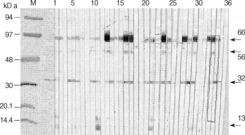
In the immunoblotting, 4 major bands were observed; 13 kDa, 32 kDa, 56 kDa, 66 kDa from 65 sera. The occurrence of at least 1 major band was denoted as being immunoblot positive (Fig. 1). Combinations of the positive band patterns and the number of corresponding sera (in parentheses) were as follows: 13 kDa (2 sera), 32 kDa (8), 56 kDa (0), 66 kDa (10), 66 + 13 kDa (1), 66 + 32 kDa (19), 66 + 56 kDa (1), 66 + 56 + 32 kDa (10), 66 + 32 + 13 kDa (4), 66 + 56 + 32 + 13 kDa (1), 32 + 13 kDa (3), and 32 + 56 kDa (7). Of these combinations, 66 kDa and 32 kDa were most frequent bands. The number of sera positive to 13 kDa was 10 (15.4%), to 32 kDa was 52 (80%), to 56 kDa was 19 (29.2%), and to 66 kDa was 46 (70.8%). The number of sera positive to both 66 kDa and 32 kDa was 34 (35.1%). With ELISA, 63 sera (65.0%) were positive to Toxocara ES. There was no significant correlation between the IgG ELISA titer and the degree of eosinophilia (r = 0.156, P = 0.156). The number of sera positive to both immunoblot and ELISA was 58 (60.0%), the number negative to both was 27 (27.8%), the number of immunoblot positive and ELISA negative was 7 (7.2%), and the number of immunoblot negative and ELISA positive was 5 (5.2%).

DISCUSSION

Although it is known that eosinophilia is associated with toxocariasis, there have been few serological studies on the relationship between toxocariasis and eosinophilia. In a previous study, 14 (93.3%) of 15 patients with eosinophilia showed positive reactions in a Toxocara serologic test [7]. In the present study, the immunoblot positive rate was 67%, and that by ELISA 65%. These are comparable with the study of Kwon et al. [8]. The difference may have been due to a difference in the subjects assessed. The present study targeted apparently healthy adults, whereas Kwon et al. [8] studied patients who visited a University Hospital.
The causes of eosinophilia are known to be mainly allergic diseases, parasitic diseases, cancer, and rheumatic diseases. Among the parasitic diseases, toxocariasis may be the most common cause of eosinophila. The sera used in the present study were from people without any clinical symptoms whose health status was regularly checked with a comprehensive medical test. The cross reaction to other helminthic infections is negligible [3]. In the above results, the seropositive rate for both the immunoblot and ELISA at 60.0% was very high in comparison with that of the previous data from apparently healthy rural adults (5%). There were 5 sera (5.2%) that were immunoblot negative and ELISA positive. These may be false positives in ELISA results. There were also 7 (7.2%) sera that were immunoblot positive but ELISA negative. These may be false negatives in ELISA or they may have originated from a low titer, although the antibody reaction is specific to certain bands. To get more accurate information on the active toxocariasis among people with eosionophilia, further comparative studies with enough numbers of the reference group are needed. Also, the possibility of cross reactions with other helminthic diseases should be eliminated.
In the present study, the immunoblotting band pattern was different from that of the previous studies. Maizel et al. [9] reported 32, 55, 70, 120 kDa and 400 kDa bands. Magnaval et al. [10] mentioned 2 groups of bands: a high molecular weight group 132, 147, and 200 kDa, and a low molecular weight group 24, 28, 30, and 35 kDa. The low molecular weight bands readily cross-react with other parasitic nematode infections [10]. Nunes et al. [11] reported 5 groups: bands larger than 205 kDa, bands near 205 kDa, 116-97 kDa, 55-50 kDa, and 35-29 kDa. In the present study, high molecular bands above 66 kDa could not be detected. The different immunoblotting band patterns may have originated from differences in the ways the antigen was acquired, different laboratory techniques, variability of the epitopes of antigens or cross reaction with other helminthic infections. To overcome this variability, selection of a specific antigen and production of recombinant antigens is necessary. Double-sandwich ELISA with Toxocara specific antigen could also be applied for better screening of toxocariasis [12].
No clinical information on the symptoms, including atopy or allergies of the serum donors, was collected, since they were apparently healthy people. To detect a current active infection of toxocariasis, IgE needs to be tested by ELISA and an assessment of eosinophil cationic protein (ECP) is required [7,13]. People that test positive may have covert toxocariasis, so that a primary health care may need to include screening for toxocariasis among those with eosinophila [14]. This would prevent misdiagnosis of toxocariasis as idiopathic hypereosinophilic syndrome or cancer, which may result in inappropriate clinical interventions [15].

ACKNOWLEDGEMENTS

This work was supported by the Korea Research Foundation Grant (KRF-2003-015-E00113).

REFERENCES

1. Magnaval JF, Michault A, Calon N, Charlet JP. Epidemiology of human toxocariasis in La Reunion. Trans R Soc Trop Med Hyg. 1994. 88:531-533. PMID: 7992328.
crossref
2. Kim YH, Huh S. Prevalence of Toxocara canis, Toxascaris leonina and Dirofilaria immitis in dogs in Chuncheon, Korea (2004). Korean J Parasitol. 2005. 43:65-67. PMID: 15951642.
crossref pmid pmc
3. Park HY, Lee SU, Huh S, Kong Y, Magnaval JF. A seroepidemio-logical survey for toxocariasis in apparently healthy residents in Gangwon-do, Korea. Korean J Parasitol. 2002. 40:113-117. PMID: 12325440.
crossref pmid pmc
4. Malla N, Aggarwal AK, Mahajan RC. A serological study of human toxocariasis in north India. Natl Med J India. 2002. 15:145-147. PMID: 12186327.

5. Logar J, Soba B, Kraut A, Stirn-Kranjc B. Seroprevalence of Toxocara antibodies among patients suspected of ocular toxocariasis in Slovenia. Korean J Parasitol. 2004. 42:137-140. PMID: 15381862.
crossref pmid pmc
6. Fillaux J, Santillan G, Magnaval JF, Jensen O, Larrieu E, Sobrino-Becaria CD. Epidemiology of toxocariasis in a steppe environment: the Patagonia study. Am J Trop Med Hyg. 2007. 76:1144-1147. PMID: 17556626.
pmid
7. Choi JH, Suh YJ, Jung JW, Song HJ, Suh CH, Huh S, Nahm DH, Park HS. Clinical significance of serum ECP and sero-prevalence of human toxocariasis in patients with eosinophilia. J Asthma Allergy Clin Immunol. 2003. 23:26-32.

8. Kwon NH, Oh MJ, Lee SP, Lee BJ, Choi DC. The Clinical impact of toxocariasis in patients with unknown eosinophilia. J Asthma Allergy Clin Immunol. 2005. 25:299-304.

9. Maizels RM, Kennedy MW, Meghji M, Robertson BD, Smith HV. Shared carbohydrate epitopes on distinct surface and secreted antigens of the parasitic nematode Toxocara canis. J Immunol. 1987. 139:207-214. PMID: 2438350.
pmid
10. Magnaval JF, Fabre R, Maurieres P, Charlet JP, de Larrard B. Application of the western blotting procedure for the immunodiagnosis of human toxocariasis. Parasitol Res. 1991. 77:697-702. PMID: 1805214.
crossref pmid
11. Nunes CM, Tundisi RN, Garcia JF, Heinemann MB, Ogassawara S, Richtzenhain LJ. Cross-reactions between Toxocara canis and Ascaris suum in the diagnosis of visceral larva migrans by western blotting technique. Rev Inst Med Trop Sao Paulo. 1997. 39:253-256. PMID: 9661302.
crossref
12. Iddawela RD, Rajapakse RP, Perera NA, Agatsuma T. Characterization of a Toxocara canis species-specific excretory-secretory antigen (TcES-57) and development of a double sandwich ELISA for diagnosis of visceral larva migrans. Korean J Parasitol. 2007. 45:19-26. PMID: 17374974.
crossref pmid pmc
13. Magnaval JF, Berry A, Fabre R, Morassin B. Eosinophil cationic protein as a possible marker of active human Toxocara infection. Allergy. 2001. 56:1096-1099. PMID: 11703226.

14. Magnaval JF, Glickman LT, Dorchies P, Morassin B. Highlights of human toxocariasis. Korean J Parasitol. 2001. 39:1-11. PMID: 11301585.
crossref pmid pmc
15. Lim JH, Lee KS. Eosinophilic infiltration in Korea: idiopathic? Korean J Radiol. 2006. 7:4-6. PMID: 16549949.
crossref
Fig. 1
Band patterns after immunoblotting of human sera with eosinophilia against Toxocara excretory-secretory antigen. M: protein maker, 1-32: Eosinophilia sera, 33-34: Positive controls, 35: Other sample serum, 36: Negative control.
kjp-46-29-g001