Berenji, Fata, and Hosseininejad: A case of Moniliformis moniliformis (Acanthocephala) infection in Iran
Cited By
Citations to this article as recorded by
An unexpected case of a Japanese wild boar (Sus scrofa leucomystax) infected with the giant thorny-headed worm (Macracanthorhynchus hirudinaceus) on the mainland of Japan (Honshu)
Koichiro Kamimura, Kenzo Yonemitsu, Ken Maeda, Seiho Sakaguchi, Aogu Setsuda, Antonio Varcasia, Hiroshi Sato
Parasitology Research.2018; 117(7): 2315.     CrossRef
Helminth fauna of small mammals from public parks and urban areas in Bangkok Metropolitan with emphasis on community ecology of infection in synanthropic rodents
Yossapong Paladsing, Kittiyaporn Boonsri, Wipanont Saesim, Bangon Changsap, Urusa Thaenkham, Nathamon Kosoltanapiwat, Piengchan Sonthayanon, Alexis Ribas, Serge Morand, Kittipong Chaisiri
Parasitology Research.2020; 119(11): 3675.     CrossRef
The first finding of Moniliformis moniliformis (Acanthocephala, Moniliformidae) in Belarus
V. V. Shimalov
Journal of Parasitic Diseases.2018; 42(2): 327.     CrossRef
Morphological characterization of Moniliformis moniliformis isolated from an Iraqi patient
Amal Khudair Khalaf, Baydaa Furhan Swadi, Hossein Mahmoudvand
Journal of Parasitic Diseases.2021; 45(1): 128.     CrossRef
A systematic review and meta-analysis on prevalence of gastrointestinal helminthic infections in rodents of Iran: An emphasis on zoonotic aspects
Yazdan Hamzavi, Mohammad Taghi Khodayari, Afshin Davari, Mohammad Reza Shiee, Seyed Ahmad Karamati, Saber Raeghi, Hadis Jabarmanesh, Helia Bashiri, Arezoo Bozorgomid
Heliyon.2024; 10(11): e31955.     CrossRef
Acanthocephalan infection probably associated with cockroach exposure in an infant with failure-to-thrive
Joel M. Andres, Joan E. English, Ellis C. Greiner
Pediatric Infectious Disease.2014; 6(2): 63.     CrossRef
Risk factors for different intestinal pathogens among patients with traveler's diarrhea: A retrospective analysis at a German travel clinic (2009–2017)
Thomas Theo Brehm, Marc Lütgehetmann, Egbert Tannich, Marylyn M. Addo, Ansgar W. Lohse, Thierry Rolling, Christof D. Vinnemeier
Travel Medicine and Infectious Disease.2020; 37: 101706.     CrossRef
Morphological and genetic description ofMoniliformis necromysisp. n. (Archiacanthocephala) from the wild rodentNecromys lasiurus(Cricetidae: Sigmondontinae) in Brazil
A.P.N. Gomes, N.A. Costa, R. Gentile, R.V. Vilela, A. Maldonado
Journal of Helminthology.2020;[Epub]     CrossRef
Characterization of the complete mitochondrial genomes of the zoonotic parasites Bolbosoma nipponicum and Corynosoma villosum (Acanthocephala: Polymorphida) and the molecular phylogeny of the order Polymorphida
Dai-Xuan Li, Rui-Jia Yang, Hui-Xia Chen, Tetiana A. Kuzmina, Terry R. Spraker, Liang Li
Parasitology.2024; 151(1): 45.     CrossRef
Effectiveness of Various Anthelmintics in the Treatment of Moniliformiasis in Experimentally Infected Wistar Rats
Dennis J. Richardson, Cheryl D. Brink
Vector-Borne and Zoonotic Diseases.2011; 11(8): 1151.     CrossRef
Potentially Zoonotic Helminthiases of Murid Rodents from the Indo-Chinese Peninsula: Impact of Habitat and the Risk of Human Infection
Kittipong Chaisiri, Praphaiphat Siribat, Alexis Ribas, Serge Morand
Vector-Borne and Zoonotic Diseases.2015; 15(1): 73.     CrossRef
Moniliformis moniliformis Infection in Two Florida Toddlers
Allison F. Messina, Frederick J. Wehle, Si Intravichit, Kenneth Washington
Pediatric Infectious Disease Journal.2011; 30(8): 726.     CrossRef
Repertory of eukaryotes (eukaryome) in the human gastrointestinal tract: taxonomy and detection methods
I. Hamad, D. Raoult, F. Bittar
Parasite Immunology.2016; 38(1): 12.     CrossRef
Human Acanthocephaliasis: a Thorn in the Side of Parasite Diagnostics
Blaine A. Mathison, Ninad Mehta, Marc Roger Couturier, Romney M. Humphries
Journal of Clinical Microbiology.2021;[Epub]     CrossRef
Morphological and Molecular Descriptions of Macracanthorhynchus ingens (Acanthocephala: Oligacanthorhynchidae) Collected from Hedgehogs in Iran
Mohsen Najjari, Amir Tavakoli Kareshk, Mohammad Darvishi, Gholamreza Barzegar, Majid Dusti, Mohammad Hasan Namaei, Ebrahim Shafaie, Rahmat Solgi, Payam Behzadi
Interdisciplinary Perspectives on Infectious Diseases.2022; 2022: 1.     CrossRef
The present status of human helminthic diseases in Iran
M. B. Rokni
Annals of Tropical Medicine & Parasitology.2008; 102(4): 283.     CrossRef
Bacterial load of German cockroach (Blattella germanica) found in hospital environment
Taha Menasria, Fatima Moussa, Souad El-Hamza, Samir Tine, Rochdi Megri, Haroun Chenchouni
Pathogens and Global Health.2014; 108(3): 141.     CrossRef
Rodent-borne diseases and their public health importance in Iran
Mohammad Hasan Rabiee, Ahmad Mahmoudi, Roohollah Siahsarvie, Boris Kryštufek, Ehsan Mostafavi, Peter J. Krause
PLOS Neglected Tropical Diseases.2018; 12(4): e0006256.     CrossRef
Zoonotic Gastrointestinal Helminths in Rodent Communities in Southern Guatemala
Wendy C. Hernández, David Morán, Federico Villatoro, Manuel Rodríguez, Danilo Álvarez
Journal of Parasitology.2020; 106(3): 341.     CrossRef
Paleoparasitology of Human Acanthocephalan Infection: A Review and New Case from Bonneville Estates Rockshelter, Nevada, U.S.A.
Katelyn McDonough, Taryn Johnson, Ted Goebel, Karl Reinhard, Marion Coe
Journal of Parasitology.2023;[Epub]     CrossRef
Parasitism of Prehistoric Humans and Companion Animals from Antelope Cave, Mojave County, Northwest Arizona
Martín H. Fugassa, Karl J. Reinhard, Keith L. Johnson, Scott L. Gardner, Mônica Vieira, Adauto Araújo
Journal of Parasitology.2011; 97(5): 862.     CrossRef
Endoparasites of Small Mammals in Edo State, Nigeria: Public Health Implications
Clement Isaac, Benjamin Igho Igbinosa, John Asekhaen Ohiolei, Catherine Eki Osimen
The Korean Journal of Parasitology.2018; 56(1): 93.     CrossRef
Imaging Features of Pediatric Pentastomiasis Infection: a Case Report
Can Lai, Xi Qun Wang, Long Lin, De Chun Gao, Hong Xi Zhang, Yi Ying Zhang, Yin Bao Zhou
Korean Journal of Radiology.2010; 11(4): 480.     CrossRef
Molecular Characterization of a New Moniliformis sp. From a Plateau Zokor (Eospalax fontanierii baileyi) in China
Guo-Dong Dai, Hong-Bin Yan, Li Li, Lin-Sheng Zhang, Zhan-Long Liu, Sheng-Zhi Gao, John Asekhaen Ohiolei, Yao-Dong Wu, Ai-Min Guo, Bao-Quan Fu, Wan-Zhong Jia
Frontiers in Microbiology.2022;[Epub]     CrossRef
Helminthofauna Diversity in Synanthropic Rodents of the Emilia-Romagna Region (Italy): Implications for Public Health and Rodent Control
Filippo Maria Dini, Carlotta Mazzoni Tondi, Roberta Galuppi
Veterinary Sciences.2024; 11(11): 585.     CrossRef
An Overview of German Cockroach, Blattella germanica, Studies Conducted in Iran
Hassan Nasirian
Pakistan Journal of Biological Sciences.2010; 13(22): 1077.     CrossRef
Acanthocephala Species of Mammals in Türkiye and A New Species Record from Foxes
Mehmet Öztürk, Şinasi Umur
Turkish Journal of Parasitology.2024; 48(1): 66.     CrossRef
First record of Moniliformis moniliformis (Bremser, 1811) (Moniliformida: Moniliformidae) in mice of the genus Apodemus kaup, 1829 (Rodentia: Muridae) in Serbia
Božana Tošić, Borislav Čabrilo, Milan Miljević, Marija Rajičić, Branka Bajić, Ivana Budinski, Olivera Bjelić-Čabrilo
Kragujevac Journal of Science.2024; 46(1): 163.     CrossRef

Abstract

Only a few cases of Acanthocephala infections have been reported in humans, and Moniliformis moniliformis is the most common species around the world. We report here a case of infection with M. moniliformis, which passed in the stool of a 2-year-old girl in Iran. The patient had abdominal pain, diarrhea, vomiting, and facial edema. According to her mother, the patient had habit of eating dirt and once a cockroach was discovered in her mouth. In stool examination, eggs of M. moniliformis were not found. She was treated with levamisole and the clinical symptoms reduced within 2 weeks. The specimen contained 2 pieces of a female worm with a total length of 148 mm lacking the posterior end. The spiral musculature of the proboscis receptacle and the shape of the trunk allowed its generic determination. Previously 2 cases of M. moniliformis infection were reported in Iran. This is the 3rd case of M. moniliformis infection in Iran.

INTRODUCTION

Moniliformis moniliformis is an endoparasite found in the intestine of its definitive host and found in most parts of the world (Ikeh et al., 1992; Roberts and Janovy, 2005). Common definitive hosts include rats, mice, hamsters, dogs, and cats. Humans may be incidental hosts, with the worm in the small intestine. The definitive host is infected by eating the beetle or cockroach (Ikeh et al., 1992).
Human infections with this parasite have been reported in Australia, Asia (Pakistan, Indonesia, Bangladesh, Japan, and Iran), Europe (Russia and Italy), America (Texas, Florida, Alaska, and Honduras), and Africa (Sudan, Nigeria, Egypt, and Madagascar) (Muller, 1975; Beaver et al., 1984; Ikeh et al., 1992; Baker et al., 2000). In this paper we report a young girl from Taybad (Khorasan-e-razavi province state), Iran, who had a history of eating a cockroach and passed a female M. moniliformis worm in her stool.

CASE RECORD

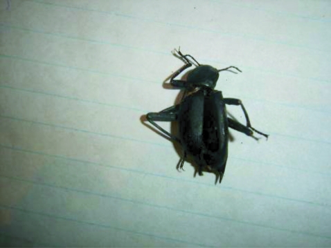
The patient is a 2-year-old girl, who according to her mother's report, passed 6 worms in the stool. The last worm was sent to our laboratory. According to her mother, the patient had habit of pica (eating dirt), and once a cockroach was discovered in her mouth by her mother (Fig. 1). The patient had abdominal pain, diarrhea, and vomiting. Moreover, she had edema in her body and more apparently in the face.
In stool examination, eggs of Moniliformis moniliformis were not found, but Giardia cysts were detected. CBC (Hb, WBC number, and percentage of eosinophils) was normal. She was treated with levamisole (3 mg/kg/day for 3 days) in lieu of pyrantel pamoate. After the treatment, the patient's clinical symptoms reduced within 2 weeks and there was no more report of passing worms.

Description of the parasite

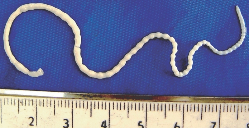
The submitted specimen contained 2 pieces of a worm with a total length of 148 mm and the posterior end was lost. The spiny head was more or less destroyed, and thus its detailed morphology was not clearly visible. Based on the size (more than 130 mm), this worm was regarded as a female. The worm was white with regular horizontal lines, which imitated segmented appearance. The spiral musculature of the proboscis receptacle and the shape of the trunk allowed its generic and specific determination (Fig. 2).

DISCUSSION

The acanthocephalans (spiny-headed worms) were formerly included in the same phylum as the nematodes (Garcia, 2001). These worms are a small group of pseudocoelomate endoparasites with a number of unique structural and biological features (Marquards et al., 2000). A prominent characteristic of the phylum, from which it receives its name, is a protrusible proboscis that is armed with recurved hooks, by which the worm attaches to the wall of the intestine of the definitive host (Marquards et al., 2000; Garcia, 2001). All members of this phylum are parasites and sexes are separate (Marquards et al., 2000).
Records of Acanthocephala infections in human are rare, but cases of infections by 7 different species have been reported (Roberts and Janovy, 2005). These species include Acanthocephalus bufonis, Corynosoma strumosum, Macracanthorhynchua hirudinaceus, Moniliformis moniliformis, Bolbsoma sp. (Counselman et al., 1989; Marquards et al., 2000; Roberts and Janovy, 2005), Macracanthorhynchua ingens (Counselman et al., 1989; Roberts and Janovy, 2005), and Acanthocephalus rauschi (Marquards et al., 2000; Roberts and Janovy, 2005). In these instances, it is probable that the man ate the paratenic host raw (Roberts, and Janovy, 2005).
Moniliformis moniliformis is found in most parts of the world. The male and female worms differ slightly in size with the male averaging 4-13 cm and the females averaging 10-27 cm (Lawlor et al., 1990). The male has a copulatory bursa (Beaver et al., 1984). Adult worms are white, and because of pitted horizontal lines on the body surface, they appear to be segmented (Beaver et al., 1984; Roberts and Janovy, 2005). The body consists of a proboscis, located on the anterior end of the worm, a neck, and a trunk (Roberts and Janovy, 2005). The cylindrical proboscis is hollowed with a thin muscular wall, and armed with 12-15 rows of recurved hooks, 7-8 hooks to each row (Beaver et al., 1984; Roberts and Janovy, 2005). We regret that the proboscis of our specimen was destroyed and not clearly visible.
Two cases of M. moniliformis infection have been reported previously in Iran (Sahba et al., 1970; Moayedi et al., 1971). The first case was an 18-mo-old child from a village of Zabol (Sistan-o-Baluchestan province state) in southeast Iran with a history of pica, whose symptoms were abdominal distention, anorexia, weakness, vomiting, and foamy diarrhea. He passed 15 M. moniliformis worms after a successful treatment with anthelmintic drugs (Sahba et al., 1970). The second case was a patient from Isfahan in central Iran that was reported by Moayedi et al. (Counselman et al., 1989; Marquards et al., 2000). In the first previously reported case in Iran, laboratory findings included leukocytosis (16,000/mm3) with high eosinophilia (24%) (Marquards et al., 2000), but in our case all laboratory findings were normal.
The symptoms of M. moniliformis infection often include abdominal pain, vomiting, severe fatigue, diarrhea, dizziness, irritability, anorexia, pallor, weight loss, weakness, somnolence, tinnitus, cough, jaundice, protuberant abdomen, retarded development in children, fever, and intermittent burning sensations around the umbilicus (Beaver et al., 1984; Counselman et al., 1989; Ikeh et al., 1992; Marquards et al., 2000; Roberts and Janovy, 2005). In our case, according to the patient's mother, the child complained of loss of appetite and abdominal pain. Moreover, the patient has edema especially in her face. However, the presence of Giardia cysts in her stool may have contributed to these symptoms.
The presence of 12-15 rows on the proboscis with 7-8 spines on each row is the main characteristic of this species (Roberts and Janovy, 2005). In the definitive hosts damage to intestinal mucosa may occur by penetration of proboscis, and is compounded by the worm's tendency to release their hold occasionally and reattach at another place. These damages may cause inflammation, ulcer, and hemorrhages in the definitive host (Marquards et al., 2000; Garcia, 2001; Roberts and Janovy, 2005).
When infected with M. moniliformis, most cockroach species move more slowly, and travel shorter distances (Moore et al., 1994; Libersat and Moore, 2000; Roberts and Janovy, 2005). This increases the likelihood of parasite's predation by the definitive host (Libersat et al., 2000).
Due to people's main occupation in this village (Khiyaban), which is animal husbandry, and poor health, lower socioeconomic condition, as well as abundance of the intermediate host of M. moniliformis, we believe that the infection may exist in other people, specially the kids of this rural area. Health education for the families particularly mothers to promote better hygiene and prevention of pica is an important preventive method.

REFERENCES

1. Beaver PC, Jung RC, Cupp EW. Clinical Parasitology. 1984. 9th ed. Philadelphia, USA: Lea & Febiger; 544-547.

2. Counselman K, Field C, Lea G, Nickol B, Neafie R. Moniliformis moniliformis from a child in Florida. Am J Trop Med Hyg. 1989. 41:88-90. PMID: 2764231.
pmid
3. Garcia LS. Diagnostics Medical Parasitology. 2001. 4th ed. Washington D.C., USA: ASM press; 477-501.

4. Ikeh Z, Anosike J, Okon E. Acanthocephalan infection in man in northern Nigeria. J Helminthol. 1992. 66:241-242. PMID: 1452999.
crossref pmid
5. Lawlor B, Read A, Keymer A, Parveen G, Crompton D. Non-random mating in a parasitic worm: mate choice by males? Anim Behav. 1990. 40:870-876.
crossref
6. Libersat F, Moore J. The parasite Moniliformis moniliformis alters the escape response of its cockroach host Periplaneta americana. J Insect Behav. 2000. 13:103-110.
crossref
7. Marquards WC, Demaree RS, Grieve RB. Parasitology and Vector Biology. 2000. 2nd ed. San Diego, USA: Harcourt academic Press; 491-499.

8. Moayedi B, Izadi M, Maleki M, Ghadirian E. Human infection with Moniliformis moniliformis (Bremser, 1811) Travassos, 1915 (syn. Moniliformis dubius). Report of a case in Isfahan, Iran. Am J Trop Med Hyg. 1971. 20:445-448. PMID: 5104317.
pmid
9. Moore J, Freehling M, Gotelli NJ. Altered behavior in two species of blattid cockroaches infected with Moniliformis moniliformis (Acanthocephala). J Parasitol. 1994. 80:220-223. PMID: 8158464.
crossref pmid
10. Muller R. Worm and Disease, a Manual of Medical Helminthology. 1975. 1st ed. London, UK: William Heinemann Medical Book Ltd; 64.

11. Roberts L, Janovy JR. Foundation of Parasitology. 2005. 7th ed. Boston, USA: McGraw-Hill; 493-509.

12. Sahba GH, Arfaa F, Rastegar M. Human infection with Moniliformis dubius (Meyer, 1931) in Iran. Trans Roy Soc Trop Med Hyg. 1970. 64:284-286. PMID: 5449054.
crossref
13. Taraschewski H. In Baker JR, Muller R, Rollinson D eds, Host-parasite interaction in Acanthocephala: a morphological approach. Advances in parasitology. 2000. USA: Academic Press; 1-179.
crossref
Fig. 1
The cockroach discovered in the patient's mouth.
kjp-45-145-g001
Fig. 2
The specimen contains 2 pieces of a female Moniliformis moniliformis with a total length of 148 mm lacking the posterior end, which passed in the stool of the patient.
kjp-45-145-g002
1