INTRODUCTION
Oculotrema hippopotami Stunkard, 1924 (Oculotrematinae Yamaguti, 1968), the only monogenean known for successful colonization on a mammal poses many unanswered questions. It was originally described from Hippopotamus amphibius Linnaeus, 1758 from the Giza Zoo (Egypt), but has also been reported from Sudan and Uganda [1], Zimbabwe [2] and South Africa [3,4]. Stunkard gave a comprehensive description of O. hippopotami, though based on highly contracted specimens [5]. Du Preez et al. [4] amended the original description of an adult by adding information on size of the parasite, particulars of egg and copulatory system structure, redefined morphological parameters of adults, and revealed the presence of a bucco-oesophageal canal, a uterine evagination, an operculated egg, the retention of marginal hooklets in the mature parasite and the ability to double its length and feed over a large area around its position of attachment. They reported a maximum of 24 parasites on a single eye and a maximum of 37 on an individual host specimen. The prevalence was found to be 75% for adults, 85% for sub-adults and 90% for the total sample [4]. In addition, Du Preez et al. and Tinsley [4,6] studied oncomiracidia and Thurston studied the larvae of O. hippopotami [7]. Sub-adults of O. hippopotami were briefly mentioned in a few publications but never were studied exclusively. Although Thurston [7,8] and Du Preez et al. [4] mentioned immature worms (sub-adults), but none provided more details about them.
Some studies have been devoted to the habitat of this monogenean, the hippopotamus eye tissues, but their histopathological changes at the place of attachment of the parasite have never been studied. The same applies to details of the distal parts of the copulatory system that have not been studied using scanning electron microscopy (SEM) [9]. To date there are no comparative studies on morphological and metrical characteristics of adults and sub-adults of O. hippopotami. In addition, this is the first study on the structural analysis of this parasite comparing adult and sub-adult forms and providing further information on this unique mammalian monogenean.
MATERIALS AND METHODS
Host animals and sample collection
A total of 6 hippopotami (Hippopotamus amphibius: 4 males and 2 females) were examined in Mpumalanga Province, South Africa in August and September 2016. The geographical coordinates for the hippopotamus examined are listed in Table 1. After collecting monogeneans, the host tissue was biopsied and fixed with 10% buffered formalin for histopathological studies. For the morphological examination, parasites were relaxed in a small Petri-dish with 0.9% saline, and while between 2 microscope slides were fixed by 70% ethanol.
Microscopical examination
Worms were stained in Mayer’s acid carmine, destained in 4% hydrochloric acid in 70% ethanol, dehydrated in ascending concentrations of ethanol (12 hr each), and cleared in 100% xylene and then in 50% Canada balsam and 50% xylene (12 hr each). Whole worms were then mounted in Canada balsam.
Scanning Electron Microscopy (SEM)
Specimens fixed and stored in 70% ethanol were processed following standard methods [11], which included critical point drying in sample baskets and mounted on SEM sample mounts (stubs) using conductive double-sided carbon tape. Samples were coated with gold and palladium for 3 min using a Polaron #3500 sputter coater (Quorum (Q150 TES) www.quorumtech.com) establishing an approximate thickness of 20 nm. Samples were placed and observed in an FEI Helios Dual Beam Nanolab 600 (FEI, Hillsboro, Oregon, USA) Scanning Electron Microscope and digital images were obtained in the Nanolab software system (GEI, Hillsboro, Oregon, USA). Samples were viewed under low vacuum conditions using 10 kV, spot size 2, 0.7 Torr using a GSE detector.
Energy dispersive X-ray analysis (EDXA)
Standard methods, similar to the SEM procedure, were used for preparation. Specimens were examined and positioned with the above SEM instrument, which was equipped with a Phoenix energy-dispersive x-ray analyzer (FEI). X-ray spot analysis and live scan analysis were performed at 16 kV with a spot size of 5 and results were recorded on charts and stored with digital imaging software attached to a computer. The TEAM (Texture and Elemental Analytical Microscopy), a modification of the EDXA system (Energy Dispersive X-ray analysis), software system (FEI) was used. The data included the weight and atom percentages of the detected elements following correction factors.
Histopathological observations
Biopsied host tissues with attached monogeneans previously fixed with 10% formalin were processed by the standard methods for paraffin-blocked specimens [12,13]. The paraffin-blocked tissue was sectioned at 4–5 microns, placed on glass slides and stained with hematoxylin and eosin (H&E). Additional sections were stained with Mallory’s trichrome to study the pathological responses to the parasite [14]. H&E as a standard stain for tissue whereas Mallory’s trichrome to differentiate granular tissue typical of parasite invasion. The prepared sections were viewed with a LSM laser (Carl Zeiss, Thornwood, New York, USA) equipped compound microscope and representative pictures were taken at varying magnifications with a digital camera. The histopathological sections were selected from a much larger collection of sections on 63 glass slides in RAH’s collection. The following abbreviations were used in histopathological studies: CF–collagenous fibers, Ep–stratified epithelium, H–hemorrhaging, N–necrosis, S–sucker.
RESULTS
Infection status with O. hippopotami
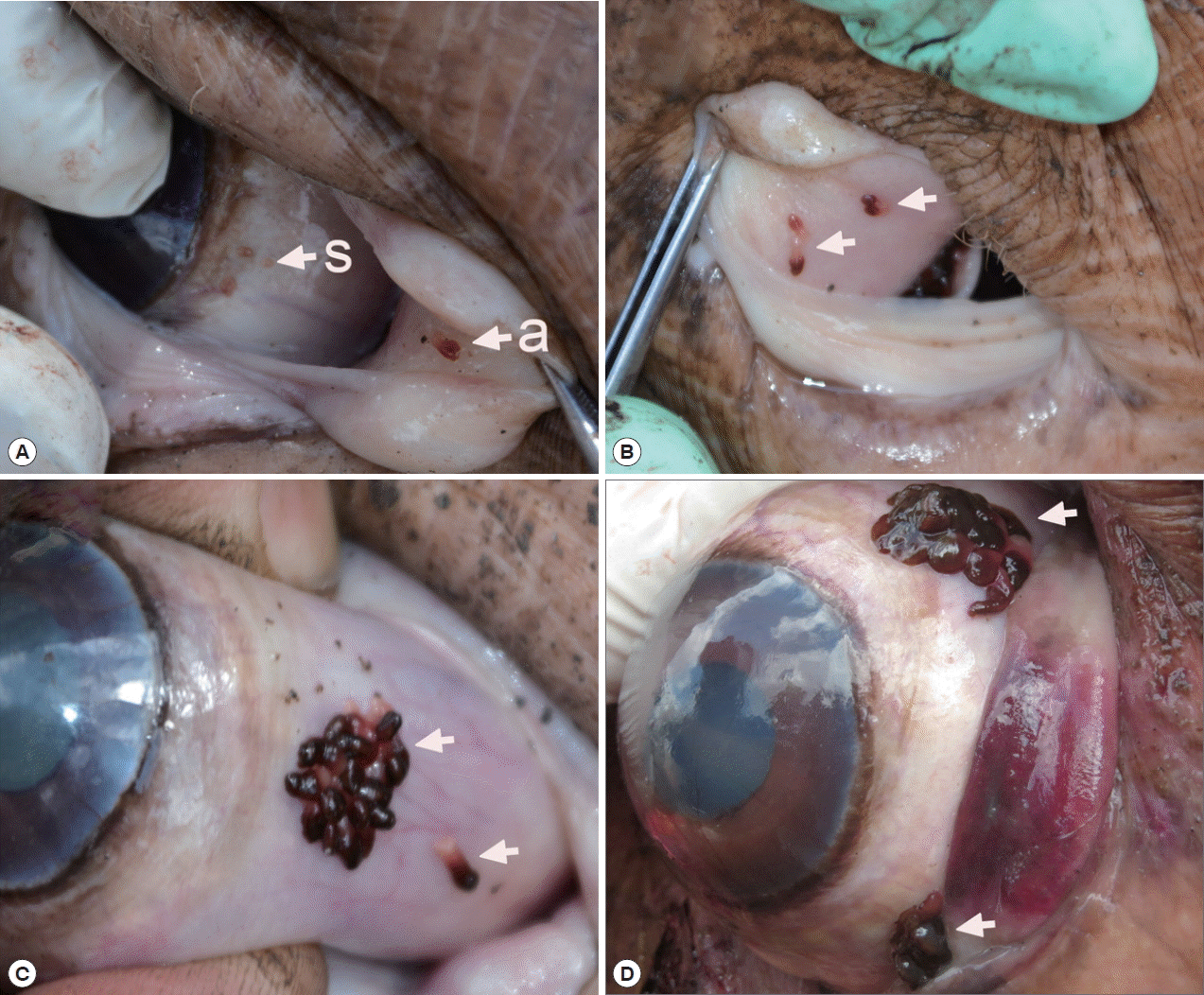
A total 5 out of 6 examined hippopotami (83.3%) were infected with O. hippopotami, intensity range 4–97 (36) (Table 1). Commonly both eyes of hippopotami were infected (Table 1). Parasites had a firm grip on host tissues and they were never found detached. Most of these parasites were found on the nictitating membrane of eyes, and seldom on the side of the eye globe (Fig. 1). Different attachment sites were seen on the host’s eyes (Fig. 1C, D) with parasites forming clusters on several cases.
Morphological characteristics of O. hippopotami
Distinguishing features between adults and sub-adults of O. hippopotami Monogeneans of 2 age groups, adults and sub-adults were recorded from the hosts. Two hosts were infected with adults only, while 1 host was uninfected (Table 1). Adults were easily distinguished from sub-adults with the naked eye: they were large, with a cherry-red anterior 2/3 of the body, collected in clusters of at least 5 or more individuals (Figs. 1, 2). Sub-adults were found separately from the cluster of adults and were noticeably smaller (Fig. 2), with an underdeveloped reproductive system. They were colorless, or white-yellowish to slightly pinkish (Fig. 1A). X-ray scans also showed major differences between the sub-adult and adult (see below). The most distinctive feature between the 2 age groups was the body size. Adults of O. hippopotami were on average 4.6 times larger than sub-adults (Fig. 2A, B; Table 2). Measurements of adults and sub-adults and comparisons with the original description and latest report [4] provided in Table 2. Sub-adults found in the present study varied in size and degree of development. The presence of a mixture of different generations on the same host for Oculotrema (in contrast to Polystoma Zeder, 1800) was a novel observation in the present study.
Morphological features of an adult O. hippopotami (Fig. 2A and Fig. 3A)
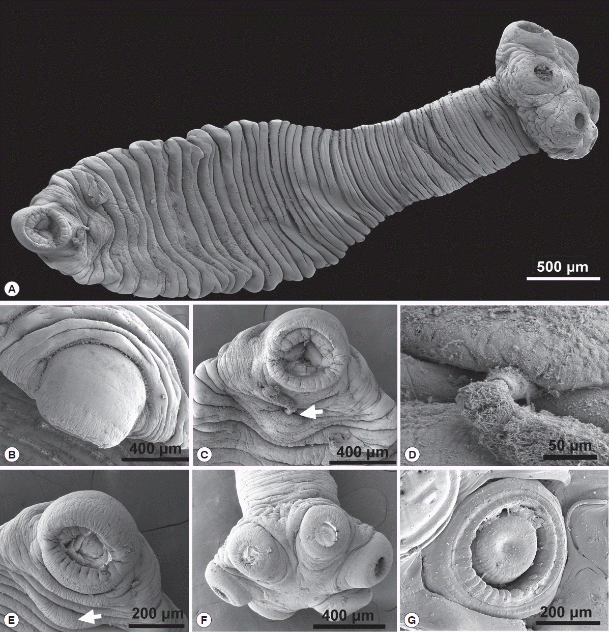
Measurements listed in Table 2. The average haptor width/haptor sucker ratio was 3.2 (Table 2) and the average body/haptor length ratio 6.5. The musculature of the oral sucker in the adult well developed, with few tissue layers (Fig. 3C). Ventral part of the body flattened, possibly due to the life-long mode of attachment of the worms. Likewise, the oral sucker opening also oriented in the same plane (Fig. 3B). The genital system is shown on Fig. 2C. The muscular cirrus with ejaculated sperms are shown in Figs. 3C, D. Arrows on Figs. 3C, E show the position of a genital papilla present in adults of O. hippopotami. Figs. 3F, G show the haptor and one of 6 well-developed suckers of an adult, designed for a very firm long-term attachment to the host tissue. The haptor width/haptor sucker ratio measured for the current study was 3.45 (3.62–3.67) in comparison with up to 4.5 of Du Preez et al. [4]. Tegument consists of plates elastically connected to each other and strands between plates. The attachment of sclerotized structures also differs structurally (see X-rays analysis part of the study). The external edges of suckers in the sub-adult form may be folded or corrugated (Figs. 4A, F, G).
Morphological features of a sub-adult O. hippopotami (Fig. 2B and Fig. 4A)
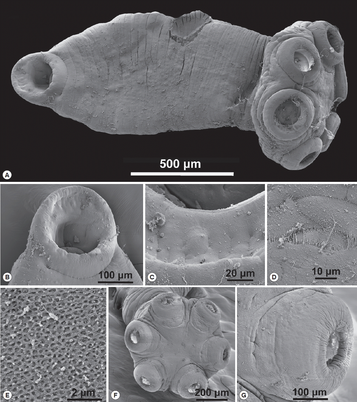
Measurements listed in Table 2. The average haptor width/haptor sucker ratio was 2.9. The average body/haptor length ratio in sub-adults was 3.25. The musculature of the oral sucker undeveloped (Fig. 4B, C for a close view). Ventral part of the body flattened. Oral sucker opening oriented at an angle to the plane of the ventral part of the body (Fig. 4B). Genital papilla not seen. Suckers not as well developed as in the adult forms. Haptor width/haptor sucker ratio was 3.01 (2.50–3.52). Reproductive organs of the smallest sub-adults were not developed. The tegument consists of plates elastically connected to each other (Fig. 4D) with strands between plates. The tegument had a honeycomb-structure (Fig. 4E). En face view of the 6 muscular suckers of O. hippopotami depicted in Fig. 4F. The position of suckers and their orientation was similar to the adult form, but the relative ratio of the sucker to the haptor and haptor to body differed (Table 2). Haptor suckers were smaller and less developed in comparison with adults (Fig. 4G).
Histopathologic findings on the effect of O. hippopotami infections in the eyes of hippopotamus
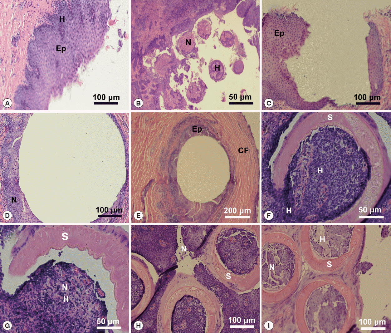
A lateral orbit of the eye socket of a hippopotamus was shown in Fig. 5A. This figure shows the surface of the eye of the host; a stratified epithelial lining with a connective tissue base below the basement membrane. There were extensive hemorrhaging (disruption of capillary vessels) and pooled blood cells as well as necrotic tissue in the area where parasites attached (Fig. 5B), which displayed a section of an infected hippopotamus eye pulled away from the surface of the eye. It displayed the 6 marks of host tissue where suckers attached. An area of the host eyes where specimens of O. hippopotami had been attached shown in Fig. 5C. Note the difference between normal host epithelium and epithelium in the invaded area (bottom left). The eye surface of the host has been altered as a result of the firm grip of parasites. Note tissue necrosis and hemorrhaging on the edges of the marking (Fig. 5D). See also Fig. 5E, showing host epithelial eye cells surrounding the point of attachment. Note the extensive encapsulation with collagenous fibers, in an attempt of the host to “wall-off” the parasite. Fig. 5F displayed the effect of a parasite sucker attaching to the host epithelium. Note the extensive tissue necrosis in the center of the attachment area of the sucker. Also note extensive necrosis caused the sucker and hemorrhaging of host tissue (Fig. 5G). The effect of parasite suckers’ attachment to the surface of the eye and the resulting necrotic tissue inside the sucker depicted in Fig. 5H. Note small capillaries scattered throughout the eye epithelium with red blood cells in the vessels. Compare this to Fig. 5I, which showed 3 of the 6 O. hippopotami suckers with a disruption of eye tissue and hemorrhaging with resulting cell necrosis. Note the corrugated nature of the sucker with specialized collagenous fibers. Compare to Fig. 5G with a single intact worm sucker surrounding host epithelial cells.
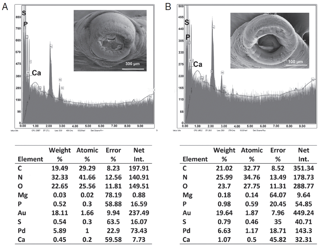
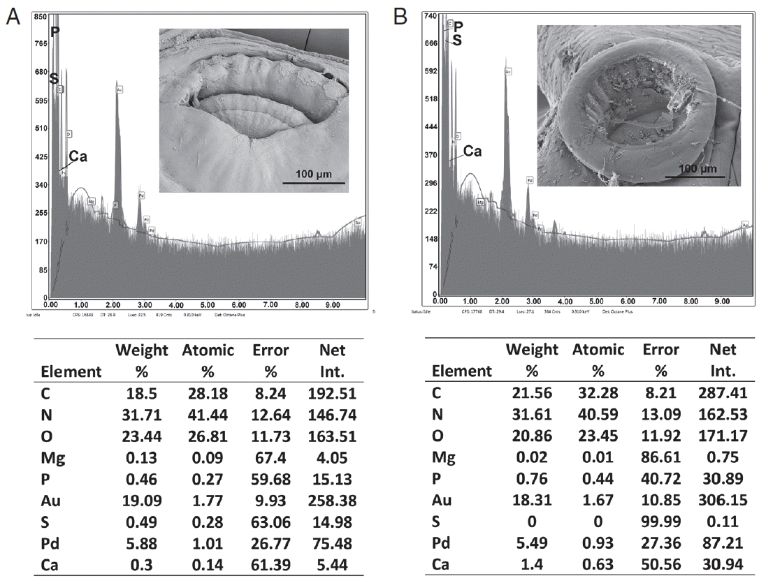
EDXA findings on the adult and sub-adult forms of O. hippopotami
Table 3 and Figs. 6, 7 contained the data for the X-ray scans of the adult and sub-adult worms. Three areas were scanned, i.e., body, mouth and sucker. Duplicate scans were done on different specimens for a qualitative evaluation to understand changes in the chemistry of the organism during its lifespan. The processing and coating chemicals (Au, Pd and Os) were omitted from the table. Note the definite lack of hardening ions (Ca, S, P) in the scanned areas of the sub-adults. The common chemical ions (C, N and O) in living protoplasm were consistent for both life stages.
DISCUSSION
Because of severe drought in August and September 2016 in northeastern South Africa and declining overall condition of hippopotami, a decision was made to control compromised animals from the Kruger National Park Associated Private Nature Reserves (APNR).
The present infection levels of O. hippopotami compare to that reported in the previous studies, and are very high and unique in Monogenea under natural conditions compared to almost all other representatives of Monogenea in general [15], and particularly Polystomatidae [16]. Five of 6 examined animals from the current study were infected. Thurston & Laws reported a prevalence of 76% on a large sample size (960 of 1,263 hippopotami were positive for O. hippopotami) [1]. Du Preez et al. [4] reported a prevalence of 90%. Several factors probably contribute to this phenomenon. Life in clusters ensures fertilization, and, consequently, the release of eggs into the water. A pronounced care for the offspring, the constant contact of the mother with the calf, and the presence of adult individuals in the proximity to the standing water ensures infection. The only serious obstacle is the mechanical influence on the worms of the eyelid during blinking, which they successfully overcome by developing a unique sclerotized suction attachment system. The degree of attachment to the tissues of the host is so strong that in a number of cases, it was practically impossible to tear off the monogeneans from the eye tissue. High levels of infection together with absence of obvious pathogenicity to the host indicate an extremely successful adaptation of this parasite to the host. The effect of the infection of this monogenean on the host does not appear to have an influence on visual acuity, since no studies indicated the presence of the O. hippopotami specimens in the anterior part of the eye or cornea.
New morphological features were revealed in the present study, such as flattened dorsal side of the body provided for the first time with SEM micrographs; the presence of genital papillae observed only in adults. In addition, differences were shown in the orientation of oral suckers between adults and sub-adults. The new information on sperm ejaculatory structures of O. hippopotami was shown with SEM micrographs. New metrical and graphical information obtained for adults and sub-adults were compared with previous studies. Finally, the metrical data on the distal part of the vas deferens is reported for the first time.
Halton and Jennings stated that specimens of O. hippopotami feed on epithelial cells and mucous [17]. Fig. 3B shows an adaptation of the whole body shape to this mode of nutrition - flattened ventral side of the body contrasting with arched dorsal side. Earlier, Moeng et al. [18] reported that the oral sucker is designed for long-term positioning of the worm, clamped between the host’s eyeball and eyelid. It was reported that the oral sucker morphology reflects the mode of feeding. The sub-terminal flat mouth of O. hippopotami was considered to scoop up the mucous and epithelial cells [18]. The musculature of oral sucker of adults is more developed than in sub-adults; compare Fig. 3C and Fig. 4B. The size of the worm suggests that increased food consumption causes greater development of the musculature of the oral sucker. Tegument structure revealed in the present study has a typical polystomatid honeycomb-structure (Fig. 4E) [19].
According to Tinsley [6], Du Preez et al. [4] and our observations, specimens of O. hippopotami do not possess a vagina, however Theunissen, mentioned its presence and marked it on a photograph (Figs. 2, 4, 7 pp 125) [9]. The cirrus of O. hippopotami has no armament [6] and because of this unarmed cirrus, hypodermal insemination that has been reported in other monogenean groups does not appear to be the case for O. hippopotami [4] and the process of insemination for this species remains unknown. Du Preez et al. [4] proposed that in O. hippopotami the sperm is introduced via the muscular cirrus directly in the genital atrium. Using SEM in the present study (Fig. 3C, D) we could show the muscular cirrus and ejaculated sperms; ejaculation possibly was triggered due the thermal or chemical effects during fixation of living worms in ethanol. Similar to the event observed in other groups of monogeneans (Diplozoidae), where placing living or recently dead monogeneans in a drop of glycerin-gelatin on a slide, triggers the ejection of the contents of the digestive tract as observed with a stereomicroscope (Rubtsova, unpublished data). The genital pore is surrounded by prominent muscular papilla (Fig. 3C, E), noted in all adult specimens, that may possibly play an important role in the process of insemination.
All infected hosts of the present study were infected with either adult worms, or a combination of adults and sub-adults. Our observations (see Fig. 1A), as well as observations of Du Preez et al. [4] (see Fig. 1B of the latter authors), demonstrate that the same individual hippopotamus can be infected by different generations of monogeneans during its lifespan. Moeng observed the same tendency [20]. Therefore, the assumption can be made that the presence of a mixture of different generations of O. hippopotami is a common occurrence in the eyes of hippopotamus as was noted during the current study in 3 of 6 studied hosts. This feature is more advantageous for the survival of the parasites. For representatives of Polystoma genus, on the contrast, it is typical to see only one age group present in the urinary bladder of one anuran amphibian host [19,21].
It was previously noted by Du Preez et al. [4], as well as seen during the present study, that O. hippopotami spend their lifespan in clusters or groups in certain part of the hippo’s eye. Sub-adults found on the same eye together with adults somehow settle away from the adult clusters. This may be related to the necessity of mutual fertilization of individuals of one cluster. Adults appear to stay together in clusters during their lifetime. Sub-adults usually found separately from the cluster of adults and may begin a new colony and not compete for the resources with parental specimens (Fig. 1A(s)), also see Fig. 1B in Du Preez et al. [4].
The size differences were the most reliable characteristics for differentiation of adult and sub-adult forms of polystomatid monogeneans [21]. Measurements of body size of sub-adults in the present study (Table 2) are comparable to those of adults described by Stunkard [5]. The presence of eggs and the level of development of the reproductive system (dimensions of uterus, ovary and testes) of Stunkard’s [5] specimens correspond to the dimensions mentioned by Du Preez et al. [4] and our data for adults. Differences in body size may be due to extremely contraction of the worms as mentioned in the original description [5].
In our study, the average body length of an adult was 3.5 times larger than that of sub-adults. Note that the length of the body for O. hippopotami is very variable due to its ability to contract. As was noted in the present study, the tegument, that has a honeycomb-structure, consists of plates elastically connected to each other (Fig. 4D) with strands between plates. This structure allows the body of O. hippopotami to stretch freely in length and shrink almost into a sphere as observed during this study. Considering the maximum measurement of body length from Du Preez et al. [4] for an adult being 32.51 mm and minimal length of sub-adults in our study is 1.2 mm, this ratio could reach 27.
The relative size of the suckers to the size of the haptor varies (3.2 in adults to 2.9 in sub-adults), but not as significantly as the ratio of the size of the haptor to the length of the body (6.8 in adults to 3.7 in sub-adults).
Most of the measurements of adults are within the variability range specified in previous publications, except for a notable difference in body length and width in the original description that is noticeably smaller than those described in Du Preez et al. [4] and the present study. We also report herein the length of a thick-walled muscular portion of the vas deferens and the diameter of the cirrus for both adults and sub-adults for the first time.
Du Preez et al. [4] mentioned the prominent bud-shaped attachment marks left after the removal of the parasite. The present study indicates a convexity of host tissue is grabbed inside the sucker. Pathological changes in the host tissues included necrosis and hemorrhage at the sites of attachment of the parasite. See Fig. 5 for pathological changes in the host tissue at attachment sites that are due to the mode of the attachment of O. hippopotami. The suction is aided by skeletal musculature and reinforced with an internal skeleton of the suckers [6]. During its evolution, O. hippopotami lost its hamuli, due to the delicate habitat, where irritation of host tissue could be disadvantageous [6]. It is quite possible that such kind of influence does not have a pronounced negative effect on the functions of the host as a whole, but mostly by putting a burden on the tissue immunity. Most likely the presence of O. hippopotami in the eye (both on the side of the globe or on nictitating membrane (but never on cornea) does not affect the visual acuity of the hippopotamus. Similar changes in attachment points are also known for other groups of parasites, for example, acanthocephalans [23].
Such pathological changes, i.e. necrosis and hemorrhage found at the host tissues, cause a slight deterioration in the quality of life of the host (parasite, obviously never is an obstacle to the eyesight acuity of the host). Thus, this parasite-host system is an example of an almost ideal mutual adaptation, and O. hippopotami can be considered one of the most successful monogeneans.
Structural analysis of different body parts of O. hippopotami in both age groups are also reported for the first time and shows qualitative differences in the presence of hardening ions (S, P, Ca) in attachment structures (oral and haptor suckers) that increases with the age of the worm. EDXA has been used to track the change in chemical ions for areas of a parasite that become hardened with age (forming chemical bonds such as sulfide bonds for amino acids, apatite formation). This allows the parasite to harden certain areas of the body for attachment to the host. In the case of Oculotrema, this tendency for suckers of haptor and in oral sucker was observed. The skeletal complex of the suckers of Oculotrema is incredibly complex, consisting of few layers of 3 annular zones [9], which provide a strong retention of the parasite, which is impeded by the constant movement of the eyelid throughout the parasite’s lifetime. Chemical compounds of these structures change with the age of the parasite, while its body grows 3–5 times bigger with a general tendency of accumulating of hardening ions (Ca, S, P) in the areas of the attachment structures. This tendency is very weak in body wall structure, where accumulation of hardening ions changes insignificantly with age (Table 3). The same tendencies were shown for the hooks of acanthocephalans [24–26].