Introduction
Amoebic enteritis (AE), also known as amoebic dysentery, is an infectious disease caused by Entamoeba histolytica, a protozoan parasite that primarily invades the intestinal tract. The infection commonly affects the cecum, colon, and rectum [1]. However, the clinical manifestations and colonoscopic findings of AE are often nonspecific and may closely resemble those of inflammatory bowel disease and other gastrointestinal disorders [2], making accurate diagnosis challenging. This study retrospectively analyzes the clinical presentations, pathological features, and special staining results of 14 patients with confirmed AE. By integrating case data with a review of the literature, the study aims to enhance the current understanding of AE and support its differentiation from other intestinal pathologies.
Case Record
Between June 2018 and May 2024, 14 cases of AE were diagnosed at Beijing Chuiyangliu Hospital. Tissue samples were fixed in 4% neutral formaldehyde, processed routinely, embedded in paraffin, sectioned at 4 μm, and stained with hematoxylin and eosin. Special stains, including periodic acid-Schiff (PAS) and hexamine silver, were applied using commercial kits (Besso Biotechnology, Zhuhai, China). The study was approved by the ethics committee of Beijing Chuiyangliu Hospital (No. 2024-009KY). Informed consent was obtained from the patients.
The clinicopathological data of the 14 patients are summarized in Table 1. All patients were male, aged 28–69 years (median age 34 years, mean age 37 years). The predominant clinical manifestations included acute abdominal pain, diarrhea, and jam-like stools, occasionally accompanied by pus, blood, and mucus. Colonoscopy revealed mucosal erosion and ulceration, although lesion locations varied. The ileocecal region was the most frequently affected site (8/14), followed by the rectum (6/14); other involved sites included the appendiceal orifice, ascending colon, transverse colon, and terminal ileum.
Notably, in case 7, diagnosis was delayed for 8 months due to the patient’s refusal to undergo follow-up colonoscopies, during which antibiotics were administered without effect. A final diagnosis of AE was confirmed via colonoscopy and histopathological examination. In case 14, the patient had been previously diagnosed with ulcerative colitis 3 years earlier but showed no improvement despite long-term medical therapy. The correct diagnosis of AE was ultimately established by colonoscopy and pathological assessment.
Endoscopic findings
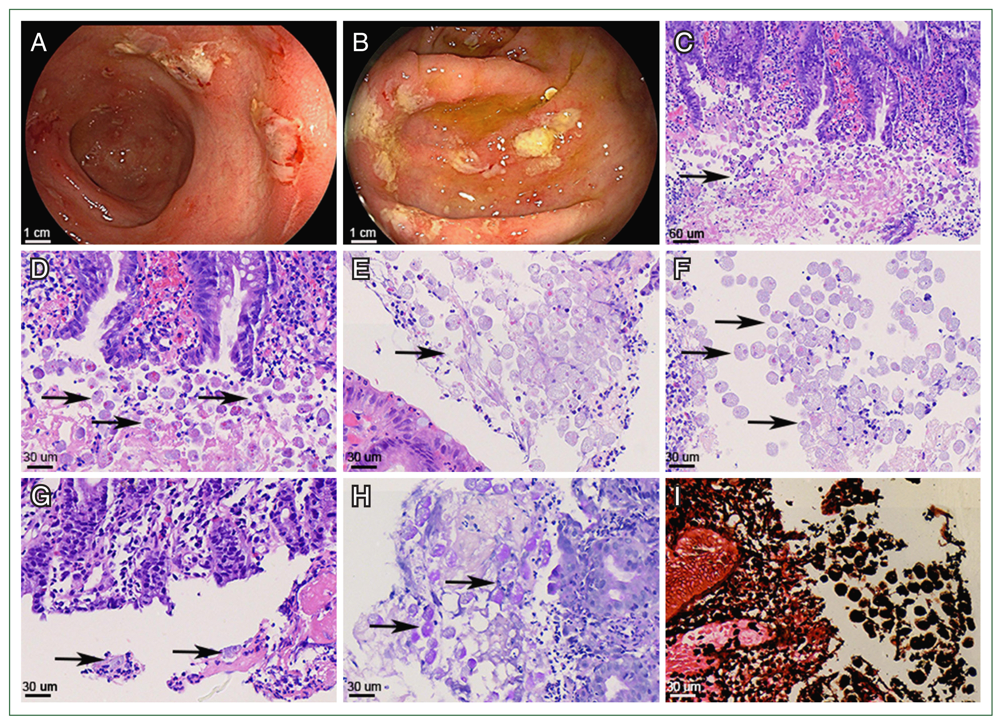
Colonoscopy revealed multiple ulcerative lesions in the colon, some of which were diffusely scattered (Fig. 1A, B). These ulcers were coated with yellowish-white exudate and blood. Larger ulcer measured approximately 3.0–3.5 cm in diameter.
Histopathological features
Microscopic examination demonstrated moderate to severe acute and chronic inflammation of the intestinal mucosa, characterized by infiltration of lymphocytes, plasma cells, and neutrophils, along with mucosal erosion or ulceration. Gray-blue, round or oval amoebic trophozoites were frequently identified within necrotic debris and exudate. These trophozoites exhibited eccentrically placed nuclei with clear nuclear membranes and prominent central nucleoli. Their foamy cytoplasm stained light pink and contained glycogen vacuoles and phagocytosed red blood cells, lymphocytes, or tissue fragments (Fig. 1C–G). PAS and hexamine silver stains were positive in cases 4 and 5 (Fig. 1H, I).
Treatment and outcomes
Nine patients received oral metronidazole (750 mg ter in die (tid)) and diloxanide (500 mg, tid) for 10 days. One patient with AE complicated by liver abscess was treated with intravenous metronidazole (500 mg every 8 h for 14 days), followed by oral metronidazole (750 mg tid for 10 days). Clinical symptoms such as abdominal pain, diarrhea, and purulent stools were significantly alleviated in these patients following treatment.
Discussion
E. histolytica infection can result in AE [3], a disease with variable clinical severity. Presentations range from mild abdominal discomfort to severe ulcerative colitis with mucus and blood (amoebic dysentery), appendicitis, or granulomatous lesions (amebomas) [4,5]. Consistent with prior studies [6,7], our data revealed that the ileocecal junction is the most frequently affected site, followed by the rectum.
E. histolytica is transmitted via the fecal-oral route, typically through ingestion of contaminated food or water, and does not require an intermediate vector. Upon entering the host, cysts undergo excystation in the terminal ileum, releasing motile trophozoites capable of mucosal invasion. These trophozoites adhere to and penetrate the intestinal epithelium, leading to mucosal damage, diarrhea, and colitis. While many infections are self-limiting, some progress to more severe disease, with trophozoites reverting to cysts and being excreted in feces, thereby perpetuating the transmission cycle [8,9]. The frequent involvement of the ileocecal region can thus be attributed to the early proliferation and invasion of trophozoites at this site. Clinical symptoms may include abdominal pain, diarrhea, jam-like stools, and mucinous bloody discharge. Accordingly, it is recommended that endoscopic evaluation in suspected AE cases pay close attention to the ileocecal and rectal regions for targeted biopsy.
Stool examination can aid in detecting amoebic cysts or trophozoites, but its diagnostic sensitivity remains suboptimal due to procedural and technical limitations [6]. In our study, 5 of the 14 patients had negative stool tests for trophozoites or cysts. Colonoscopy is a valuable diagnostic tool, particularly in the early stages when lesions appear as scattered mucosal erosions [7]. As the disease progresses, classic AE may present as flask-shaped ulcers with narrow necks and broad bases, accompanied by mucosal congestion, edema, and inflammatory exudates. Lesions may also appear as ulcers with a yellowish-white purulent coating and surrounding necrotic tissue [10]. However, with increased antibiotic use and improved host immunity, these characteristic lesions have become less common. In our cohort, all 14 patients exhibited ulcerative changes in the ileocecal region, colon, and rectum. Clinically, these lesions were initially misdiagnosed as nonspecific ulcerative colitis. Notably, 1 patient (case 14) had a 3-year history of presumed ulcerative colitis that was unresponsive to treatment; pathological reevaluation ultimately confirmed AE. This is a diagnostic challenge that shows how difficult it is to differentiate amoebic colitis from other ulcerative colitis based on colonoscopic findings alone.
Histopathological examination of intestinal biopsies remains the gold standard for AE diagnosis. The identification of trophozoites in biopsy specimens confirms the presence of amoebic colitis. Morphologically, trophozoites are round with a vesicular nucleus and cytoplasm containing glycogen vacuoles, ingested erythrocytes, lymphocytes, and tissue debris. Despite their diagnostic significance, trophozoites may be sparse in some biopsy samples due to sampling error, potentially resulting in false negatives.
The histopathological changes in AE, including cryptitis and crypt abscesses, may resemble those of other chronic inflammatory conditions, necessitating careful differential diagnosis. The differential diagnosis includes chronic ulcerative colitis, characterized by continuous and diffuse mucosal erosions with multiple superficial ulcers observed on colonoscopy. Histological findings in these cases typically demonstrate persistent chronic inflammation, crypt architectural distortion, and crypt abscesses [11,12]. Crohn’s disease primarily affects the terminal ileum and exhibits segmental or skip-pattern involvement. Early lesions manifest as aphthous ulcers that progressively enlarge and coalesce into longitudinal or fissuring ulcers, ultimately creating a cobblestone mucosal appearance. Histopathological examination reveals mucosal ulceration accompanied by noncaseating epithelioid granulomas [13,14]. Intestinal tuberculosis is often associated with concurrent pulmonary or systemic tuberculosis. Colonoscopic findings in these patients commonly include mucosal congestion, edema, and horizontally oriented ulcers with irregular “mouse-bite” margins. Histologically, transmural granulomatous inflammation may be observed, sometimes accompanied by central caseous necrosis, surrounded by thick lymphocytic sheaths that may coalesce into nodular patches. Acid-fast staining confirms the presence of Mycobacterium tuberculosis [15–17]. Additionally, intestinal amebiasis must be differentiated from gastrointestinal malignancies, such as adenocarcinomas and lymphomas, through detailed histopathological evaluation and adjunctive diagnostic tools.
Following treatment with metronidazole and adjunctive anti-inflammatory therapy (diclofenac), 9 patients experienced substantial clinical improvement, such as resolution of abdominal pain, diarrhea, hematochezia, and purulent stools. These outcomes highlight the effectiveness of accurate diagnosis and appropriate therapy for AE. In contrast, delayed or incorrect diagnoses can prolong morbidity. For example, case 7 initially declined colonoscopy and received empirical antibiotic therapy for 8 months without improvement. A subsequent colonoscopy and biopsy confirmed AE, and the patient responded promptly to targeted treatment. Similarly, case 14 had been misdiagnosed with ulcerative colitis for 3 years. Following colonoscopic and pathological reassessment, AE was correctly diagnosed, and the patient achieved full clinical recovery with anti-amoebic therapy.
A key limitation of this study is the lack of comparative evaluation of endoscopic and histological features before and after treatment, which could have provided additional insights into the disease course and therapeutic response.
In classic cases, AE can be readily diagnosed based on characteristic clinical features such as chronic abdominal pain, diarrhea, and jam-like stools. However, in atypical or non-classic presentations, diagnosis requires high clinical suspicion and targeted endoscopic biopsy from the affected intestinal segments. Accurate histopathological identification of trophozoites—using conventional microscopy and special stains such as PAS and hexamine silver—is essential for definitive diagnosis. Early recognition and appropriate treatment of AE are crucial for favorable patient outcomes and for preventing unnecessary treatment delays or misdiagnosis with other chronic inflammatory or neoplastic intestinal diseases.


