Abstract
A 26-year-old man residing in a village of Thai Nguyen Province, North Vietnam, visited the Thai Nguyen Provincial Hospital in July 2008. He felt a bulge-sticking pain in his left eye and extracted 5 small nematode worms by himself half a day before visiting the hospital. Two more worms were extracted from his left eye by a medical doctor, and they were morphologically observed and genetically analyzed on the mitochondrial cytochrome c oxidase 1 gene. The worms were 1 male and 1 female, and genetically identical with those of Thelazia callipaeda. By the present study, the presence of human T. callipaeda infection is first reported in Vietnam.
-
Key words: Thelazia callipaeda, oriental eyeworm, eye, human case, Vietnam
INTRODUCTION
Zoonotic parasites are widespread in the world, especially in Asian countries, including Vietnam.
Thelazia callipaeda Railliet and Henry, 1910 (Nematoda: Thelaziidae) is a nematode parasite in the genus
Thelazia [
1]. This nematode is zoonotic and parasitic in the eyes as implied by its name "oriental eyeworm" or "eyeworm". It was reported for the first time from a dog in Pakistan in 1910, and later shown to be widespread in China, France, Germany, India, Indonesia, Italy, Japan, Korea, the Netherlands, Russia, Switzerland, Taiwan, Myanmar, and Thailand [
2]. The final hosts include dogs and cats, but occasionally rabbits, monkeys, raccoons, dogs, foxes, wolves, and humans can also serve as the final host [
3]. Intermediate hosts are insects, such as flies. It is relatively well recognized that drosophilid flies but not
Musca domestica are the vector hosts for
T. callipaeda [
4-
6]. Also in Japan, 3 species of the genus
Amiota (Drosophilidae), namely,
Amiota okadai,
A. magna, and
A. nagatai, have been identified [
3].
The adult worm is parasitic in the conjunctival sac of a final host, and gives larvae continually by ovoviviparity. When a fly licks the tear in the eye of a final host, including humans, the larvae enter the conjunctival sac, and become adults in 1 month after 2 molts [
1,
3]. Symptoms of
T. callipaeda infection include conjunctivitis, excessive watering, visual impairment, and ulcers or scarring of the cornea [
7]. In some cases, the only symptom is the presence of worms obscuring the host's vision as a floater [
7].
With regard to the nematode infection in the human eyes in Vietnam,
Dirofilaria repens was recently reported from the human conjunctiva [
8]. However,
T. callipaeda infection has never been reported in Vietnam. In this study, we report for the first time a case of human
T. callipaeda infection in Vietnam which was verified by both morphology and molecular analysis using the mitochondrial cytochrome
c oxidase 1 (
cox1) gene.
CASE RECORD
The patient was a 26-year old male residing in the Cao Phong village, Hop Tien commune, Dong Hy district, Thai Nguyen Province of mountainous North Vietnam. In July 2008, he felt a bulge-sticking pain in his left eye and extracted 5 small nematode worms by himself but he did not keep them. This symptom did not disappear after collection of worms himself. Then, half a day later, he visited the Thai Nguyen Provincial Hospital and a medical doctor collected 2 more worms. These worms were identified morphologically and also by a molecular method. The morphology of the worms was not so good, but they were thin, long, and cylindrical with milkish-white color. One was a male, 10 mm in length and 0.4 mm in width, with a curled tail end, and the other was a female, 15 mm in length and 0.5 mm in width, with a slender tail end. The female worm had a scalariform buccal cavity, a long muscular esophagus and a conical tail, and the vulva opening at the anterior portion of the esophago-intestinal junction. Comparison with the figure described as
T. callipaeda in Miyazaki [
3] showed that these worms were
T. callipaeda (Nematoda: Thelaziidae).
To support the morphological diagnosis, PCR analysis was performed on the
cox1 gene with the primer of THELF-THELR for the Vietnamese
Thelazia. Total 627 nucleotide and 209 amino acid sequences of the
cox1 gene were compared with those of the previously known strains or isolates reported in GenBank (
Table 1). There were 7 nucleotide differences in the Vietnamese
T. callipaeda, but no changes in the amino acids at the changed places. The Vietnamese isolate had high homologies (98-99%) with 8
T. callipaeda isolates from China in GenBank (No. AM042549-AM042556) and 1 from Italy (No. AJ544882) [
9]. However, the homology of our isolate with
Thelazia gulosa (No. AJ544881) from Italy was low, 86% (
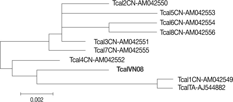
Table 1). A phylogenetic analysis between the Vietnamese
T. callipaeda and standard strains or isolates in the world showed that
T. callipaeda from Vietnam and
T. callipaeda from China and Italy is an identical group (
Fig. 1).
DISCUSSION
The first human case of thelaziasis was reported in China in 1917, and later from India, Thailand, Korea, Russia, and Japan [
3]. In Japan, there were 30 cases until 1981 [
3]. In Korea, 39 human cases were reported until 2011, and a total of 146 adult worms were collected from the patients [
10]. However, in Vietnam, this is the first time when
T. callipaeda infection is reported from a human patient. The worms were parasitic in the conjunctival sac of an eye of a 26-year-old man. He felt a bulge-sticking pain in his left eye and no other symptoms. A total of 7 worms were extracted, but 5 worms collected by the patient himself were lost and only 2 were available. Although the worm morphology was not so good, they were identified as 1 male (with a curled tail end) and 1 female (with a straight, slender tail end), and could be identified as
T. callipaeda. The flies of the family Drosophilidae, intermediate hosts for
T. callipaeda, are very common in Vietnam. This zoonotic disease can be transmitted from animals to humans.
In order to support the morphological diagnosis, the nucleotide and amino acid sequences of the cox1 gene of our Vietnamese worms were compared with those of 9 isolates of T. callipaeda reported in GenBank (8 from China and 1 from Italy). The results showed that there were 7 places of nucleotide differences in our isolate but no changes in the amino acid sequence at the changed places. Thus, the amino acid homology between the Vietnamese isolate and 8 other isolates in the world was 100%, and only 1 isolate, number 4 (GenBank no. AM042552), revealed 99% homology with our isolate (difference in 1 amino acid, i.e., phenylalanine). Therefore, our worms were identified by morphology and molecular methods as T. callipaeda.
ACKNOWLEDGMENTS
The authors acknowledge the funds supported from the National Foundation for Science and Technology Development (NAFOSTED) in Vietnam (No. 106.12-2011.13 to Nguyen Van De), and cooperation of researchers from the Hanoi Medical University (HMU), Institute of Biotechnology (IBT) and the Thai Nguyen Hospital of Vietnam.
References
- 1. Otranto D, Lia RP, Buono V, Traversa D, Giangaspero A. Biology of Thelazia callipaeda (Spirurida, Thelaziidae) eyeworms in naturally infected definitive hosts. Parasitology 2004;129:627-633.
- 2. Otranto D, Dutto M. Human thelaziasis, Europe. Emerg Infect Dis 2008;14:647-649.
- 3. Miyazaki I. An Illustrated Book of Helminthic Zoonoses. 1991, Tokyo, Japan. International Medical Foundation of Japan; pp 362-368.
- 4. Otranto D, Cantacessi C, Testini G, Lia RP. Phortica variegata as an intermediate host of Thelazia callipaeda under natural conditions: evidence for pathogen transmission by a male arthropod vector. Int J Parasitol 2006;36:1167-1173.
- 5. Otranto D, Lia RP, Testini G, Milillo P, Shen JL, Wang ZX. Musca domestica is not a vector of Thelazia callipaeda in experimental or natural conditions. Med Vet Entomol 2005;19:135-139.
- 6. Wang ZX, Hu Y, Shen JL, Wang KC, Wang HY, Jiang BL, Zhao P, Wang ZC, Ding W, Wang F, Xia XF. Longitudinal investigation and experimental studies on thelaziasis and the intermediate host of Thelazia callipaeda in Guanghua county of Hubei province. Zhonghua Liu Xing Bing Xue Za Zhi 2003;24:588-590.
- 7. Zakir R, Zhong-Xia Z, Chioddini P, Canning CR. Intraocular infestation with the worm, Thelazia callipaeda. Br J Ophthalmol 1999;83:1194-1195.
- 8. De NV, Le TH, Chai JY. Dirofilaria repens in Vietnam: detection of 10 eye and subcutaneous tissue infection cases identified by morphology and molecular methods. Korean J Parasitol 2012;50:137-141.
- 9. Otranto D, Testini G, De Luca F, Hu M, Shamsi S, Gasser RB. Analysis of genetic variability within Thelazia callipaeda (Nematoda; Thelazioidea) from Europe and Asia by sequencing and mutation scanning of the mitochondrial cytochrome c oxidase subunit 1 gene. Mol Cell Probes 2005;19:306-313.
- 10. Sohn WM, Na BK, Yoo JM. Two cases of human thelaziasis and brief review of Korean cases. Korean J Parasitol 2011;49:265-271.
Fig. 1A phylogeny tree of Thelazia callipaeda from Vietnam and other parts of the world in GenBank using the cox1 gene. TcalVN08: a sample of T. callipaeda from Vietnam; Tcal (1, 2, 3, 4, 5, 6, 7, 8) CN: samples from China (No: AM042549-AM042556); TcalTA: a sample from Italy (No: AJ544882). A phylogenetic analysis between the Vietnamese T. callipaeda and standard strains or isolates in the world showed that T. callipaeda from Vietnam and those from China and Italy is an identical group.

Table 1.The results of cox1 gene analysis on the Vietnamese isolate of Thelazia callipaeda compared with other known isolates in GenBank
Table 1.
|
GenBank No. |
Item |
Detected point |
% Comparison |
Homology (%) |
|
AM042552.1 |
Thelazia callipaeda mitochondrial partial CO1 gene for cytochrome oxidase subunit I, haplotype h4 |
1,125 |
100 |
99 |
|
AM042552.1 |
Thelazia callipaeda mitochondrial partial CO1 gene for cytochrome oxidase subunit I, haplotype h7 |
1,114 |
100 |
98 |
|
AM042552.1 |
Thelazia callipaeda mitochondrial partial CO1 gene for cytochrome oxidase subunit I, haplotype h3 |
1,114 |
100 |
98 |
|
AM042549.1 |
Thelazia callipaeda mitochondrial partial CO1 gene for cytochrome oxidase subunit I, haplotype h1 |
1,114 |
100 |
98 |
|
AJ5444882.1 |
Thelazia callipaeda mitochondrial partial CO1 gene for cytochrome oxidase subunit I |
1,114 |
100 |
98 |
|
AM042550.1 |
Thelazia callipaeda mitochondrial partial CO1 gene for cytochrome oxidase subunit I, haplotype h2 |
1,109 |
100 |
98 |
|
AM042556.1 |
Thelazia callipaeda mitochondrial partial CO1 gene for cytochrome oxidase subunit I, haplotype h8 |
1,107 |
99 |
98 |
|
AM042554.1 |
Thelazia callipaeda mitochondrial partial CO1 gene for cytochrome oxidase subunit I, haplotype h6 |
1,107 |
99 |
98 |
|
AM042553.1 |
Thelazia callipaeda mitochondrial partial CO1 gene for cytochrome oxidase subunit I, haplotype h5 |
1,108 |
100 |
99 |
|
AJ544881.1 |
Thelazia gulosa mitochondrial partial CO1 gene for cytochrome oxidase subunit I |
699 |
100 |
86 |
Citations
Citations to this article as recorded by

- Clinical and parasitological significance of thelaziosis in dogs and cats
Milan Hadzi-Milic, Andjelka Lesevic, Petar Krivokuca, Nemanja Jovanovic, Tamara Ilic
Veterinarski glasnik.2025; 79(1): 20. CrossRef - The Vector-Borne Zoonotic Nematode Thelazia callipaeda in the Eastern Part of Europe, with a Clinical Case Report in a Dog in Poland
Leszek Rolbiecki, Joanna N. Izdebska, Marta Franke, Lech Iliszko, Sławomira Fryderyk
Pathogens.2021; 10(1): 55. CrossRef - A Case of Human Thelaziasis and Review of Chinese Cases
Shi Nan Liu, Fang Fang Xu, Wen Qing Chen, Peng Jiang, Jing Cui, Zhong Quan Wang, Xi Zhang
Acta Parasitologica.2020; 65(3): 783. CrossRef - A human corneal ulcer caused by Thelazia callipaeda in Southwest China: case report
Xiaoxing Wei, Bo Liu, Yijian Li, Ke Wang, Lixia Gao, Yuli Yang
Parasitology Research.2020; 119(10): 3531. CrossRef - Thelaziosis due to Thelazia callipaeda in Europe in the 21st century—A review
Beatriz do Vale, Ana Patrícia Lopes, Maria da Conceição Fontes, Mário Silvestre, Luís Cardoso, Ana Cláudia Coelho
Veterinary Parasitology.2019; 275: 108957. CrossRef - Human Thelaziasis: Emerging Ocular Pathogen in Nepal
Ranjit Sah, Shusila Khadka, Mahesh Adhikari, Reema Niraula, Apoorva Shah, Anadi Khatri, Suzanne Donovan
Open Forum Infectious Diseases.2018;[Epub] CrossRef - Morphological and Mitochondrial Genomic Characterization of Eyeworms (Thelazia callipaeda) from Clinical Cases in Central China
Xi Zhang, Ya L. Shi, Zhong Q. Wang, Jiang Y. Duan, Peng Jiang, Ruo D. Liu, Jing Cui
Frontiers in Microbiology.2017;[Epub] CrossRef - Coinfection with Herpes Zoster Ophthalmicus and Oriental Eye Worm in a Rural Woman: The First Report of an Unusual Case
Kyung Sik Seo, Hye Min Lee, Ho Joon Shin, Joong Sun Lee
Annals of Dermatology.2014; 26(1): 125. CrossRef

