Abstract
This study aimed to determine the prevalence of intestinal helminth parasitic infections and associated risk factors for the human infection among the people of Samarkand, Uzbekistan. Infection status of helminths including Echinococcus granulosus was surveyed in domestic and wild animals from 4 sites in the Samarkand region, Uzbekistan during 2015–2018. Fecal samples of each animal were examined with the formalin-ether sedimentation technique and the recovery of intestinal helminths was performed with naked eyes and a stereomicroscope in total 1,761 animals (1,755 dogs, 1 golden jackal, and 5 Corsac foxes). Total 658 adult worms of E. granulosus were detected in 28 (1.6%) dogs and 1 (100%) golden jackal. More than 6 species of helminths, i.e., Taenia hydatigena, Dipylidium caninum, Diplopylidium nolleri, Mesocestoides lineatus, Toxocara canis, and Trichuris vulpis, were found from 18 (1.0%) dogs. Six (T. hydatigena, Toxascaris leonina, Alaria alata, Uncinaria stenocephala, D. caninum, and M. lineatus) and 2 (D. nolleri and M. lineatus) species of helminths were also detected from 5 Corsac foxes and 1 golden jackal, respectively. Taeniid eggs were found in 2 (20%) out of 10 soil samples. In the present study, it was confirmed that the prevalences of helminths including E. granulosus are not so high in domestic and wild animals. Nevertheless, the awareness on the zoonotic helminth infections should be continuously maintained in Uzbekistan for the prevention of human infection.
-
Key words: Echinococcus granulosus, dog, wild animal, helminthic parasite, Samarkand, Uzbekistan
Human cystic echinococcosis (CE) is generally an endemic disease, mainly occurring in pastoral areas worldwide. The most important pathogen in humans is believed to be
Echinococcus granulosus sensu stricto (s.s.), a synanthropic cestode that uses domestic dogs as definitive hosts and mainly sheep as intermediate hosts [
1–
3]. Humans become infected accidentally by ingesting eggs derived from the feces of infected dogs. The adult worm of
E. granulosus lives in the jejunum and duodenum of dogs and other canine carnivores (coyote, wolf, fox, jackal, etc.). The larval stage (hydatid cyst) is found in humans and herbivorous animals (typical intermediate hosts are sheep, goats, cattle, horses, and wild ungulates) [
4,
5].
CE is a re-emerging disease in the former Soviet Republics of central Asia. There has also been a parallel increase in cystic echinococcosis in farm livestock and increases in the prevalence of infections in the dog population [
6]. Agricultural land in central Asia is semi-arid mountain pasture, with a predominance of pasture-based livestock production, which provides good conditions for the transmission of
Echinococcus species in the livestock reservoir. Since the collapse of the Soviet Union in 1991, CE in central Asia has emerged as a major zoonosis with substantial increases in the incidence in humans, caused, according to Torgerson et al. [
7], by the privatization of large collective farms, the abandonment of centralized slaughtering and meat processing facilities, and few resources available for veterinary services. Uzbekistan has long been considered endemic for
E. granulosus in pigs, sheep, and cattle; however, there is little data on the prevalence of
Echinococcus infections in the country. The prevalence of CE in sheep in Uzbekistan increased from 45% to 62% between 1990 and 2002 [
8]. In Uzbekistan, a few studies have investigated the risk factors for
E. granulosus infection in intermediate and definitive animal hosts [
6,
8]. We herein aim to ascertain prevalence of
E. granulosus and other helminthes infection in dogs, fox and jackal in the Samarkand region.
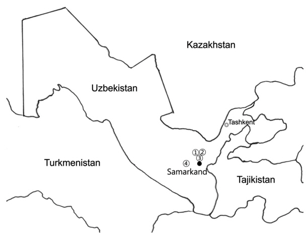
We conducted collaborative projects to control echinococcosis in the Samarkand Region of Uzbekistan (2014–2018) under the title of “Capacity Building of Infectious and Parasitic Diseases Control in the Republic of Uzbekistan”, supported by the Korean International Cooperation Agency (KOICA). Fecal samples were collected from dogs or hunted animals in Komchik, Chaparashli, O’rta Saydov, and Nurobod of the Samarkand Region (
Fig. 1). We examined a total of 1,761 dogs and wild animals (1,749 domestic dogs [
Canis lupus familiaris]: 582 from Komchik, 507 from O’rta Saydov, 463 from Chaparashli, and 197 from Nurobod); 5 stray dogs, 4 from Komchik, 2 from Chaparashli; 5 Corsac foxes (
Vulpes corsac), 2 from Chaparashli and 3 from O’rta Saydov; and one golden jackal (
Canis aureus) from O’rta Saydov for
Echinococccus granulosus infections. These samples were transferred to the laboratories of the Isaev Research Institute of Medical Parasitology in Sarmarkand for examination. We used formalin-ether sedimentation and direct fecal smears to detect the presence of helminth eggs. Stray dogs and wild animals were caught and dissected for the investigation of parasite infections. To protect the individuals involved in the necropsy, the procedures were performed according to the 1981 FAO/UNEP/WHO Guidelines [
9]. Following the transfer of animals to the lab, their small intestines were removed, opened, and examined in a dissecting pan containing water. The mucosa was scraped with a scalpel. We examined mucosal scrapings and intestinal contents under a stereomicroscope. We used soil surveys around houses of residents with probable
Echinococcus cysts (9 people from Chaparashli and 5 people from O’rta Saydov). We collected soil samples from 10 sites around each house using a soil sampler and examined the soil for helminth eggs. The sediment from each site was equally divided and examined by floatation with a saturated salt solution [
10] and Petri dish plates for helminth eggs. The sediment in the Petri dishes was diluted in saline and examined under a stereomicroscope to look for the presence of helminth eggs.
A total of 1,761 dogs and wild animals were examined. We found 658 adult worms of
Echinococcus granulosus in house and stray dogs (Collection rate: 1.60% [28/1,755]) and a golden jackal (100% [1/1]) (
Table 1). However, we found no
E. granulosus adult worms in the Corsac foxes (0% [0/5]). Stray dogs showed high infection rates (66.7% [4/6]: 50% [1/2] from Chaparashli and 75% [3/4] from Komchik). However, infection rate in domestic dogs (1.37% [24/1,749]; 0.43% [2/463] from Chaparashli, 0.86% [5/582] from Komchik, 7.11% [14/197] from Nurobod and 0.59% [3/507] from O’rta Saydov) was lower at 3 collection sites (Chaparashli, Komchik, and O’rta Saydov), but not in the Nurobod site. It is assumed that the low echinococcosis infection rate in domestic dogs from the 3 collection sites has resulted from a regulation requiring veterinarians to administer anti-helminthic drugs every 3 months to dogs. Aminjanov and Aminjanov (2004) have been studied a total of 240 village dogs and 279 farm dogs in Uzbekistan [
8]. Of these, 56 farm dogs (20.1%) and 19 village dogs (7.9%) were infected. The farm dogs were significantly more infected than the village dogs. There are an estimated 1.5 million dogs in Uzbekistan, and 75% of the households own dogs. That is, there is one dog for every 15 persons. That many dogs might represent a considerable biomass of parasites and pose a high risk for human infections [
11]. In this study, the jackal was first identified as a definitive host for
E. granulosus in Uzbekistan. Therefore, new control strategies to prevent the transmission of
E. granulosus to sheep are needed. Dogs either live with herds of sheep or look after the house or farm. Meanwhile, stray dogs roam freely and live on food garbage. In addition to stray dogs, other carnivores, such as jackals and foxes, particularly in mountainous areas, may enter human houses and farms in search of food. These carnivores may consume the infected organs of slaughtered animals, which sometimes are left behind around private abattoirs in small villages [
12]. We also collected helminth parasites from the intestines of house and stray dogs, corsac foxes, and a golden jackal. A total of 749 parasites were collected from 28 house dogs and stray dogs, including 7 parasites (Collection rate: 7.14% [2/28])
Taenia hydatigena, 124 (57.14% [16/28])
Dipylidium caninum, 3 (7.14% [2/28])
Diplopylidium nolleri, 10 (10.71% [2/28])
Mesocestoides lineatus, 500 (100% [28/28])
Echinococcus granulosus, 54 (28.57% [8/28])
Toxocara canis, and 51 (39.29% [11/28])
Trichocephalus vulpis. We collected 48 parasites from 5 Corsac foxes, including 3 (20.00% [1/5])
Taenia hydatigena, 4 (40.00% [2/5])
Toxascaris leonina, 24 (60.00% [3/5])
Alaria alata, 5 (20.00% [1/5])
Uncinaria stenocephala, 5 (40.00% [2/5])
Dipylidium caninum, and 4 (40.00% [2/5])
Mesocestoides lineatus. We collected 163 parasites from 1 golden jackal, including 2
Diplopylidium nolleri, 3
Mesocestoides lineatus, and 158
E. granulosus (
Table 2). Humans are infected by the ingestion of
E. granulosus eggs in contaminated food, water, and soil, or through direct contact with animal hosts. The soil surveys around houses of residents with probable
Echinococcus cysts done by ultrasonographic investigation found taeniid eggs at 2 houses. Of the 15 households surveyed, only 1 house had no dogs. The eggs of other tapeworm species identified in dogs were
Toxocara canis, Dipylidium caninum, and
Taenia hydatigena.
Echinococcus granulosus eggs are morphologically indistinguishable from those of other taeniid cestode, and the release of eggs is variable and inconsistent. PCR techniques have been reported to be useful where the presence of the parasite in the dog population is relatively low, as well as for discriminating
Echinococcus from other taeniid infections in dogs [
13–
15].
Wild carnivores, including jackals, wolves, and probably foxes, have been found to be infected with Echinococcosis adult worms, demonstrating the co-existence of a domestic and sylvatic cycle [
12,
16–
18]. In Uzbekistan, the distribution of reservoir hosts is important for understanding the epidemiology of echinococcosis, as well as the potential impact on human health. Physical contact with stray dogs or accidental contact with wild canid feces is risk factors for echinococcosis. Periodic mass treatment of dogs with anti-helmintics, such as praziquantel or albendazole, the prohibition of giving raw infected viscera to dogs, and adequate inspection of abattoirs, as well as educational measures to change human practices that facilitate hydatid disease transmission, have been reported to be effective in the control of echinococcosis [
19–
21]. This investigation of
E. granulosus infections in stray dogs and golden jackals confirmed them to be potential reservoir hosts for human infections in the Samarkand Region. The results suggest that a control program for reservoir hosts is also necessary to prevent echinococcosis in humans and livestock in Uzbekistan.
Notes
-
CONFLICT OF INTEREST
The authors declare no conflict of interest related to this study.
ACKNOWLEDGMENTS
This work was supported by the Korean International Cooperation Agency (KOICA) under the title of “Capacity Building of Infectious Diseases Control in the Republic of Uzbekistan” in 2014–2018 (no. p2014-00076-1).
Fig. 1Survey region in the Samarkand (1, Komchik; 2, O’rta Saydov; 3, Chaparashli; 4, Nurobod).

Table 1Infection status of domestic and wild animals collected in the Samarkand region from 2015 to 2018
Table 1
|
Animals/no. of animal examined |
Parasites |
Collection sites |
|
House dogs/1,749 |
Echinococcus granulosus
Toxocara canis
Trichocephalus vulpis
|
Chaparashli, Komchik, Nurobod, O’rta Saydov |
|
Stray dogs/6 |
Echinococcus granulosus
Dipylidium caninum
Diplopylidium nolleri
Mesocestoides lineatus
Taenia hydatigena
Toxocara canis
Trichocephalus vulpis
|
Chaparashli, Komchik |
|
Golden jackal/1 |
Echinococcus granulosus
Diplopylidium nolleri
Mesocestoides lineatus
|
Komchik |
|
Corsac foxes/5 |
Alaria alata (Diplostomatidae)
Dipylidium caninum
Taenia hydatigena
Toxascaris leonina
Uncinaria stenocephala
Mesocestoides lineatus
|
Chaparashli, O’rta Saydov |
Table 2Helminth parasites isolated from the intestine of dogs and wild animals captured in the Samarkand region from 2015 to 2018
Table 2
|
Animals |
Parasites |
|
Taenia hydatigena
|
Toxascaris leonina
|
Alaria alata
|
Uncinaria stenocephala
|
Dipylidium caninum
|
Diplopylidium nolleri
|
Mesocestoides lineatus
|
Echinococcu granulosus
|
Toxocara canis
|
Trichocephalus vulpis
|
|
House dogs |
0 |
0 |
0 |
0 |
0 |
0 |
0 |
200 |
12 |
21 |
|
Stray dogs |
7 |
0 |
0 |
0 |
124 |
3 |
10 |
300 |
42 |
30 |
|
Golden jackal |
0 |
0 |
0 |
0 |
0 |
2 |
3 |
158 |
0 |
0 |
|
Corsac foxes |
3 |
4 |
24 |
5 |
6 |
0 |
4 |
0 |
0 |
0 |
References
- 1. Eckert J, Deplazes P. Biological, epidemiological, and clinical aspects of echinococcosis: a zoonosis of increasing concern. Clin Microbiol Rev 2004;17:107-135.
- 2. Moro PL, Schantz PM. Cystic echinococcosis in the Americas. Parasitol Int 2006;55(suppl):181-186.
- 3. Ito A, Nakao M, Lavikinen A, Hoberg E. Cystic echinococcosis: Future perspectives of molecular epidemiology. Acta Trop 2017;165:3-9.
- 4. Thompson RCA, Lymbery AJ.
Echinococcus and Hydatid Disease. Wallingford, UK. CAB International; 1995, p 477.
- 5. Dalimi A, Sattari A, Motamedi G. A study on intestinal helminthes of dogs, foxes and jackals in the western part of Iran. Vet Parasitol 2006;142:129-133.
- 6. Torgerson PR, Oguljahan B, Muminov AE, Karaeva RR, Kuttubaev OT, Aminjanov M, Shaikenov B. Present situation of cystic echinococcosis in Central Asia. Parasitol Int 2006;55(suppl):207-212.
- 7. Torgerson PR, Shaikenov BS, Rysmukhambetova AT, Ussenbayev AE, Abdybekova AM, Burtisurnov KK. Modelling the transmission dynamics of Echinococcus granulosus in dogs in rural Kazakhstan. Parasitology 2003;126:417-424.
- 8. Aminjanov M, Aminjanov S. Echinococcosis and research in Uzbekistan. In Torgerson P, Shaikenov B eds, Echinococcosis in Central Asia: Problems and Solutions. Almaty, Kazakhstan. Dauir Publishing House; 2004, pp 13-19.
- 9. Eckert J, Gemmell MA, Matyas Z, Soulsby EJI. Guidelines for Surveillance, Prevention and Control of Echinococcosis/Hydatidosis. 2nd ed. Geneva, Switzerland. World Health Organization; 1981, p 147.
- 10. Eslami A. Recovery of cestods eggs from the village courtyard soil in Iran. Vet Parasitol 1996;10:95-96.
- 11. Moro O, Schantz PM. Echinococcosis: a review. Int J Infect Dis 2009;13:125-133.
- 12. Dalimi A, Motamedi G, Hosseini M, Mohammadian B, Malaki H, Ghamari Z, Ghaffari Far F. Echinococcosis/hydatidosis in western Iran. Vet Parasitol 2002;105:161-171.
- 13. Christofi G, Deplazes P, Christofi N, Tanner I, Economides P, Eckert J. Screening of dogs for Echinococcus granulosus coproantigen in a low endemic situation in Cyprus. Vet Parasitol 2002;104:299-306.
- 14. Stefanić S, Shaikenov BS, Block S, Deplazes P, Dinkel A, Torgerson PR, Mathis A. Polymerase chain reaction for detection of patent infections of Echinococcus granulosus (“sheep strain”) in naturally infected dogs. Parasitol Res 2004;92:347-351.
- 15. Varcasia A, Garippa G, Scala A. The diagnosis of Echinococcus granulosus in dogs. Parasitologia 2004;46:409-412.
- 16. Sadjjadi SM. Present situation of echinococcosis in the Middle East and Arabic North Africa. Parasitol Int 2006;55(suppl):197-202.
- 17. Wang Z, Wang X, Liu X. Echinococcosis in China, a review of the epidemiology of Echinococcus spp. Ecohealth 2008;5:115-126.
- 18. Abdybekova AM, Torgerson PR. Frequency distributions of helminths of wolves in Kazakhstan. Vet Parasitol 2012;184:348-351.
- 19. Heath DD, Jensen O, Lightowlers MW. Progress in control of hydatidosis using vaccination—a review of formulation and delivery of the vaccine and recommendations for practical use in control programmes. Acta Trop 2003;85:133-143.
- 20. Moro PL, Schantz PM. Echinococcosis: historical landmarks and progress in research and control. Ann Trop Med Parasitol 2006;100:703-714.
- 21. Craig PS, McManus DP, Lightowlers MW, Chabalgoity JA, Garcia HH, Gavidia CM, Gilman RH, Gonzalez AE, Lorca M, Naquira C, Nieto A, Schantz PM. Prevention and control of cystic echinococcosis. Lancet Infect Dis 2007;7:385-394.
Citations
Citations to this article as recorded by

- Molecular identification and phylogenetic positioning of nematodes Toxocara canis, T. cati (Ascarididae) and Toxascaris leonina (Toxocaridae) from domestic and wild carnivores in the Fergana Valley, Uzbekistan
A. E. Kuchboev, A. G. Sotiboldiyev, B. K. Ruziev, A. A. Safarov
Biosystems Diversity.2025; 33(3): e2538. CrossRef - High-Quality Chromosome-Level Genome Assembly of the Corsac Fox (Vulpes corsac) Reveals Adaptation to Semiarid and Harsh Environments
Zhihao Zhang, Tian Xia, Shengyang Zhou, Xiufeng Yang, Tianshu Lyu, Lidong Wang, Jiaohui Fang, Qi Wang, Huashan Dou, Honghai Zhang
International Journal of Molecular Sciences.2023; 24(11): 9599. CrossRef - Time series modeling of animal bites
Fatemeh Rostampour, Sima Masoudi
Journal of Acute Disease.2023; 12(3): 121. CrossRef - Diagnostic tools for the detection of taeniid eggs in different environmental matrices: A systematic review.
Ganna Saelens, Lucy Robertson, Sarah Gabriël
Food and Waterborne Parasitology.2022; 26: e00145. CrossRef - Fleas from the Silk Road in Central Asia: identification of Ctenocephalides canis and Ctenocephalides orientis on owned dogs in Uzbekistan using molecular identification and geometric morphometrics
Georgiana Deak, Alisher Safarov, Xi Carria Xie, Runting Wang, Andrei Daniel Mihalca, Jan Šlapeta
Parasites & Vectors.2022;[Epub] CrossRef - Control of cystic echinococcosis in the Middle Atlas, Morocco: Field evaluation of the EG95 vaccine in sheep and cesticide treatment in dogs
Fatimaezzahra Amarir, Abdelkbir Rhalem, Abderrahim Sadak, Marianne Raes, Mohamed Oukessou, Aouatif Saadi, Mohammed Bouslikhane, Charles G. Gauci, Marshall W. Lightowlers, Nathalie Kirschvink, Tanguy Marcotty, María-Gloria Basáñez
PLOS Neglected Tropical Diseases.2021; 15(3): e0009253. CrossRef - Co-infection of Trichuris vulpis and Toxocara canis in different aged dogs: Influence on the haematological indices
I. V. Saichenko, A. A. Antipov, T. I. Bakhur, L. V. Bezditko, S. S. Shmayun
Biosystems Diversity.2021; 29(2): 129. CrossRef - Spread and seasonal dynamics of dogs helminthiasis in BilaTserkva district
I. Saichenko
Naukovij vìsnik veterinarnoï medicini.2021; (1(165)): 119. CrossRef - Monitoring of parasitic diseases of dogs
Bogdan Morozov, Andrii Berezovskyi
EUREKA: Health Sciences.2021; (4): 109. CrossRef - An epizootic situation is in relation to the nematodosiss of gastroenteric channel of dogs
I. Saichenko, A. Antipov
Naukovij vìsnik veterinarnoï medicini.2020; (1(154)): 54. CrossRef
