Abstract
Psoriasis is a chronic inflammatory seronegative disease closely associated with ankylosing spondylitis and inflammatory bowel disease. This study aimed to investigate the anti-inflammatory effects of candidate metabolites, such as D-proline, L-iditol, and propionylcarnitine, identified in Clonorchis sinensis preparations, in in vitro and in vivo models. None of the 3 metabolites exhibited cytotoxicity within the tested concentration range. In LPS-stimulated RAW 264.7 cells, D-proline and L-iditol significantly reduced the expression of pro-inflammatory cytokines, and in an imiquimod-induced psoriasis-like mouse model, L-iditol significantly reduced disease severity and histopathological scores, whereas the effects of other metabolites were inconsistent. These results suggest that L-iditol, a candidate metabolite identified in Clonorchis sinensis formulations, exhibits anti-inflammatory effects in both in vitro and in vivo environments and may have therapeutic potential for inflammatory skin diseases such as psoriasis.
-
Key words: Clonorchis sinensis, D-proline, L-iditol, propionylcarnitine, psoriasis, anti-inflammation
Introduction
Psoriasis is a chronic, immune-mediated skin disorder characterized by hyperproliferation of keratinocytes and infiltration of immune cells, particularly those associated with Th1 and Th17 responses [
1,
2]. The disease affects approximately 0.6% to 4.8% of the global population and is driven by a complex interplay between genetic predisposition and environmental triggers such as stress, trauma, and infections [
3-
5]. At the molecular level, psoriasis is sustained by elevated levels of pro-inflammatory cytokines including tumor necrosis factor (TNF)-α, interleukin (IL)-6, IL-17, and IL-1β, which collectively promote epidermal thickening, immune activation, and sustained inflammation [
6].
Recent advances in psoriasis management have markedly expanded therapeutic options, ranging from novel topical agents to highly specific biologics. Newly approved topicals such as tapinarof (aryl hydrocarbon receptor agonist) [
7] and roflumilast (PDE4 inhibitor) [
8] provide steroid-sparing alternatives for mild-to-moderate disease, while oral small molecules including deucravacitinib, a selective TYK2 inhibitor, have shown durable efficacy in moderate-to-severe cases [
9]. Yet, tapinarof may trigger local irritation (e.g., folliculitis, contact dermatitis) and data on head-and-neck or intertriginous site use are limited [
7]; roflumilast, although well tolerated in short-term trials, lacks robust long-term real-world evidence [
8,
10]; and deucravacitinib, despite strong clinical efficacy, raises concerns about infection risk and long-term safety requiring ongoing pharmacovigilance [
9,
11]. Biologic therapies targeting key cytokine pathways—including TNF‑α, IL‑12/23 (ustekinumab), IL‑17 (secukinumab, ixekizumab, brodalumab, and bimekizumab) and IL‑23 (guselkumab, risankizumab, and tildrakizumab)—have revolutionized clinical outcomes [
12,
13]. In addition, the IL‑36R inhibitor spesolimab was recently approved for generalized pustular psoriasis [
14]. Nonetheless, despite these advances, several fundamental limitations remain: high cost and limited global accessibility, loss of efficacy or treatment switching due to immunogenicity, long-term safety concerns including increased infection risk, and specific contraindications such as inflammatory bowel disease (IBD) exacerbation with IL‑17 blockade or psychiatric warnings with brodalumab [
12,
13]. Furthermore, conventional systemic agents (e.g., methotrexate, cyclosporine, acitretin) continue to be restricted by organ toxicity [
13]. Collectively, these challenges highlight the ongoing need for safer, more affordable, and broadly effective therapeutic strategies.
Psoriasis is one of the ‘seronegative’ diseases in addition to ankylosing spondylitis (AS) and IBD. They are closely related clinically and often co-occur in patients and families, and a previous study highlighted shared genetic pathways and pleiotropic genes (genes influencing multiple traits) among these conditions [
15]. Similar with IBD and AS, for psoriasis, current treatment strategies—including biologics and small molecule inhibitors—have significantly improved disease control, challenges such as high cost, immunogenicity, and partial response rates highlight the need for novel anti-inflammatory agents. Insights into the mechanisms that helminths use to modulate specific immune cells may give clues about their ability to protect against inflammatory diseases and several autoimmune diseases [
16]. Reducing the inflammation associated with IBD like Crohn's and colitis has been demonstrated by
Trichuris suis therapy [
17]. Although helminth therapy has generated substantial interest in modulating excessive immune responses, concerns prevail around the implications of pathogenic effects caused by infection with live pathogens, particularly at high doses [
18]. Therefore, identification of protective active helminth-derived molecules to substitute treatment with whole worm could circumvent this issue [
19].
Clonorchis sinensis (CS) have garnered attention for their ability to modulate host immune responses through secreted proteins and metabolites. Previous studies from our group demonstrated that crude somatic proteins (CSp) and excretory/secretory proteins (CS-ESP) derived from CS exert therapeutic effects in a mouse model of AS, primarily through downregulation of pro-inflammatory mediators [
20,
21]. To identify functionally relevant metabolites, untargeted LC-MS/MS-based metabolomic analysis of CS preparations, including CSp and CS-ESP, was performed. Among the identified metabolites, D-proline, L-iditol, and propionylcarnitine were selected from the top 10 most abundant compounds in each protein fraction based on their relative abundance, consistent detection across samples, and potential biological relevance. Although these metabolites have been implicated in various metabolic and regulatory processes, their roles in modulating inflammatory responses in psoriasis have not been previously investigated. Therefore, this study aimed to evaluate the anti-inflammatory effects of these candidate metabolites in both in vitro and in vivo models and to provide initial insight into their potential as immunomodulatory agents in chronic inflammatory diseases.
Methods
Ethics statement
All animal experiments were approved by the Chonnam National University Animal Care and Use Committee (CNU IACUC-H-2018-35) and conducted in accordance with institutional guidelines.
CS protein extraction
The CS metacercariae were collected from the second intermediate host,
Pseudorasbora herzi, caught in a stream in Korea, after artificial digestion of the fish [
21,
22]. Briefly, metacercariae were orally administered to rabbits (New Zealand White, both genders: 2.2–2.4 kg; Koatech). Adult CS were recovered from the rabbits after 3 months. Preparation of CSp or CS-ESP was performed as previously reported [
20,
21]. For obtaining CSp, harvested adult worms were washed thoroughly in sterile 1× phosphate-buffered saline (Oatech) and homogenized on ice in a lysis buffer containing protease inhibitors. The homogenate was centrifuged at high speed to remove debris, and the resulting supernatant containing crude proteins was collected. For CS-ESP preparation, live adult worms were incubated in RPMI-1640 medium (Oatech) supplemented with antibiotics at 37°C in 5% CO
2 for 24–48 h. The culture supernatant containing CS-ESP was collected, centrifuged to remove particulate debris, filtered, and concentrated using centrifugal filter units. Both CSp and CS-ESP samples were aliquoted and stored at -80°C until further analysis. Protein concentration was measured using the Pierce BCA Protein Assay Kit (Thermo Scientific).
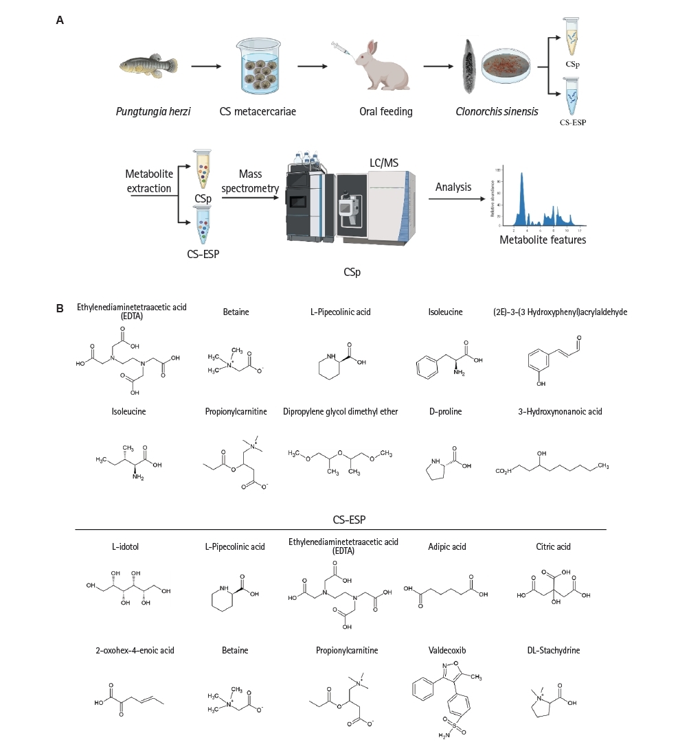
To extract metabolites, CSp and CS-ESP samples were treated with 80% methanol and incubated at -80°C for 1 h. After centrifugation at 4,000× g for 15 min, the supernatant was collected, evaporated to dryness using a SpeedVac, and reconstituted in 100 μl of LC-MS mobile phase. LC-MS/MS analysis was conducted at Ebiogene using a Q Exactive Orbitrap mass spectrometer (Thermo Scientific) with an electrospray ionization source. Chromatographic separation was performed using a 2-column system: a C18 trapping column (1.8 μm, 2.1×5 mm) followed by an Eclipse Plus C18 RRHD analytical column (1.8 μm, 2.1×50 mm). The mobile phases were 0.1% formic acid in water (solvent A) and 0.1% formic acid in 80% acetonitrile (solvent B), and a linear gradient was applied over 26 min at 0.2 mL/min. MS data were acquired over the m/z range of 100–1,000. Spectral data were processed using Compound Discoverer 3.3 (Thermo Fisher Scientific) with the "Untargeted Metabolomics with Statistics Detect Unknowns" workflow. Metabolite annotation was based on mzCloud spectral matching. Confidence levels were assigned per metabolomics standards initiative criteria: level 2 for metabolites with <10 ppm mass error and mzCloud scores >80, and level 3 for ChemSpider matches with <5 ppm error. Redundant features were filtered by peak intensity to retain unique metabolite signals (
Fig. 1A,
B).
LC–MS/MS–based metabolomic analysis was performed to identify significantly altered metabolites. Metabolites were initially filtered based on annotation confidence and subsequently prioritized according to their relative signal intensity (peak area) and consistent detection across samples. Due to the exploratory nature of this study and limitations of the available dataset, additional parameters such as fold change and background signal filtering were not systematically applied. Based on these criteria, D-proline (858919, Merck), L-iditol (I6035, Merck) and propionylcarnitine (AG007VN1, ChemSpace US) were selected for further experimental validation. A summary of the top candidate metabolites and their LC-MS/MS parameters is provided in
Supplementary Table S1. Commercially available compounds with identical molecular structures to the selected metabolites were purchased and used for subsequent in vitro and in vivo experiments (
Fig. 1).
RAW 264.7 cells were cultured in RPMI-1640 medium (Oatech) supplemented with 10% fetal bovine serum (Gibco) and 1% penicillin-streptomycin (Gibco) under standard culture conditions at 37°C in a humidified atmosphere containing 5% CO
2. For the cell viability assay, cells were seeded at a density of 1×10⁵ cells per well in a 96-well plate and allowed to adhere overnight. The following day, the cells were treated with varying concentrations of 3 different compounds: D-proline, L-iditol and propionylcarnitine at 1, 10, 50, and 100 μg/ml 20 μl of Cell Titer 96 AQueous One Solution Reagent (MTS reagent, G3580, Promega) was added to each well and the cells were further incubated for 2 h. Absorbance was then measured at 490 nm using a Reader 96-well microplate reader (Molecular Devices) to determine cell viability based on metabolic activity. All samples were measured in triplicate [
21].
To determine the experimental concentrations of the selected candidate metabolites, preliminary concentration-screening experiments were performed following cell viability assessment. Cells were treated with increasing concentrations of each metabolite to identify a concentration range without cytotoxic effects. Based on these results, a single concentration that showed no cytotoxicity and demonstrated reproducible anti-inflammatory effects was selected and used for all subsequent in vitro experiments, including cytokine expression analysis. Detailed concentration-screening data are provided in the
Supplementary Figs. S1-
S3.
RAW 264.7 cells were seeded at a density of 5×10⁵ cells per well in a 6-well plate and allowed to adhere overnight. To investigate the anti-inflammatory effects of selected compounds, cells were co-cultured for 24 h in the presence of LPS (4 μg/ml) to induce an inflammatory response. The treatment groups included D-proline (50 μg/ml), L-iditol (10 μg/ml), and propionylcarnitine (5 μg/ml), each added to the culture medium along with LPS. Following the incubation period, total RNA was extracted from the cells using a standard RNA isolation protocol. Complementary DNA was synthesized from the extracted RNA using a PrimeScript 1st stand complementary DNA Synthesis kit (Takara). Real-time quantitative PCR was subsequently performed to evaluate the mRNA expression levels of inflammation-related cytokines and enzymes, including IL-1β, TNF-α, IL-6, inducible nitric oxide synthase (iNOS) and cyclooxygenase-2 (COX2) (
Table 1). The relative gene expression levels were analyzed to assess the modulatory effects of each compound on LPS-induced inflammation in RAW 264.7 cells. All quantifications were normalized versus endogenous GAPDH. The relative quantitation value of each target gene after normalizing to GAPDH as compared with the calibrator for that target was calculated using 2
-ΔΔCt. All samples were measured in triplicate.
Male SKG mice (8–10 weeks old) of the BALB/c strain were reared in a specific pathogen-free environment and randomly assigned to 6 experimental groups. In the initial preliminary experiment, the study was conducted with a negative control (NC) group (
n=5), a positive control (PC) group (
n=10), a dexamethasone (DEX) group (
n=5), D-proline (
n=5), L-iditol (
n=5), and propionylcarnitine (
n=5). Subsequently, in the additional experiment, propionylcarnitine was excluded as it did not show a significant effect, and the study was conducted with NC (
n=5), PC (
n=9), DEX (
n=4), D-proline (
n=11), and L-iditol (
n=11). The dorsal hair of the mice was completely removed, and 5% imiquimod (IMQ) cream was topically applied to the shaved area daily from day 1 to day 7 to induce psoriasis-like skin inflammation. From day 3 to day 7, D-proline (50 mg/kg), L-iditol (10 mg/kg), and propionylcarnitine (5 mg/kg) were topically co-applied with IMQ. In addition, DEX (1 mg/kg) was administered intraperitoneally once daily from day 3 to day 7. Psoriasis Area and Severity Index (PASI) scores were measured daily from day 1 to day 7 before topical application of IMQ to the dorsal skin of mice. Two independent observers evaluated skin condition in a randomized and blinded manner. Erythema (redness), induration (thickness) and desquamation (scale) were each scored on a scale from 0 to 4 (0, none; 1, slight; 2, moderate; 3, severe; 4, very severe). The total PASI score was calculated as the sum of these 3 items [
23]. The scores from the observers were averaged to derive the final value.
After the mice were sacrificed, dorsal skin tissues were carefully excised and immediately fixed in 10% formalin for 24 h at room temperature to preserve tissue morphology. Following fixation, the samples underwent a graded ethanol series for dehydration, were cleared in xylene, and then embedded in paraffin to prepare tissue blocks. The paraffin-embedded tissues were sectioned at a thickness of 10 μm using a microtome. The sections were mounted on glass slides and stained with hematoxylin and eosin to evaluate histopathological changes associated with psoriasis. Histological evaluation was performed by 4 independent observers using the Psoriasis Histopathological Score (PHS) in a randomized, blinded manner. Inflammatory cell infiltration, incomplete keratosis, hyperkeratosis, epidermal thickness, and Monroe microabscesses were each scored on a scale of 0 to 3 (0, none; 1, mild; 2, moderate; 3, severe). The total PHS score was calculated as the sum of these individual items [
24]. The scores from the observers were averaged to derive the final value.
Statistical analyses were performed using GraphPad Prism 10 (GraphPad Software). Normality of the data was assessed using the Shapiro-Wilk test. For normally distributed data, comparisons among groups were performed using one-way or two-way ANOVA followed by Bonferroni multiple comparisons test. For data that did not meet normality assumptions, non-parametric tests were used as appropriate: the Friedman test was applied for overall comparisons of repeated measures within the same group, followed by the Wilcoxon signed-rank test for post-hoc pairwise comparisons. The Kruskal-Wallis test was used for independent group comparisons. P<0.05 was considered to indicate a statistically significant difference: *P<0.05, **P<0.01, ***P<0.001, and ****P<0.0001.
Results
Cell viability of D-proline, L-iditol, and propionylcarnitine in RAW 264.7 macrophages
Cell viability and anti-inflammatory effects were confirmed for each candidate substance in RAW 264.7 cells (
Figs. 2-
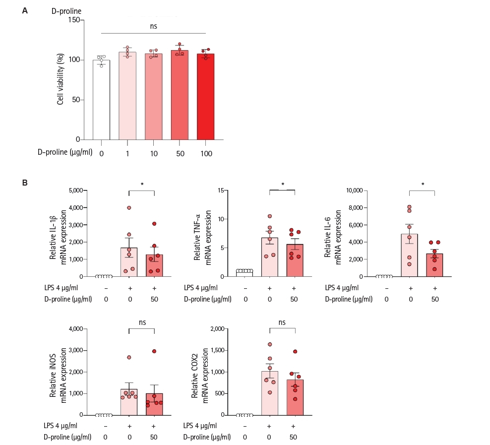
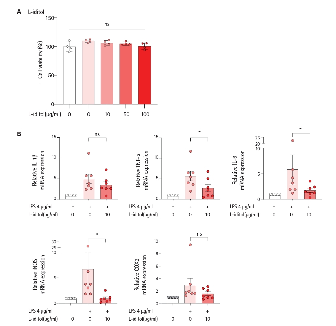
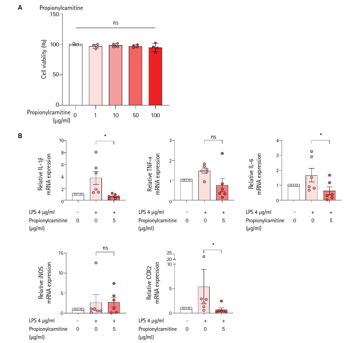
4). To determine the cytotoxicity of the selected candidate metabolites identified in CS preparations, RAW 264.7 murine macrophages were treated with increasing concentrations of D-proline, L-iditol, or propionylcarnitine. Cell viability was assessed after 24 h using the MTS reagent. As shown in
Figs. 2A,
3A, and
4A, none of the compounds exhibited significant cytotoxic effects at their respective treatment concentrations. Viability remained comparable to the untreated control group, even at the highest tested doses. These results indicate that all 3 metabolites are well tolerated by macrophages under the experimental conditions and are suitable for further investigation of their functional effects.
To determine the optimal experimental concentration of the selected candidate metabolites, preliminary dose-response experiments were performed. RAW 264.7 cells were treated with 1, 10, 50, and 100 μg/ml of each metabolite, concentrations that did not affect cell viability. The concentrations used in these experiments were pre-screened to ensure they did not induce cytotoxicity, with cell viability maintained at levels comparable to the untreated control (as shown in
Figs. 2A,
3A, and
4A of the main manuscript), and inflammatory cytokine expression levels were measured (
Figs. 2B,
3B,
4B). Among the tested concentrations, D-proline showed reduced inflammatory cytokine levels at 50 and 100 μg/ml, L-iditol at 10 and 50 μg/ml, and propionylcarnitine at 1, 5, and 10 μg/ml. Based on these results, additional preliminary experiments were conducted for each metabolite to determine a single optimal concentration. Finally, a single concentration for each metabolite that did not affect cell viability and showed consistent anti-inflammatory effects in the preliminary screening was selected and used for all subsequent in vitro and in vivo experiments.
To evaluate the anti-inflammatory properties of D-proline, L-iditol, and propionylcarnitine, RAW 264.7 cells were pre-stimulated with LPS to induce a robust inflammatory response and then treated with each metabolite at their effective non-toxic concentrations. Quantitative real-time PCR was performed to analyze the mRNA expression levels of key inflammatory mediators. Treatment with D-proline (50 μg/ml) significantly reduced the expression of IL-1β, TNF-α, and IL-6 compared to LPS-only controls (Wilcoxon signed-rank test,
W=-21.00,
n=6 pairs,
P=0.031). Mean±SD values were as follows: IL-1β, 1,678±1,377 versus 1,295±1,034; TNF-α, 6.78±2.75 versus 5.66±2.31; IL-6, 4,967±2,740 versus 2,673±1,216 (
Fig. 2B). However, treatment with D-proline could not show statistically significant changes in the mRNA expression levels of iNOS and COX2, respectively.
Treatment with L-iditol (10 μg/ml) significantly reduced the expression of TNF-α (
W=-28.00, 5.67±3.12 versus 2.75±2.38,
P=0.016), IL-6 (
W=-26.00, 5.81±7.48 versus 1.75±1.06,
P=0.031), iNOS (
W=-28.00, 6.68±8.76 versus 0.97±0.76,
P=0.016), compared to LPS-only (
Fig. 3B). However, treatment with L-iditol could not show statistically significant changes in the mRNA expression levels of IL-1β and COX2, respectively.
Treatment with propionylcarnitine (5 μg/ml) significantly reduced the expression of IL-1β, IL-6 and COX2 compared to LPS-only controls (Wilcoxon signed-rank test,
W=-21.00,
n=6 pairs,
P=0.031). Mean±SD values were as follows: IL-1β, 3.76±2.76 versus 0.65±0.37; IL-6, 1.68±1.14 versus 0.63±0.66; COX2, 5.39±8.53 versus 0.72±0.87 (
Fig. 4B). However, treatment with propionylcarnitine could not show statistically significant changes in the mRNA expression levels of TNF-α and iNOS, respectively. Collectively, these data demonstrate that all 3 metabolites can suppress the expression of inflammatory genes in activated macrophages, indicating their potential as immunomodulatory compounds.
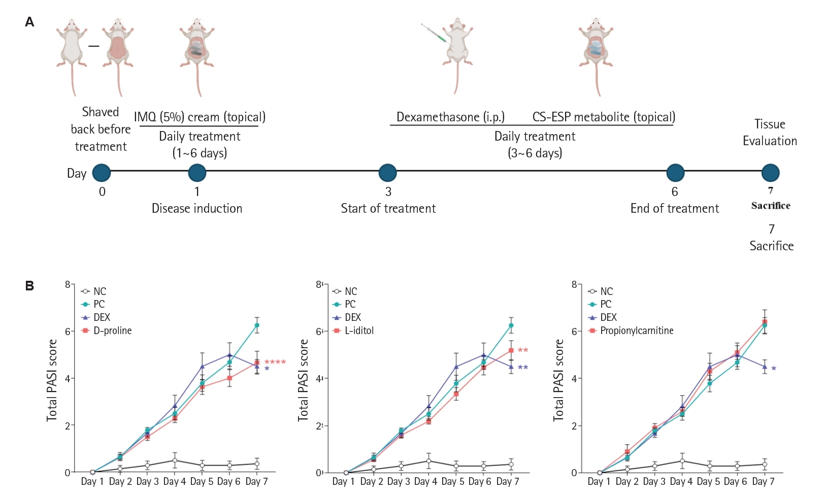
To evaluate the therapeutic effect of the candidate metabolites, an IMQ-induced psoriasis mouse model was constructed (
Fig. 5A). Evaluation of clinical skin severity based on the total PASI score revealed that treatment with specific candidate metabolites identified in CS preparations attenuated psoriatic inflammation to varying degrees (
Fig. 5B). A high PASI score (mean±SEM, 6.25±0.33) was yielded in the PC groups, showing pronounced erythema, epidermal thickening, and scaling. This confirmed the successful induction of psoriasis-like pathology. At the end of the experiment, the DEX group showed the lower PASI score than the PC groups (4.50±0.29 versus PC,
P=0.004), supporting its effectiveness as a systemic anti-inflammatory agent. Treatment of D-proline or L-iditol exhibited the significantly lower PASI scores than the PC groups (4.66±0.49 and 5.19±0.42 versus PC,
P<0.0001 and
P=0.002). However, treatment of propionylcarnitine did not show significant change in the PASI score compared to that of PC group. Collectively, we found that D-proline and L-iditol harbored notable anti-inflammatory activity in vivo and may contribute as therapeutics in psoriatic skin inflammation. In support of these observations, two-way ANOVA revealed a significant treatment effect, primarily driven by D-proline and L-iditol groups (
F(6, 252)=68.36,
P<0.0001).
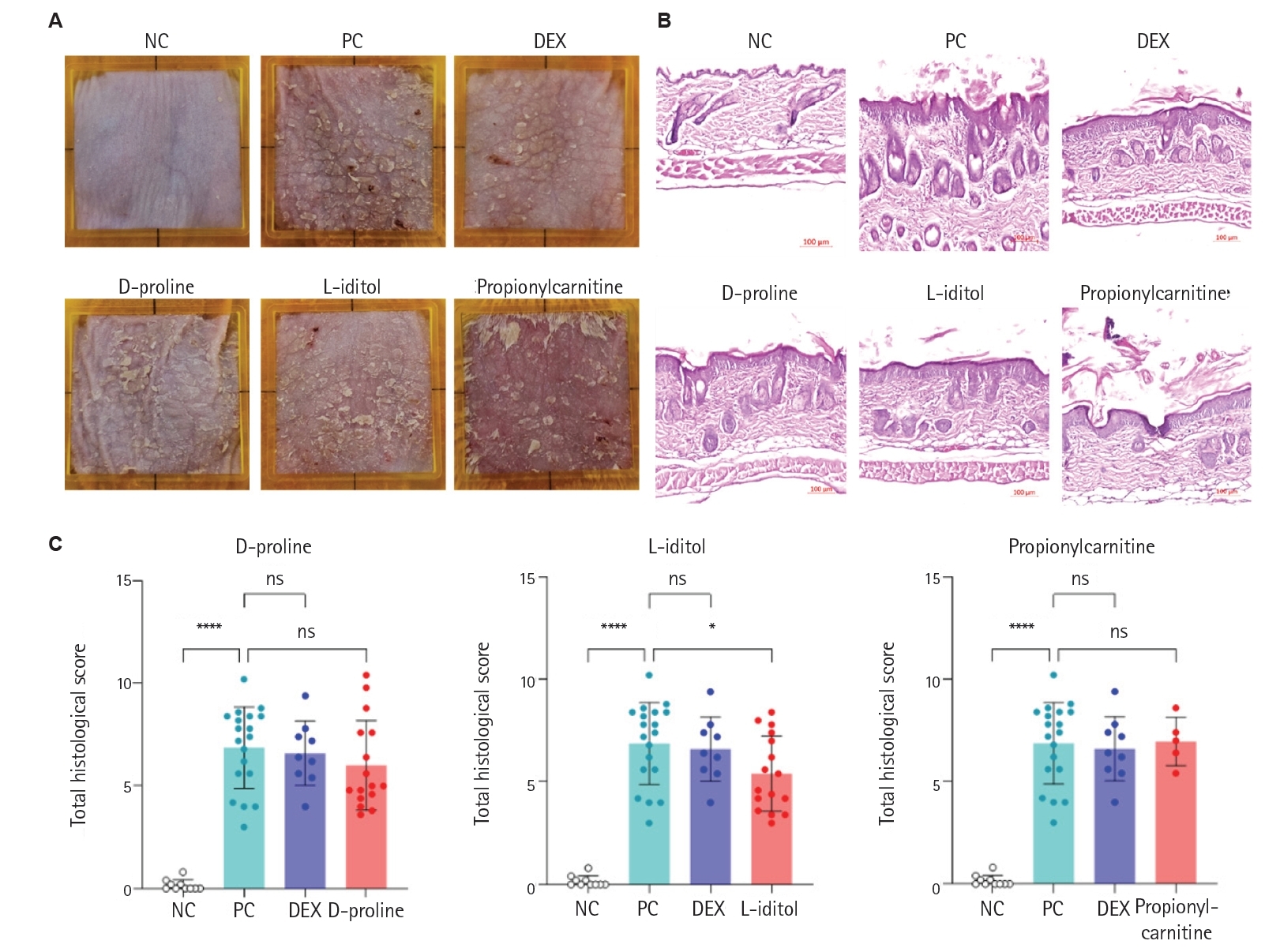
The anti-psoriatic effects of candidate metabolites identified in CS preparations were further supported by visual and histological evaluations of dorsal skin on day 7 (
Fig. 6A-
C). Mice in the PC group exhibited pronounced erythema and substantial keratin accumulation compared to the NC, clearly confirming the successful induction of psoriasis-like skin pathology. In contrast, the DEX group displayed visibly reduced redness and keratin buildup, consistent with its known anti-inflammatory efficacy. Among the metabolite-treated groups, both D-proline and L-iditol led to a reduction in skin redness relative to the PC group. However, propionylcarnitine showed inconsistent results, with decreased keratin deposition but persistent erythema in several cases (
Fig. 6A). Histopathological examination of skin tissues (
Fig. 6B) further corroborated these clinical observations. The PC group displayed marked dermal thickening, dense immune cell infiltration, and increased epidermal thickness, accompanied by evident hyperkeratosis and parakeratosis. While both D-proline and L-iditol treatments showed partial improvements, L-iditol led to a more substantial reduction in dermal thickness and inflammatory infiltration compared to D-proline, suggesting a more pronounced histological benefit. In contrast, propionylcarnitine did not improve keratin-related changes despite a slight reduction in dermal thickness. Quantitative analysis using the PHS supported these findings (
Fig. 6C). The PC group exhibited significantly elevated PHS compared to NC (mean±SD, 6.87±0.46 for the PC group versus 0.16±0.08 for the NC group,
P<0.0001), confirming the severity of induced pathology. Among the treatment groups, only L-iditol showed a statistically significant reduction in PHS (5.41±0.46 versus PC,
P=0.038), highlighting its potential as a promising candidate for mitigating psoriatic histopathology. Collectively, these results demonstrate a divergence between clinical and histological efficacy; while both D-proline and L-iditol attenuated clinical severity (PASI), L-iditol provided a more robust and consistent anti-inflammatory effect at the tissue level, as evidenced by the significant improvement in PHS.
Discussion
In this study, we identified and evaluated the anti-inflammatory potential of the candidate metabolites identified in CS preparations—D-proline, L-iditol, and propionylcarnitine. When tested in vitro, all 3 compounds showed no cytotoxicity at their effective concentrations, and subsequent assays confirmed their ability to suppress key pro-inflammatory mediators in LPS-stimulated macrophages. In vivo, among the tested metabolites, L-iditol emerged as the most promising candidate due to its consistent efficacy in both clinical (PASI) and histological (PHS) assessments. Although D-proline attenuated visual skin severity, it did not lead to a significant improvement in the composite histopathological score, highlighting the superior immunomodulatory potential of L-iditol in this psoriasiform model. The anti-psoriatic efficacy of L-iditol was particularly notable, as these compounds not only reduced visual erythema and keratin buildup but also mitigated histological features such as dermal thickening and immune cell infiltration. These improvements were comparable to or approaching those observed with dexamethasone, a known systemic immunosuppressant. Although propionylcarnitine showed partial effects by reducing keratin deposition, it did not lead to a statistically significant reduction in PASI or PHS scores, suggesting a more limited therapeutic potential in this model.
Previous studies have demonstrated that CS-derived fractions, including CSp [
20] and CS-ESP [
21], exert significant anti-inflammatory effects in various inflammatory disease models, suggesting that parasite-derived components can modulate host immune responses. Building upon these findings, our group previously performed proteomic analyses of CS-ESP and demonstrated that several proteins identified through this approach also exhibited anti-inflammatory activity [
21]. These results indicate that the immunomodulatory properties of parasite-derived products are not restricted to a single molecular class but may arise from multiple molecular layers, including proteins and other bioactive components. In this context, the present study further expanded the analytical scope to the metabolomic level. Using LC-MS/MS–based metabolomic profiling, we identified candidate metabolites identified in CS preparations and subsequently evaluated their anti-inflammatory effects. Our findings demonstrate that selected parasite-derived metabolites significantly modulate inflammatory responses, supporting the concept that metabolites contribute to the overall anti-inflammatory activity previously observed for CSp and CS-ESP. The anti-inflammatory effects observed in this study are consistent with previous findings regarding metabolite-mediated immune modulation. Proline has been reported to inhibit inflammatory responses, including a reduction in TNF-α levels, under inflammatory conditions [
25]. Although direct evidence for L-iditol is limited, structurally related metabolites such as myo-inositol have been shown to inhibit pro-inflammatory cytokines, including TNF-α, IL-1β, and IL-6 [
26]. Additionally, carnitine-related metabolites have been reported to exhibit anti-inflammatory effects by reducing cytokine production and regulating macrophage activation [
27]. These findings support the potential for the identified metabolites to modulate key inflammatory mediators associated with psoriasis.
The differential expression patterns of cytokines observed in response to LPS stimulation may reflect distinct regulatory mechanisms and expression kinetics. It is well known that cytokines such as TNF-α are rapidly induced, whereas others, including IL-6 and IL-1β, may exhibit delayed or sustained expression depending on transcriptional regulation [
28-
31]. Due to these temporal variations, measurements taken at a specific time point (e.g., 24 h) may show significant reductions in certain cytokines while others remain less affected. Psoriasis is driven by a complex inflammatory network, in which the TNF-α, IL-6, and IL-23/IL-17 axis play central roles [
5,
32,
33]. In this study, candidate metabolites identified in CS preparations significantly reduced the expression of key pro-inflammatory cytokines, including TNF-α and IL-6, in activated macrophages. Given that TNF-α amplifies IL-17-mediated responses and IL-6 facilitates Th17 differentiation, these results suggest a potential modulatory effect on the broader psoriatic inflammatory milieu. However, as this study focused primarily on cytokine expression in LPS-stimulated macrophages and phenotypic changes in an acute IMQ model, direct evidence for the modulation of IL-23/IL-17 signaling or keratinocyte-specific pathways was not obtained. Therefore, while the reduction of early-stage inflammatory mediators provides a plausible basis for the observed therapeutic effects, the definitive involvement of the Th17 axis remains to be further validated through protein-level analysis and targeted mechanistic studies.
The current study has several limitations. First, although the tested metabolites exhibited anti-inflammatory effects, the specific molecular targets and signaling pathways involved were not directly identified. The involvement of the Th17 pathway could not be clarified directly based on the current data. because IL-17 expression was not evaluated. To elucidate the protein-level dynamics and downstream signaling, additional validation using ELISA or Western blot is warranted in future studies. Second, due to the small sample size of the propionylcarnitine administration group, in vivo results should be interpreted cautiously. The lack of significant effects in PASI and PHS may be due to insufficient statistical power, as the corresponding compound was excluded from subsequent experiments following the initial preliminary study. Third, the IMQ-induced psoriasis model used in this study primarily reflects an acute inflammatory response characterized by rapid onset and cytokine-driven keratinocyte hyperproliferation. This model may not fully capture the complex, long-term pathophysiology of chronic human psoriasis, which involves persistent T-cell memory and systemic comorbidities. Therefore, while our results demonstrate the potent anti-inflammatory potential of the candidate metabolites, further studies using chronic or genetic psoriasis models are warranted to evaluate their long-term therapeutic efficacy and safety. Finally, metabolite identification was performed based on LC-MS/MS spectral matching (metabolomics standards initiative levels 2–3) without verification using orthogonal analysis methods [
5]. Although LC-MS/MS–based annotation are widely used [
34], it remains a major bottleneck in metabolomics research due to limitations in confirming structural identity [
34,
35]. Therefore, these results must be interpreted cautiously. Nevertheless, the use of commercially available high-purity compounds for subsequent functional validation partially compensates for the identification uncertainty by confirming the biological activity of the predicted molecular structures.
In conclusion, our data highlights the potential of specific candidate metabolites identified in CS preparations, particularly L-iditol, as candidates for anti-inflammatory drug development. Despite the limitations, these findings contribute to a growing body of evidence supporting the role of helminth-derived molecules in immune modulation and encourage further exploration into their therapeutic applications in chronic inflammatory diseases such as psoriasis.
Notes
-
Data availability
Data will be made available on request.
-
Author contributions
Conceptualization: Won EJ, Kim TJ. Data curation: Lee YJ, Kim MJ, Yu SM, Won EJ, Kim TJ. Formal analysis: Lee YJ. Funding acquisition: Ryu JH, Won EJ, Kim TJ. Investigation: Lee YJ. Methodology: Lee YJ, Kim MJ, Yu SM, Won EJ, Kim TJ. Project administration: Won EJ, Kim TJ. Resources: Won EJ, Kim TJ. Software: Won EJ, Kim TJ. Supervision: Ryu JH, Won EJ, Kim TJ. Writing – original draft: Lee YJ, Won EJ, Kim TJ. Writing – review & editing: Kim MJ, Yu SM, Ryu JH, Won EJ, Kim TJ.
-
Conflict of interest
The authors have no conflicts of interest to declare.
-
Funding
This study was supported by the grants from the National Research Foundation of Korea (NRF) funded by the Ministry of Education, Science and Technology (NRF-2022R1C1C1002741 and RS-2024-00410713) and the grants of Chonnam National University Hospital Biomedical Research Institute (BCRI26054 and BCRI25056).
Supplementary information
Fig. 1.Workflow of metabolite analysis of candidate metabolites from Clonorchis sinensis crude somatic proteins (CSp) and excretory/secretory proteins (CS-ESP). (A) Schematic representation of the overall experimental workflow, including the extraction of CSp and CS-ESP from Clonorchis sinensis, followed by metabolite extraction for subsequent profiling. Both protein fractions were subjected to methanol-based extraction procedures to isolate small molecule metabolites suitable for LC-MS/MS analysis. (B) Comparison of the top 10 most abundant metabolites identified in the CSp and CS-ESP samples based on untargeted LC-MS/MS profiling. Metabolite abundance was ranked according to signal intensity, and key metabolites were selected for further functional evaluation based on biological relevance and literature support.
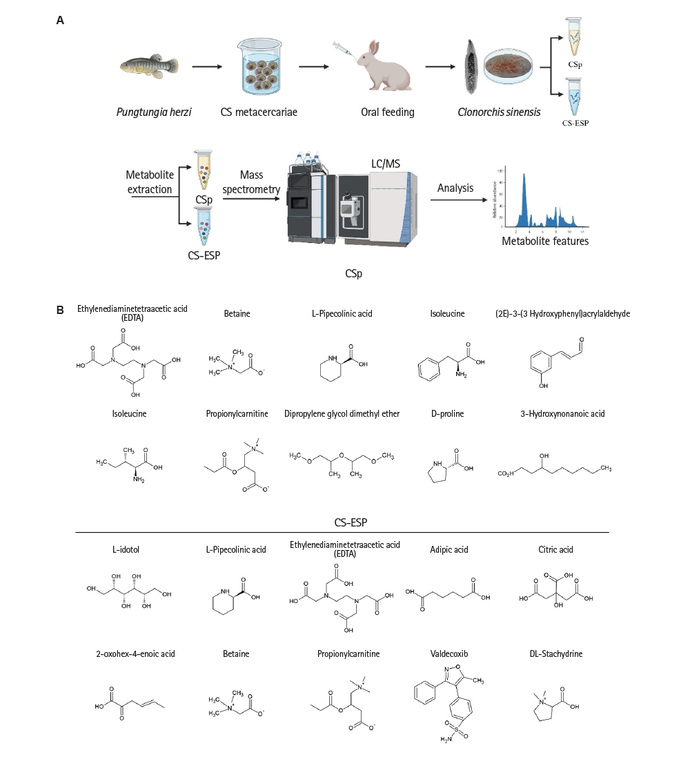
Fig. 2.Anti-inflammatory effect of D-proline in vitro. (A) Effect of D-proline on cell viability in RAW 264.7 macrophages. Cells were treated with D-proline for 24 h, and viability was assessed using an MTS assay. D-proline treatment did not result in any significant cytotoxicity at the tested concentrations, indicating it is well tolerated by RAW 264.7 cells under these conditions. (B) Gene expression analysis of inflammatory markers in RAW 264.7 cells treated with 4 μg/ml LPS and 50 μg/ml D-proline for 24 h. RT-PCR was performed to evaluate the expression levels of key pro-inflammatory genes. D-proline treatment led to a reduction in the expression of several LPS-induced genes, suggesting its potential anti-inflammatory effect. Statistical analysis was performed using the Friedman test with multiple comparisons, followed by Wilcoxon signed-rank test for pairwise comparisons. Data are presented as mean±SD (n=6). Statistical analysis was performed using the Kruskal-Wallis test followed by Dunn’s multiple comparisons test. *P<0.05. Each symbol represents an individual biological replicate. IL, interleukin; TNF, tumor necrosis factor; iNOS, inducible nitric oxide synthase; COX, cyclooxygenase; ns, not significant.
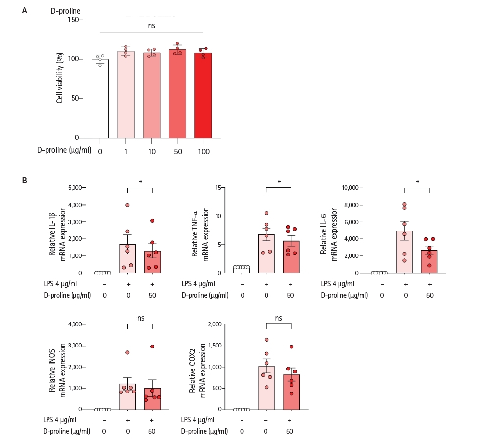
Fig. 3.Anti-inflammatory effect of L-iditol in vitro. (A) Effect of L-iditol on cell viability in RAW 264.7 macrophages. Cells were treated with L-iditol for 24 h, and viability was assessed using an MTS assay. L-iditol treatment did not result in any significant cytotoxicity at the tested concentrations, indicating it is well tolerated by RAW 264.7 cells under these conditions. (B) Gene expression analysis of inflammatory markers in RAW 264.7 cells treated with 4 μg/ml LPS and 10 μg/ml L-iditol for 24 h. RT-PCR was performed to evaluate the expression levels of key pro-inflammatory genes. L-iditol treatment led to a reduction in the expression of several LPS-induced genes, suggesting its potential anti-inflammatory effect. Statistical analysis was performed using the Friedman test with multiple comparisons, followed by Wilcoxon signed-rank test for pairwise comparisons. Data are presented as mean±SD (n=7). Statistical analysis was performed using the Kruskal-Wallis test followed by Dunn’s multiple comparisons test. *P<0.05. Each symbol represents an individual biological replicate. IL, interleukin; TNF, tumor necrosis factor; iNOS, inducible nitric oxide synthase; COX, cyclooxygenase; ns, not significant.
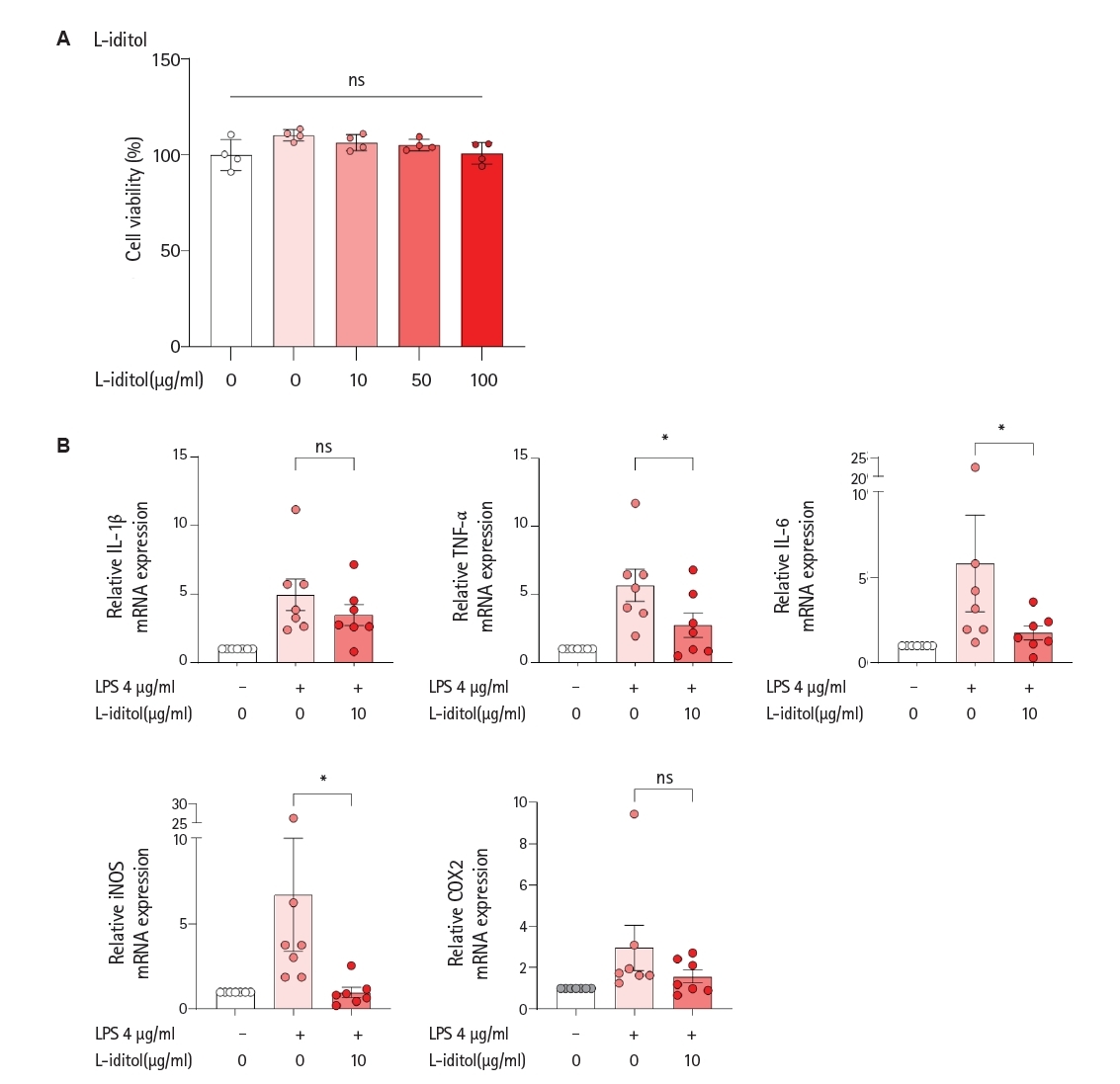
Fig. 4.Anti-inflammatory effect of propionylcarnitine in vitro. (A) Effect of propionylcarnitine on cell viability in RAW 264.7 macrophages. Cells were treated with propionylcarnitine for 24 h, and viability was assessed using an MTS assay. Propionylcarnitine treatment did not result in any significant cytotoxicity at the tested concentrations, indicating it is well tolerated by RAW 264.7 cells under these conditions. (B) Gene expression analysis of inflammatory markers in RAW 264.7 cells treated with 4 μg/ml LPS and 5 μg/ml propionylcarnitine for 24 h. RT-PCR was performed to evaluate the expression levels of key pro-inflammatory genes. Propionylcarnitine treatment led to a reduction in the expression of several LPS-induced genes, suggesting its potential anti-inflammatory effect. Statistical analysis was performed using the Friedman test with multiple comparisons, followed by Wilcoxon signed-rank test for pairwise comparisons. Data are presented as mean± SD (n=6). Statistical analysis was performed using the Kruskal-Wallis test followed by Dunn’s multiple comparisons test. *P<0.05. Each symbol represents an individual biological replicate. ns, not significant; IL, interleukin; TNF, tumor necrosis factor; iNOS, inducible nitric oxide synthase; COX, cyclooxygenase.
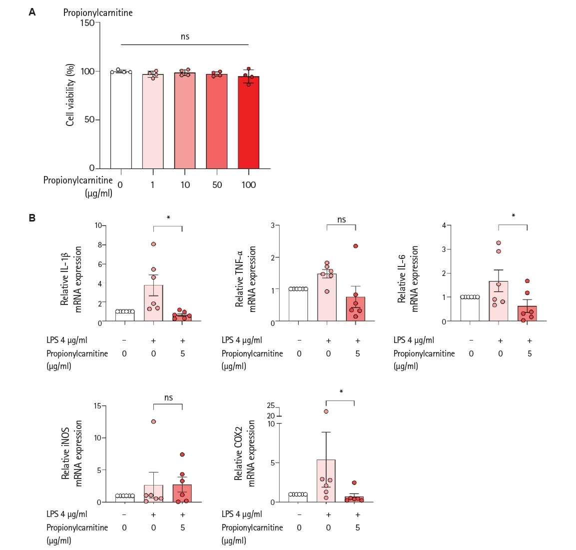
Fig. 5.Psoriasis-like skin inflammation model and treatment in SKG mice. (A) Experimental timeline for psoriasis induction and treatment. Imiquimod (IMQ) cream was applied daily to the shaved dorsal skin of SKG mice from day 1 to day 6. From day 3 to 6, mice received either intraperitoneal dexamethasone (DEX) or topical IMQ mixed with candidate metabolites identified in Clonorchis sinensis preparations (D-proline, L-iditol, or propionylcarnitine). (B) Psoriasis Area and Severity Index (PASI) scores, reflecting erythema, thickness, and scaling, were assessed daily before IMQ application to evaluate treatment efficacy. Statistical analysis was performed using two-way ANOVA with Bonferroni’s multiple comparisons test. NC, negative control group; PC, positive control group. *P<0.05, **P<0.01, ***P<0.001, ****P<0.0001.
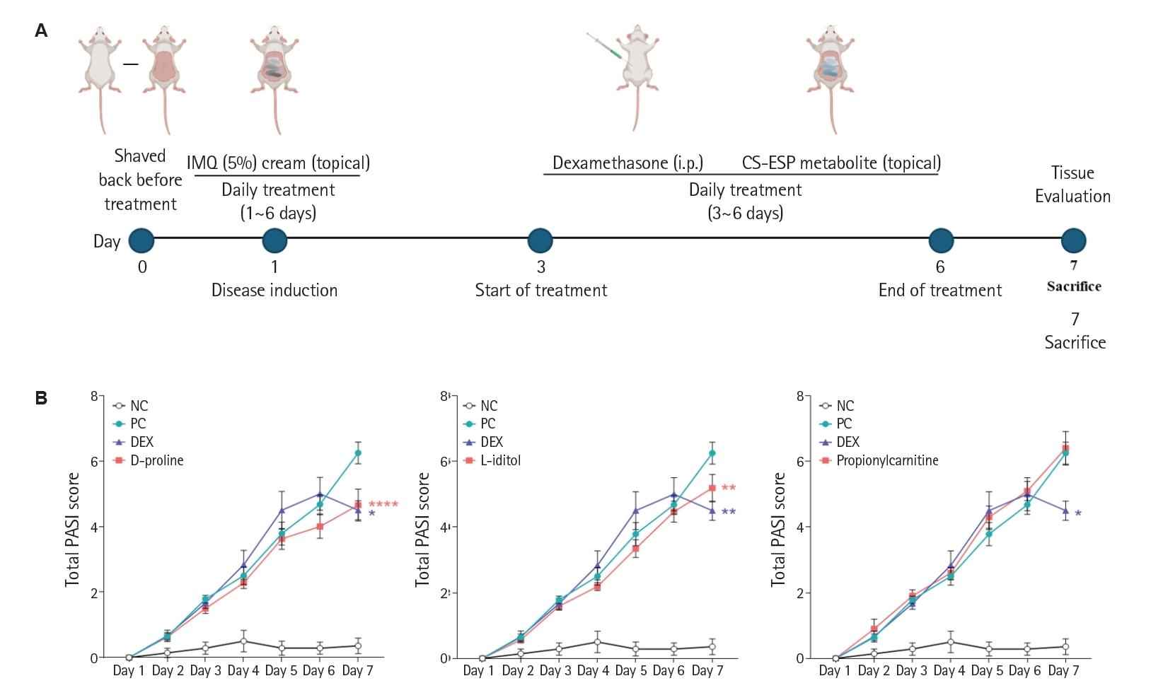
Fig. 6.Anti-inflammatory effects of candidate metabolites identified in Clonorchis sinensis preparations in the imiquimod-induced psoriasis model. (A) Representative images of dorsal skin from SKG mice taken on day 7. The positive control group (PC, imiquimod only) showed marked erythema and keratin buildup, while dexamethasone (DEX), D-proline, and L-iditol treatments reduced redness; propionylcarnitine reduced keratin but not erythema. (B) Hematoxylin and eosin staining of dorsal skin sections revealed dermal thickening, immune cell infiltration, and keratin abnormalities in the PC, which were alleviated by DEX and partially by D-proline and L-iditol. L-iditol showed greater improvement than D-proline, while propionylcarnitine had limited effect. (C) Psoriasis Histopathological Score was significantly increased in the PC versus negative control group (NC) and significantly reduced only in the L-iditol. Statistical analysis was performed using one-way ANOVA with Bonferroni’s multiple comparisons test. ns, not significant. *P<0.05, **P<0.01, ***P<0.001, ****P<0.0001.
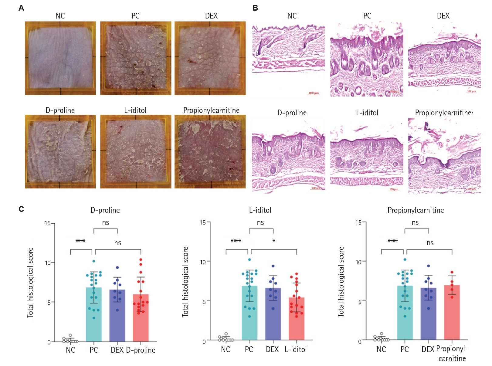
Table 1.Primer sequences for cytokine gene expression analysis
Table 1.
|
Target primer |
Oligonucleotide sequence (5' → 3') |
|
GAPDH |
Forward |
CAACTTTGGCATTGTGGAAGG |
|
Reverse |
ACACATTGGGGGTAGGAACAC |
|
TNF-α |
Forward |
TCTTGTGTTTCTGAGTAGTTGT |
|
Reverse |
CCTTTACTCTGACCCCTTTATT |
|
IL-1β |
Forward |
ATGGTGAAGTCAATTATGTCCT |
|
Reverse |
ACAAGATAGAAGTCAAGAGCAA |
|
IL-6 |
Forward |
AGGTAGCTATGGTACTCCAG |
|
Reverse |
GCACTTGCAGAAAACAATCT |
|
iNOS |
Forward |
GGAAATAGAAACAACAGGAACC |
|
Reverse |
CATTGTTGGTGGCATAAAGTAT |
|
COX2 |
Forward |
AGAAGGGTTCCCAATTAAAGAT |
|
Reverse |
AACAGAAAAACTGTTTCGAAGT |
References
- 1. Wu M, Dai C, Zeng F. Cellular mechanisms of psoriasis pathogenesis: a systemic review. Clin Cosmet Investig Dermatol 2023;16:2503-15. https://doi.org/10.2147/CCID.S420850
- 2. van der Fits L, Mourits S, Voerman JS, et al. Imiquimod-induced psoriasis-like skin inflammation in mice is mediated via the IL-23/IL-17 axis. J Immunol 2009;182:5836-45. https://doi.org/10.4049/jimmunol.0802999
- 3. Brożyna AA, Slominski RM, Nedoszytko B, Zmijewski MA, Slominski AT. Vitamin D signaling in psoriasis: pathogenesis and therapy. Int J Mol Sci 2022;23:8575. https://doi.org/10.3390/ijms23158575
- 4. Ryan S. Psoriasis: characteristics, psychosocial effects and treatment options. Br J Nurs 2010;19:820-825. https://doi.org/10.12968/bjon.2010.19.13.48856
- 5. Zhou X, Chen Y, Cui L, Shi Y, Guo C. Advances in the pathogenesis of psoriasis: from keratinocyte perspective. Cell Death Dis 2022;13:81. https://doi.org/10.1038/s41419-022-04523-3
- 6. Davidovici BB, Sattar N, Prinz J, et al. Psoriasis and systemic inflammatory diseases: potential mechanistic links between skin disease and co-morbid conditions. J Invest Dermatol 2010;130:1785-96. https://doi.org/10.1038/jid.2010.103
- 7. Lebwohl MG, Stein Gold L, Strober B, et al. Phase 3 trials of tapinarof cream for plaque psoriasis. N Engl J Med 2021;385:2219-29. https://doi.org/
- 8. Lebwohl MG, Stein Gold L, Strober B, et al. Trial of roflumilast cream for chronic plaque psoriasis. N Engl J Med 2020;383:229-39. https://doi.org/10.1056/NEJMoa2103629
- 9. Strober B, Thaci D, Sofen H, et al. Deucravacitinib versus placebo and apremilast in moderate to severe plaque psoriasis: efficacy and safety results from the 52-week, randomized, double-blinded, phase 3 Program fOr Evaluation of TYK2 inhibitor psoriasis second trial. J Am Acad Dermatol 2023;88:40-51. https://doi.org/10.1016/j.jaad.2022.08.061
- 10. Stein Gold L, Adam DN, Albrecht L, et al. Long-term safety and effectiveness of roflumilast cream 0.3% in adults with chronic plaque psoriasis: a 52-week, phase 2, open-label trial. J Am Acad Dermatol 2024;91:273-80. https://doi.org/10.1016/j.jaad.2024.03.030
- 11. Armstrong AW, Lebwohl M, Warren RB, et al. Safety and efficacy of deucravacitinib in moderate to severe plaque psoriasis for up to 3 years: an open-label extension of randomized clinical trials. JAMA Dermatol 2025;161:56-66. https://doi.org/10.1001/jamadermatol.2024.4688
- 12. Armstrong AW, Read C. Pathophysiology, clinical presentation, and treatment of psoriasis: a review. JAMA 2020;323:1945-60. https://doi.org/10.1001/jama.2020.4006
- 13. Griffiths CEM, Armstrong AW, Gudjonsson JE, Barker JNWN. Psoriasis. Lancet 2021;397:1301-15. https://doi.org/10.1016/S0140-6736(20)32549-6
- 14. Bachelez H, Choon SE, Marrakchi S, et al. Trial of spesolimab for generalized pustular psoriasis. N Engl J Med 2021;385:2431-40. https://doi.org/10.1056/NEJMoa2111563
- 15. Ellinghaus D, Jostins L, Spain SL, et al. Analysis of five chronic inflammatory diseases identifies 27 new associations and highlights disease-specific patterns at shared loci. Nat Genet 2016;48:510-8. https://doi.org/10.1038/ng.3528
- 16. Kuijk LM, van Die I. Worms to the rescue: can worm glycans protect from autoimmune diseases? IUBMB Life 2010;62:303-12. https://doi.org/10.1002/iub.304
- 17. Summers RW, Elliott DE, Urban JF Jr, Thompson RA, Weinstock JV. Trichuris suis therapy for active ulcerative colitis: a randomized controlled trial. Gastroenterology 2005;128:825-32. https://doi.org/10.1053/j.gastro.2005.01.005
- 18. Navarro S, Pickering DA, Ferreira IB, et al. Hookworm recombinant protein promotes regulatory T cell responses that suppress experimental asthma. Sci Transl Med 2016;8:362ra143. https://doi.org/10.1126/scitranslmed.aaf8807
- 19. Ahangari Maleki M, Malek Mahdavi A, Soltani-Zangbar MS, Yousefi M, Khabbazi A. Randomized double-blinded controlled trial on the effect of synbiotic supplementation on IL-17/IL-23 pathway and disease activity in patients with axial spondyloarthritis. Immunopharmacol Immunotoxicol 2023;45:43-51. https://doi.org/10.1080/08923973.2022.2112220
- 20. Lee YJ, Kim MJ, Jo S, et al. Clonorchis sinensis-derived protein attenuates inflammation and new bone formation in ankylosing spondylitis. Front Immunol 2021;12:615369. https://doi.org/10.3389/fimmu.2021.615369
- 21. Kim MJ, Yoo HM, Lee YJ, et al. Clonorchis sinensis excretory/secretory proteins ameliorate inflammation in rheumatoid arthritis and ankylosing spondylitis. Parasit Vectors 2025;18:85. https://doi.org/10.1186/s13071-025-06677-3
- 22. Hong SJ, Seong KY, Sohn WM, Song KY. Molecular cloning and immunological characterization of phosphoglycerate kinase from Clonorchis sinensis. Mol Biochem Parasitol 2000;108:207-16. https://doi.org/10.1016/s0166-6851(00)00220-6
- 23. Jabeen M, Boisgard AS, Danoy A, et al. Advanced characterization of imiquimod-induced psoriasis-like mouse model. Pharmaceutics 2020;12:789. https://doi.org/10.3390/pharmaceutics12090789
- 24. Karamani C, Antoniadou IT, Dimou A, et al. Optimization of psoriasis mouse models. J Pharmacol Toxicol Methods 2021;108:107054. https://doi.org/10.1016/j.vascn.2021.107054
- 25. Obayashi Y, Arisaka H, Yoshida S, Mori M, Takahashi M. Proline protects liver from D-galactosamine hepatitis by activating the IL-6/STAT3 survival signaling pathway. Amino Acids 2012;43:2371-80. https://doi.org/10.1007/s00726-012-1317-8
- 26. Seif M, Dahabiyeh L, Al-Hunaiti A, et al. Anti-inflammatory activity and metabolite profiling of myo-inositol in LPS-stimulated macrophages. PLoS One 2026;21:e0341193. https://doi.org/10.1371/journal.pone.0341193
- 27. Bollati C, Tosi M, d'Adduzio L, et al. Antioxidant and anti-inflammatory activity of a new formulation of slow-release amino acids in human intestinal Caco-2 cells. Antioxidants (Basel) 2025;14:271. https://doi.org/10.3390/antiox14030271
- 28. DeForge LE, Remick DG. Kinetics of TNF, IL-6, and IL-8 gene expression in LPS-stimulated human whole blood. Biochem Biophys Res Commun 1991;174:18-24. https://doi.org/10.1016/0006-291x(91)90478-p
- 29. Wong S, Schwartz RC, Pestka JJ. Superinduction of TNF-alpha and IL-6 in macrophages by vomitoxin (deoxynivalenol) modulated by mRNA stabilization. Toxicology 2001;161:139-49. https://doi.org/10.1016/s0300-483x(01)00331-6
- 30. Hobbs S, Reynoso M, Geddis AV, Mitrophanov AY, Matheny RW Jr. LPS-stimulated NF-κB p65 dynamic response marks the initiation of TNF expression and transition to IL-10 expression in RAW 264.7 macrophages. Physiol Rep 2018;6:e13914. https://doi.org/10.14814/phy2.13914
- 31. Zimmermann M, Aguilera FB, Castellucci M, et al. Chromatin remodelling and autocrine TNFα are required for optimal interleukin-6 expression in activated human neutrophils. Nat Commun 2015;6:6061. https://doi.org/10.1038/ncomms7061
- 32. Baliwag J, Barnes DH, Johnston A. Cytokines in psoriasis. Cytokine 2015;73:342-50. https://doi.org/10.1016/j.cyto.2014.12.014
- 33. Goodman WA, Levine AD, Massari JV, et al. IL-6 signaling in psoriasis prevents immune suppression by regulatory T cells. J Immunol 2009;183:3170-6. https://doi.org/10.4049/jimmunol.0803721
- 34. Theodoridis G, Gika H, Raftery D, et al. Ensuring fact-based metabolite identification in liquid chromatography-mass spectrometry-based metabolomics. Anal Chem 2023;95:3909-16. https://doi.org/10.1021/acs.analchem.2c05192
- 35. Wang X, Liu Y, Jiang C, et al. TidyMass2: advancing LC-MS untargeted metabolomics through metabolite origin inference and metabolic feature-based functional module analysis. Nat Commun 2026;17:1755. https://doi.org/10.1038/s41467-026-68464-7