Abstract
Dirofilaria spp. are mosquito-borne filarial nematodes that primarily infect mammals. Here, we describe a filarial nematode infection in a female little egret (Egretta garzetta) from Jeju, Korea. Necropsy revealed multiple slender nematodes (4–6 cm) in the subcutaneous tissues of both legs. Histopathological examination demonstrated granulomatous inflammation with intralesional parasites. Morphologically, the nematodes exhibited a thick multilayered cuticle with prominent longitudinal ridges, consistent with members of the genus Dirofilaria. Molecular analysis based on 18S rRNA and mitochondrial c oxidase subunit I (cox1) gene sequences indicated that the parasite was most closely related to D. repens; however, the level of sequence divergence precluded definitive species-level identification. Phylogenetic analysis further demonstrated that the isolates clustered within the Dirofilaria clade and were clearly separated from avian-associated filarioid nematodes. This study provides molecular and histopathological evidence of a Dirofilaria sp. infection in an avian host and supports incidental infection under natural exposure conditions.
-
Key words: Avian filariasis, Dirofilaria sp., granulomatous inflammation, little egret, subcutaneous tissue
Introduction
Dirofilaria spp. are mosquito-borne filarial nematodes that primarily infect mammals, particularly canines [
1].
Dirofilaria immitis is the causative agent of heartworm disease and resides in the pulmonary artery and right ventricle of the host [
2]. In contrast,
D. repens typically inhabits the subcutaneous tissues and is associated with subcutaneous dirofilariasis [
3].
In canines,
D. repens infection is usually asymptomatic; however, it is of significant zoonotic importance because of its ability to infect humans. In humans, adult worms may localize to the periorbital region or form subcutaneous nodules [
4].
To date, infections caused by
Dirofilaria spp. have been primarily documented in mammalian hosts, including dogs, cats, and humans. In contrast, filarioid infections in avian species are typically associated with other genera, such as
Splendidofilaria,
Eulimdana,
Lemdana, and
Diplotriaena, which inhabit various tissues depending on the species [
5]. Reports of
Dirofilaria infection in birds are extremely limited, and species-level identification in such cases remains uncertain.
In this study, we describe a filarial nematode infection detected in the subcutaneous tissues of a female little egret (Egretta garzetta) from Jeju, Korea. Based on morphological characteristics and molecular analyses, the parasite was identified as a member of the genus Dirofilaria and was most closely related to D. repens. However, the available data did not allow definitive species-level identification. This case provides morphological and molecular evidence of a Dirofilaria sp. infection in an avian host and offers insight into host–parasite interactions in an unusual host under natural exposure conditions.
Case Report
On April 7, 2025, the Jeju Wildlife Rescue Center, Korea, rescued an adult female little egret of unknown age in an exhausted and ataxic state. At rescue, the bird was severely emaciated (body weight, 260 g; body condition score, 1/5) and exhibited moderate dehydration and hypothermia. No external trauma, fractures, dislocations, or respiratory distress were observed.
Hematological examination revealed anemia (packed cell volume, 24%; reference range, 40%–60%) and markedly decreased total protein (approximately 1 g/dl; reference range, 3.5–5.5 g/dl), indicating hypoproteinemia.
The prognosis of the bird was poor, and it died the following day. To determine the cause of death, a necropsy was performed at the Department of Veterinary Pathology, College of Veterinary Medicine, Jeju National University, Korea. Ethical approval was not required because the bird was rescued from the wild and died naturally.
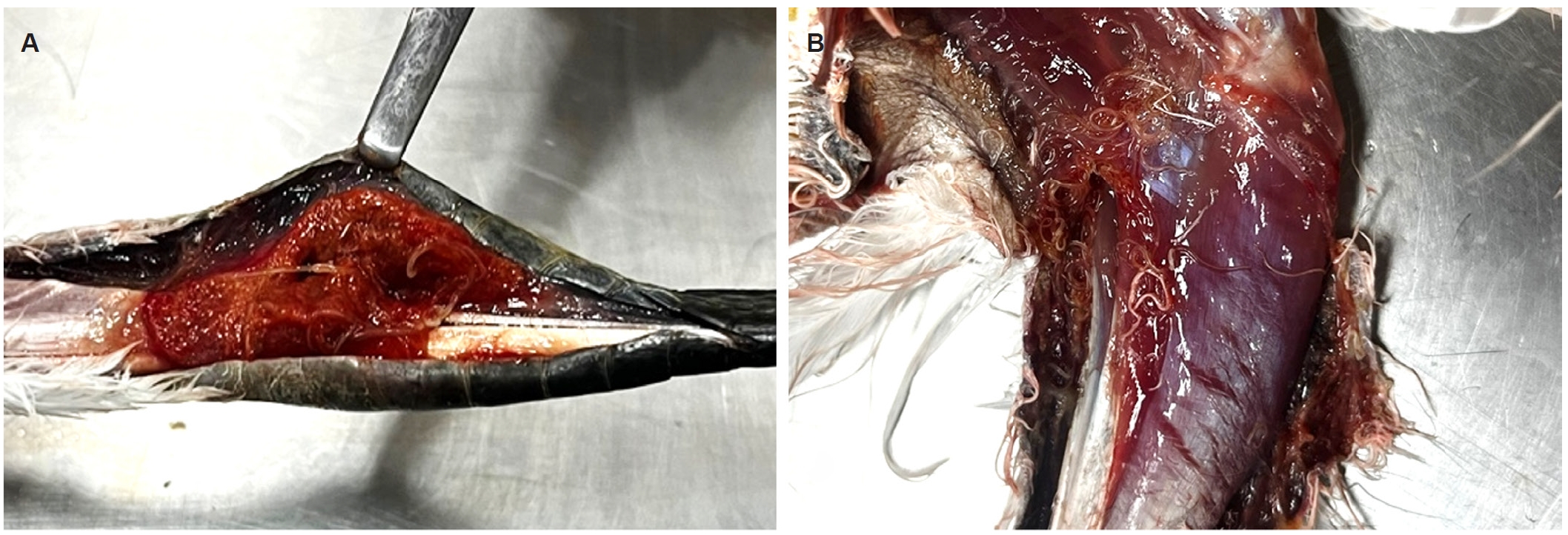
Gross examination revealed a 2-cm hemorrhagic lesion in the right pectoral muscle and a perforating lesion in the upper region of the left pectoral muscle. Multiple pulmonary hemorrhages were observed in the lungs. No distinct nodular lesions were observed externally in the legs; however, upon incision, approximately 10 slender, thread-like nematodes (4–6 cm in length) were identified within the subcutaneous tissues of both legs (
Fig. 1A,
B).
Tissue samples were collected and immediately fixed in 10% neutral-buffered formalin, routinely processed, embedded in paraffin, sectioned at 3–4 μm, and stained with hematoxylin and eosin for histopathological examination. Recovered parasites were fixed in 70% ethanol and examined under a light microscope for morphological identification.
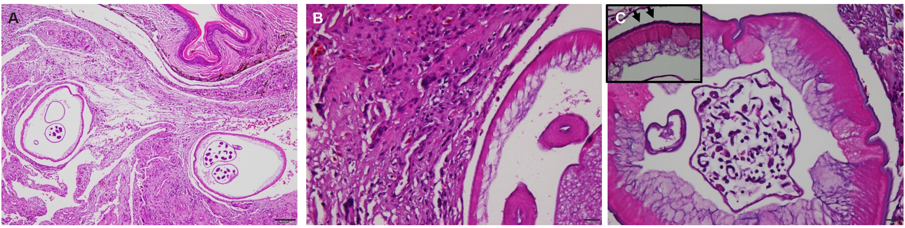
Histopathological examination revealed moderate-to-severe fibrous connective tissue proliferation and granulomatous inflammation in the subcutaneous tissues of the legs (
Fig. 2A). The lesions were characterized by infiltration of numerous macrophages, lymphocytes, as well as multinucleated giant cells surrounding intralesional parasitic organisms in the affected subcutaneous areas (
Fig. 2B). No microfilariae were observed in the examined tissue sections. Moderate-to-severe pulmonary hemorrhage was observed in the lungs.
Histological examination revealed transverse sections of a filarial nematode characterized by a thick, multilayered eosinophilic cuticle with prominent, regularly spaced external longitudinal ridges (
Fig. 2C). Beneath the cuticle, a thin hypodermis and well-developed polymyarian–coelomyarian somatic musculature were observed. Within the pseudocoelomic cavity, reproductive structures containing numerous microfilariae were present, indicating a gravid female worm.
Gross morphological examination revealed a slender, elongated filarial nematode with a cylindrical body and tapering anterior and posterior ends. The worm was white-yellowish in color and measured approximately 6.0 cm in length and 0.5 mm in diameter. Microscopic examination of the worm surface further demonstrated distinct, parallel longitudinal ridges separated by shallow grooves.
For molecular identification, genomic DNA was extracted from 4 parasite samples using the Clear-S Quick DNA Extraction Kit (Invirustech) according to the manufacturer’s instructions. PCR amplification was performed using an AccuPower PCR Premix Kit (Bioneer). The 18S rRNA gene was amplified using the primer set 18S.631f/18S.1825r, yielding an amplicon of approximately 1,127–1,155 bp [
6]. Additionally, the mitochondrial cytochrome c oxidase subunit I (
cox1) gene was amplified using specific primers, resulting in an approximately 680-bp PCR product [
7].
DNA sequencing was performed by Macrogen using the same primer sets. Sequence similarity was analyzed by comparison with reference sequences deposited in GenBank database (National Center for Biotechnology Information). Multiple sequence alignments were performed using Clustal Omega version 1.2.1 and refined using BioEdit version 7.2.5. Phylogenetic analysis was conducted using MEGA software version 6.0 based on the maximum-likelihood method with the Kimura two-parameter model. Bootstrap analysis with 1,000 replicates was performed to assess the reliability of the resulting phylogenetic tree.
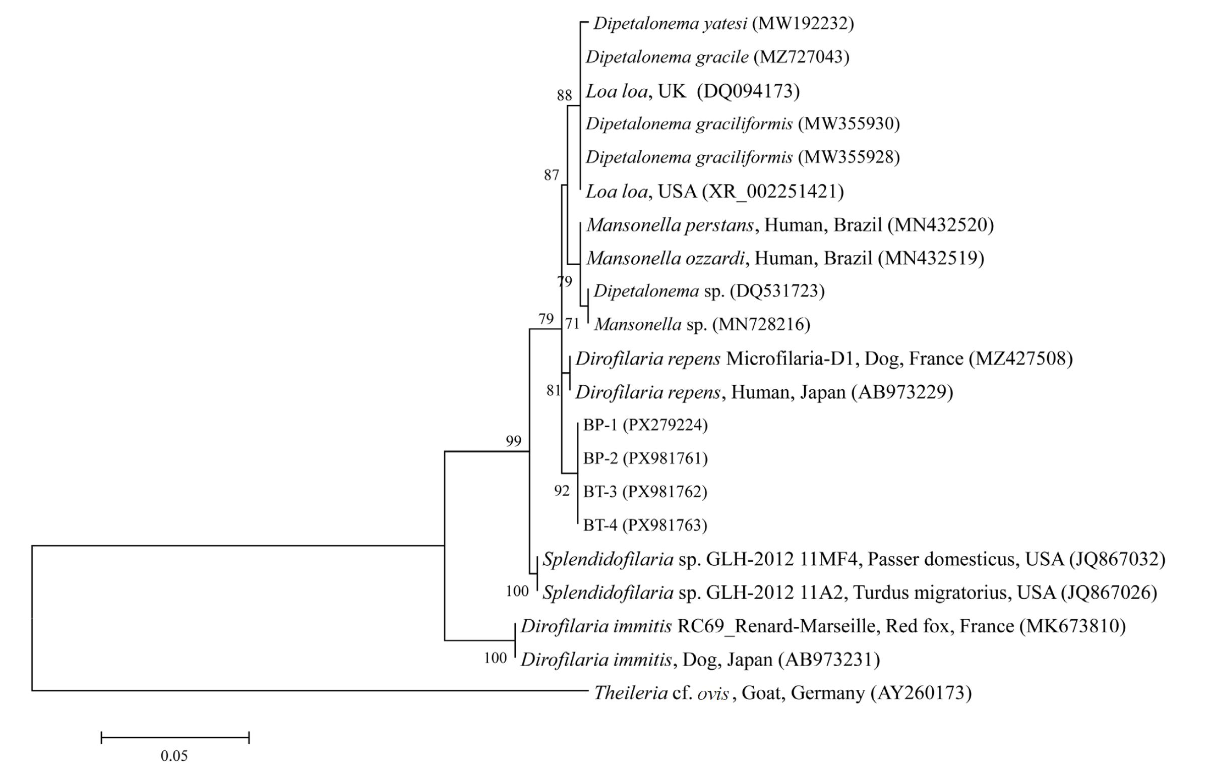
The 18S rRNA sequences obtained in this study shared the highest sequence identity (99.2%) with
D. repens sequences deposited in GenBank. However, similarly high sequence identities were also observed with other filarial genera, including
Mansonella spp. (99.0%–99.2%),
Dipetalonema spp. (98.6%–99.1%), and
Loa loa (99.0%), indicating that the 18S rRNA gene has limited discriminatory power for species-level identification. The sequences generated in this study have been deposited in GenBank under accession numbers PX279224 and PX981761–PX981763 (
Dirofilaria sp. 18S rRNA) (
Fig. 3).
The
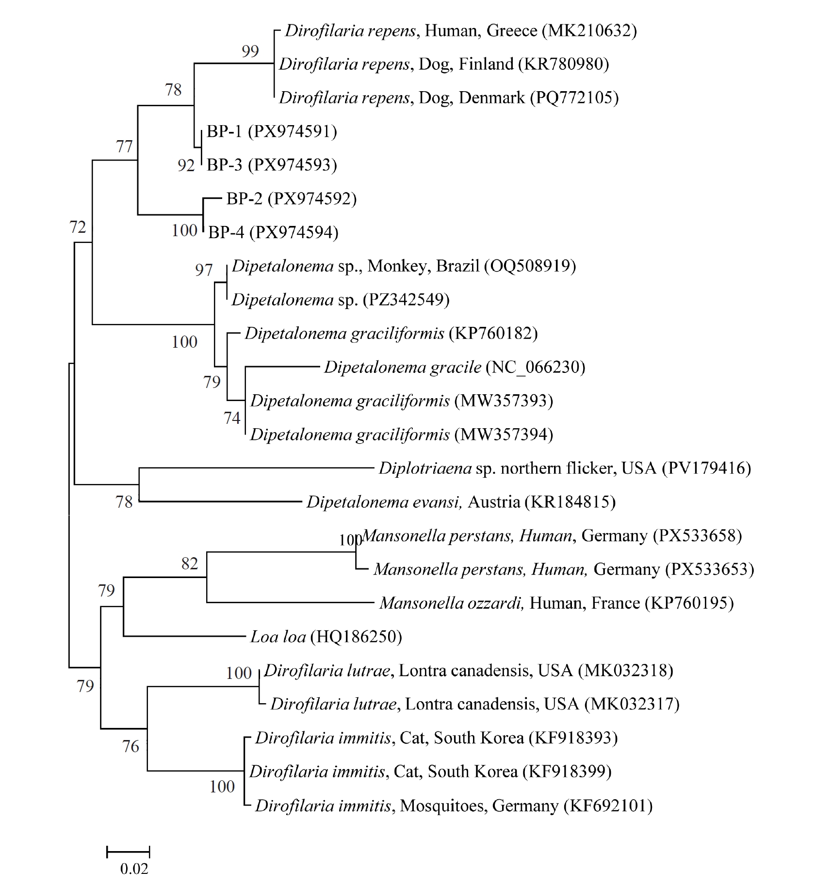
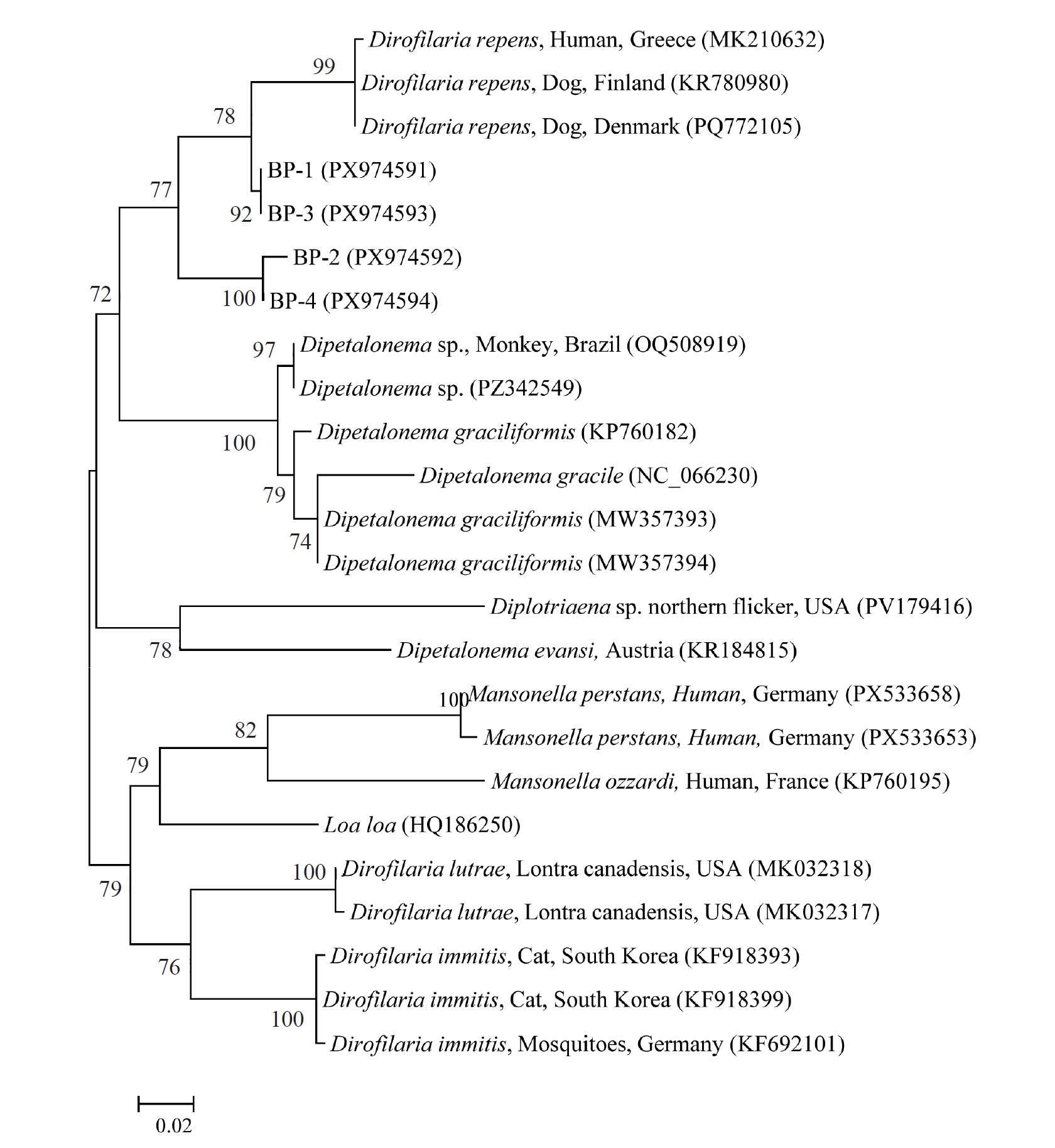
cox1 gene sequences obtained in this study showed 95.1%–100% identity among themselves and 90.1%–94.7% identity with previously reported
D. repens cox1 sequences, representing the highest similarity among the compared taxa. Additional sequence similarity analyses revealed identities of 84.3%–86.9% with
Mansonella spp., 88.0%–89.2% with
L. loa, and 72.4%–90.2% with
Dipetalonema spp. The sequences generated in this study have been deposited under accession numbers PX974591–PX974594 (
Dirofilaria sp.
cox1) (
Fig. 4).
Discussion
To our knowledge, this study represents the first case of a filarial nematode infection most closely related to
D. repens identified in an avian host. To date, reports of
Dirofilaria infection in birds have been extremely limited, with only a single case of
D. immitis infection in Humboldt penguins in Japan [
8]. In the present case, filarial nematodes detected in a little egret from Jeju Island, Korea, exhibited prominent longitudinal ridges on their surface, which are suggestive of morphological features described for
D. repens [
9], although these findings are not sufficient for definitive species-level identification. The parasites were localized within the subcutaneous tissues and were associated with granulomatous inflammation, indicating an active host tissue response.
Although no distinct external nodular lesions were observed, subcutaneous nodular lesions were identified upon incision, corresponding to the location of the parasites. The cause of death was primarily attributed to traumatic injury, including pectoral muscle damage and pulmonary hemorrhage. Clinical findings such as severe emaciation, anemia, and hypoproteinemia may have been influenced, at least in part, by parasitism, and this possibility cannot be entirely excluded. Therefore, while trauma was considered the primary cause of death, it may have represented the final event in a multifactorial process. Accordingly, the filarial infection was considered incidental with respect to the immediate cause of death, although it may have contributed to the overall condition of the bird.
Molecular analyses provided additional insight into the taxonomic identity of the parasite. The 18S rRNA gene showed high sequence similarity not only with D. repens but also with other filarial genera, including Mansonella, Dipetalonema, and Loa, reflecting the highly conserved nature of this marker and its limited utility for species-level discrimination. In contrast, the mitochondrial cox1 gene demonstrated that the sequences obtained in this study were most closely related to D. repens among the available reference sequences, although the observed sequence divergence (90.1%–94.7%) was relatively high. Taken together, these findings indicate that the parasite cannot be conclusively identified as D. repens, but rather represents a Dirofilaria sp. that is most closely related to D. repens. Furthermore, phylogenetic analyses based on both 18S rRNA and cox1 genes consistently placed the sequences obtained in this study within the Dirofilaria clade and clearly separated them from other filarial genera, including Mansonella, Dipetalonema, and Loa. In addition, all 18S rRNA sequences were identical, and 3 out of 4 cox1 sequences shared an identical haplotype, supporting the presence of a single parasite lineage rather than mixed infection or contamination.
Several genera of filarioid nematodes, including
Splendidofilaria,
Eulimdana,
Lemdana, and
Diplotriaena spp., are known to infect avian species, typically inhabiting subcutaneous, connective, muscular, vascular, or respiratory tissues [
5]. Based on host range and tissue tropism, filarial infections in birds are generally attributed to avian-specific filarioids. However, in the present study, both morphological and molecular findings consistently support the classification of the parasite as belonging to the genus
Dirofilaria, as the sequences were clearly placed within the
Dirofilaria clade and distinctly separated from avian-associated filarioid nematodes. These findings suggest that birds may be incidentally exposed to mammalian filarial parasites under natural ecological conditions.
The little egret is both a resident and migratory species in East Asia and is commonly observed on Jeju Island. Therefore, it is unclear whether the individual in this study was a local resident or a migratory bird. Considering the prepatent period of Dirofilaria spp., the geographic origin of infection cannot be determined. This uncertainty should be considered when interpreting the epidemiological context of this case.
Little egrets primarily inhabit wetland environments, such as rice paddies, rivers, fishponds, and intertidal mudflats [
10], where exposure to mosquito vectors is common. As
Dirofilaria spp. are transmitted by mosquitoes via infective third-stage larvae (L3), such ecological conditions may allow incidental exposure of birds. Although avian species are unlikely to support full parasite development, natural exposure followed by limited tissue invasion can occur, as demonstrated in this case.
This study has several limitations. Because the parasitic infection was initially considered an incidental finding during necropsy, detailed parasitological characterization, including sex and developmental stage, was not systematically performed. Furthermore, although a gravid female worm was identified, microfilariae were not observed in tissue sections, and no blood-based examination was performed. Therefore, the host competence of this species could not be determined. Additional morphological examinations and analyses using supplementary molecular markers will be required to further clarify the taxonomic status of this parasite.
Notes
-
Data availability
Data supporting the findings of this study are available from the corresponding author upon reasonable request.
-
Author contributions
Conceptualization: Jung JY. Formal analysis: Go E, Lee YJ. Investigation: Go E, Lee YJ, Nam DY, Kim B, Woo HM, Choi D. Methodology: Go E, Lee YJ. Resources: Jang J, Yun Y. Supervision: Jung JY. Writing - original draft: Go E, Lee YJ. Writing - review & editing: Kim JH, Jung JY, Seo MG.
-
Conflict of interest
Min-Goo Seo serves as an editor of Parasites, Hosts and Diseases but had no involvement in the decision to publish this article. No other potential conflicts of interest relevant to this study were reported.
-
Funding
This work was supported by a grant from the National Institute of Wildlife Disease Control and Prevention, funded by the Ministry of Environment of the Republic of Korea (NIWDC-2024-SP-02).
Fig. 1.Gross findings. (A) Subcutaneous nodular lesion of the leg identified after incision, containing numerous nematodes. (B) Numerous white filarial nematodes observed in the subcutaneous tissues of the leg.
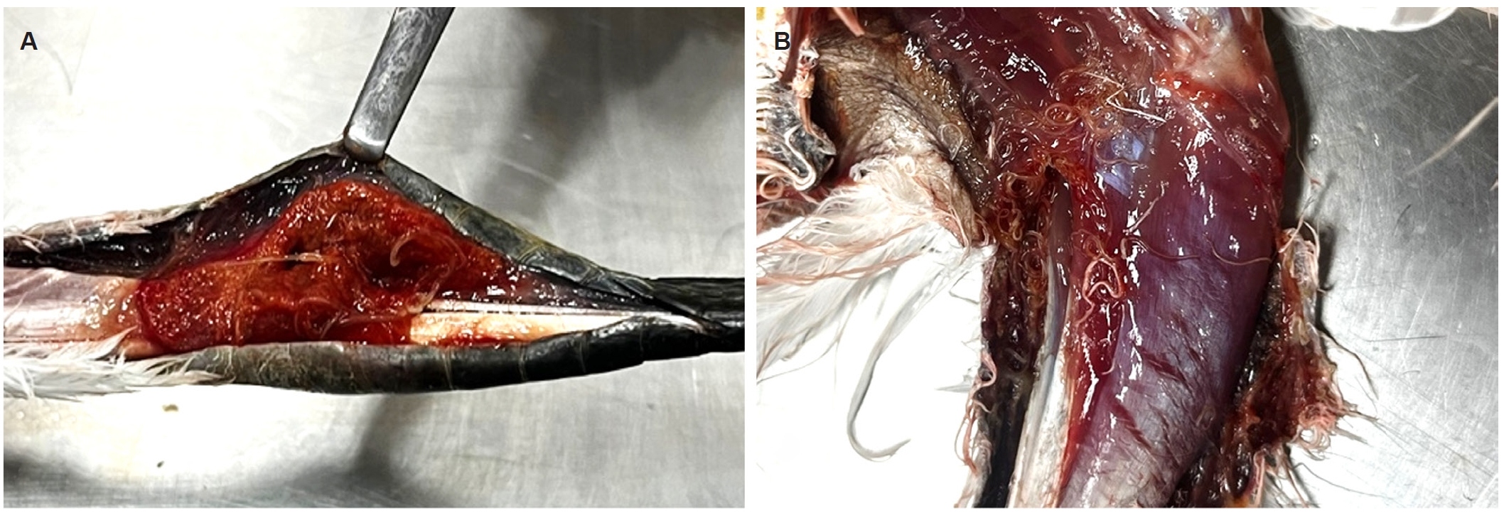
Fig. 2.Histopathological findings. All sections were stained with hematoxylin and eosin. (A) Subcutaneous lesion showing moderate to severe fibrous connective tissue proliferation with granulomatous inflammation and intralesional parasitic organisms. Scale bar=100 μm. (B) Multinucleated giant cells (arrows) with surrounding macrophages and lymphocytes forming a granulomatous reaction around the parasite. Scale bar=20 μm. (C) Transverse section of a filarial nematode showing a thick, multilayered eosinophilic cuticle. A thin hypodermis and well-developed polymyarian–coelomyarian somatic musculature are present. The inset highlights prominent external longitudinal ridges (arrows). Scale bar=20 μm.
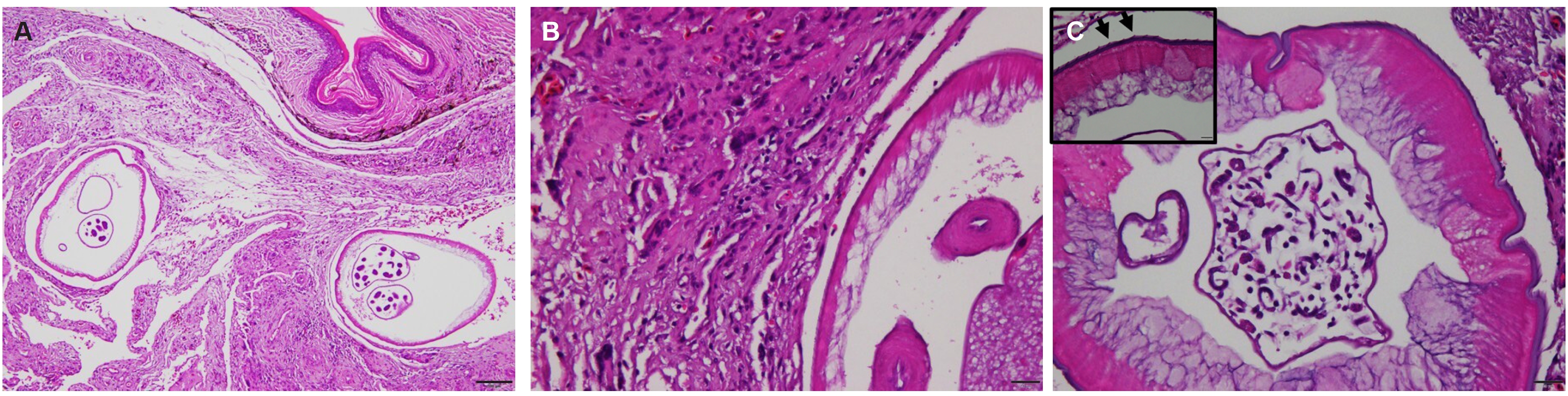
Fig. 3.Phylogenetic analysis of filarial nematodes based on 18S rRNA gene sequences. The maximum-likelihood tree shows that the sequences obtained in this study (BP-1–BP-4) cluster within the Dirofilaria clade and are most closely related to Dirofilaria repens, while clearly separated from other filarioid nematodes, including avian-associated species. Taxon labels indicate the species, host, geographic origin, and GenBank accession number; accession numbers are shown in parentheses. Statistical support was evaluated using 1,000 bootstrap replicates under the Kimura two-parameter model. The scale bar indicates 0.02 nucleotide substitutions per site. Theileria cf. ovis was used as the outgroup.
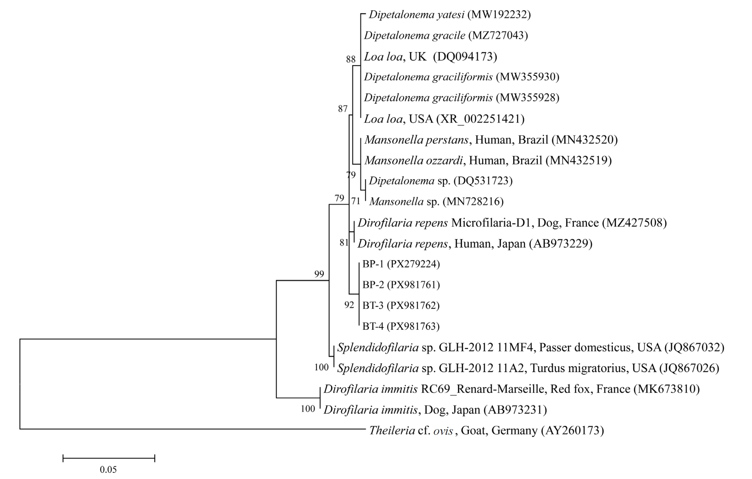
Fig. 4.Phylogenetic analysis of filarial nematodes based on mitochondrial cytochrome c oxidase subunit I (cox1) gene sequences. The maximum-likelihood tree shows that the sequences obtained in this study (BP-1–BP-4) cluster within the Dirofilaria clade and are most closely related to Dirofilaria repens, while clearly separated from other filarioid nematodes, including avian-associated species. Taxon labels indicate the species, host, geographic origin, and GenBank accession number; accession numbers are shown in parentheses. Statistical support was evaluated using 1,000 bootstrap replicates under the Kimura two-parameter model. The scale bar indicates 0.05 nucleotide substitutions per site. Diplotriaena sp. was used as the outgroup.
References
- 1. Lee SE, Kim HC, Chong ST, Klein TA, Lee WJ. Molecular survey of Dirofilaria immitis and Dirofilaria repens by direct PCR for wild caught mosquitoes in the Republic of Korea. Vet Parasitol 2007;148:149-55. https://doi.org/10.1016/j.vetpar.2007.04.015
- 2. McCall JW, Genchi C, Kramer LH, Guerrero J, Venco L. Heartworm disease in animals and humans. Adv Parasitol 2008;66:193-285. https://doi.org/10.1016/S0065-308X(08)00204-2
- 3. Albanese F, Abramo F, Braglia C, et al. Nodular lesions due to infestation by Dirofilaria repens in dogs from Italy. Vet Dermatol 2013;24:255-e56. https://doi.org/10.1111/vde.12009
- 4. Simón F, Siles-Lucas M, Morchón R, et al. Human and animal dirofilariasis: the emergence of a zoonotic mosaic. Clin Microbiol Rev 2012;25:507-44. https://doi.org/10.1128/CMR.00012-12
- 5. Bartlett CM. Filarioid nematodes. In: Atkinson CT, Thomas NJ, Hunter DB, editors. Parasitic diseases of wild birds. Wiley-Blackwell; 2008. p. 439-62.
- 6. Laidoudi Y, Ringot D, Watier-Grillot S, Davoust B, Mediannikov O. A cardiac and subcutaneous canine dirofilariosis outbreak in a kennel in central France. Parasite 2019;26:72. https://doi.org/10.1051/parasite/2019073
- 7. Folmer O, Black M, Hoeh W, Lutz R, Vrijenhoek R. DNA primers for amplification of mitochondrial cytochrome c oxidase subunit 1 from diverse metazoan invertebrates. Mol Mar Biol Biotechnol 1994;3:294-9.
- 8. Sano Y, Aoki M, Takahashi H, et al. The first record of Dirofilaria immitis infection in a Humboldt penguin, Spheniscus humboldti. J Parasitol 2005;91:1235-7. https://doi.org/10.1645/GE-3492-RN.1
- 9. Suzuki J, Kobayashi S, Okata U, et al. Molecular analysis of Dirofilaria repens removed from a subcutaneous nodule in a Japanese woman after a tour to Europe. Parasite 2015;22:2. https://doi.org/10.1051/parasite/2015002
- 10. Pang CC, Sung YH, Chung YT, et al. Spatial ecology of little egret (Egretta garzetta) in Hong Kong uncovers preference for commercial fishponds. PeerJ 2020;8:e9893. https://doi.org/10.7717/peerj.9893


 , You-Jeong Lee2,†