Abstract
Sarcocystis spp. are common protozoan parasites infecting a wide range of intermediate hosts, including wild cervids. In this study, we investigated the occurrence and molecular characteristics of Sarcocystis infection in wild Korean roe deer (Capreolus pygargus tianschanicus) from Jeju Island, Korea. Sarcocysts were identified in the heart and skeletal muscle of 18 out of 40 examined animals (45.0%) based on histopathological examination. The cysts were oval to spherical in shape, measuring 42.41–340.29 μm in length and 31.62–129.76 μm in width, and were not associated with significant inflammatory responses. Molecular analysis targeting the 18S rRNA gene yielded 2 representative sequences, which showed 99.82% identity and high similarity to Sarcocystis linearis, as well as closely related species including S. taeniata and S. grueneri. Phylogenetic analysis placed these sequences within a clade of ungulate-associated Sarcocystis spp., although species-level resolution was not achieved due to the conserved nature of the marker. These findings provide baseline data on Sarcocystis infection in wild roe deer from Jeju Island and highlight the limitations of 18S rRNA-based identification for closely related Sarcocystis species.
-
Key words: Sarcocystis spp., roe deer, molecular characterization, 18S rRNA, Korea
Sarcocystis spp. are obligate intracellular protozoan parasites with a 2-host life cycle involving carnivorous definitive hosts and herbivorous intermediate hosts [
1,
2]. These parasites are widely distributed and infect a variety of domestic and wild animals, including cervids, in which they form intramuscular cysts [
1]. Although infections in intermediate hosts are typically subclinical,
Sarcocystis spp. are of veterinary and ecological importance due to their high prevalence and complex host–parasite relationships [
3,
4].
Cervids, including roe deer (
Capreolus spp.), are well-recognized intermediate hosts for multiple
Sarcocystis species [
4,
5]. Previous studies have demonstrated a high diversity of
Sarcocystis spp. in roe deer populations, particularly in Europe [
3,
6]. However, molecular identification of closely related species remains challenging due to the limited resolution of commonly used genetic markers such as the 18S rRNA gene.
In Korea, studies on
Sarcocystis infection have primarily focused on domestic animals, and information on wildlife infections remains limited [
7-
10]. In particular, data on wild Korean roe deer (
Capreolus pygargus tianschanicus) are lacking despite their ecological importance and high population density on Jeju Island. Therefore, the present study aimed to investigate the occurrence and histopathological characteristics of
Sarcocystis infection in wild Korean roe deer from Jeju Island and to molecularly characterize the detected parasites using 18S rRNA gene sequences.
Between 2018 and 2025, a total of 40 wild Korean roe deer (C. pygargus) were submitted for postmortem examination. Among these, 18 animals were selected for the present study based on the histopathological identification of intramuscular sarcocysts in the heart and/or skeletal muscle tissues. These 18 cases were subsequently included for further histological and molecular analyses. Information regarding sex, estimated age, and body weight was collected from the necropsy records. The age of each animal was estimated using a combination of body weight, radiographic assessment of physeal closure, and antler development in males. Ethical approval for animal experimentation was not required for this study because all samples were obtained postmortem from wild roe deer that had died naturally or were found dead.
At necropsy, tissue samples including heart and skeletal muscle were collected and fixed in 10% neutral-buffered formalin. The samples were routinely processed, embedded in paraffin, sectioned at 3–4 μm, and stained with hematoxylin and eosin. Histological sections were examined for the presence, distribution, and morphological characteristics of sarcocysts. The location of cysts within muscle fibers, cyst shape, and associated inflammatory responses were evaluated. When present, cyst size was measured using a light microscope.
DNA was extracted from formalin-fixed, paraffin-embedded (FFPE) tissue sections of heart and skeletal muscle obtained from the 18 histologically positive roe deer using the Exgene FFPE Tissue DNA isolation kit (GeneAll), according to the manufacturer’s instructions. The extracted DNA concentration ranged from 18.8 to 380 ng/μl.
Nested PCR targeting the 18S rRNA gene of
Sarcocystis spp. was performed based on previously described methods [
11]. Briefly, 2 rounds of amplification were carried out using the primer sets SarcoFext/SarcoRext and SarcoFint/SarcoRint, respectively, yielding an expected amplicon size of 639 bp. To improve species-level identification, additional PCR targeting the mitochondrial cytochrome c oxidase subunit 1 (
cox1) gene was performed on selected samples that yielded high-quality 18S rRNA amplicons suitable for sequencing, as previously described [
12]. Each PCR run included a negative control to monitor potential contamination.
PCR amplification targeting the 18S rRNA gene yielded positive bands in all 18 histologically positive samples. Based on amplification quality and DNA integrity, 2 PCR products were selected for Sanger sequencing. Sequencing was performed by Cosmogenetech. Forward and reverse reads were assembled to generate consensus sequences using Geneious Prime version 2025.2.2 [
13], with low-quality regions trimmed prior to analysis.
For molecular identification, the obtained sequences were compared with reference sequences available in the GenBank database using BLAST [
14]. For phylogenetic analysis, previously published
Sarcocystis sequences were retrieved from GenBank and aligned with the sequences generated in this study based on the SSU rRNA gene. Sequence alignment was performed using Clustal W implemented in MEGA X version 10.1.7 [
15]. Phylogenetic reconstruction was conducted using Bayesian inference (BI). The best-fit nucleotide substitution model was selected using jModelTest version 2.1.10 according to the Bayesian information criterion, and the HKY+I+G model was applied. Bayesian analysis was performed in MrBayes version 3.2.6 for 10,000,000 generations [
16]. The resulting phylogenetic tree was visualized using FigTree version 1.4.4. Representative sequences from related taxa were included as outgroups.
A total of 18 wild Korean roe deer with histologically confirmed
Sarcocystis infection were included in this study. Detailed case information is summarized in
Supplementary Table S1. The study population consisted of 9 males and 9 females, with no apparent sex predilection. Age information was available for a subset of animals, with a mean estimated age of approximately 2.3 years. Body weights ranged from 2.14 to 27.3 kg (mean 18.59 kg).
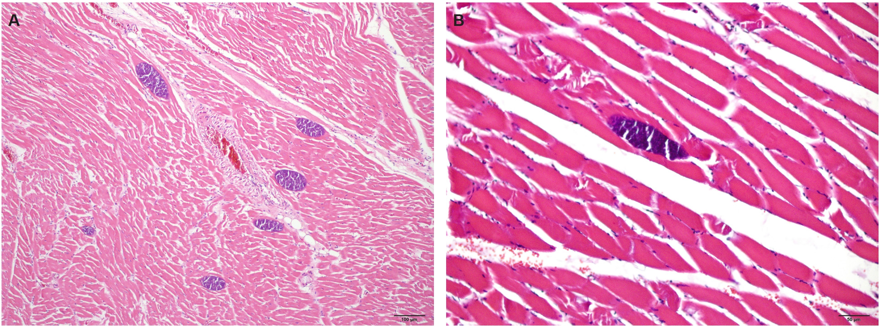
Histopathological examination revealed multiple intramuscular sarcocysts in cardiac and skeletal muscle tissues (
Fig. 1A,
B). The cysts were located within the sarcoplasm of muscle fibers and were oval to spherical in shape. Cyst size ranged from 42.41 to 340.29 μm in length (mean 134.07 μm) and 31.62 to 129.76 μm in width (mean 59.65 μm). Each cyst contained numerous bradyzoites and was surrounded by a thin, smooth cyst wall, although the detailed cyst wall structure was not clearly discernible. No inflammatory response was observed in the surrounding muscle tissue. Based on these morphological features, the observed cysts were consistent with
Sarcocystis spp., although species-level identification could not be determined by histological examination alone.
Nested PCR targeting the SSU rRNA gene yielded positive amplification in all 18 samples. Sequencing was attempted for all PCR products; however, high-quality sequences were obtained from only 2 samples. Additional PCR targeting the
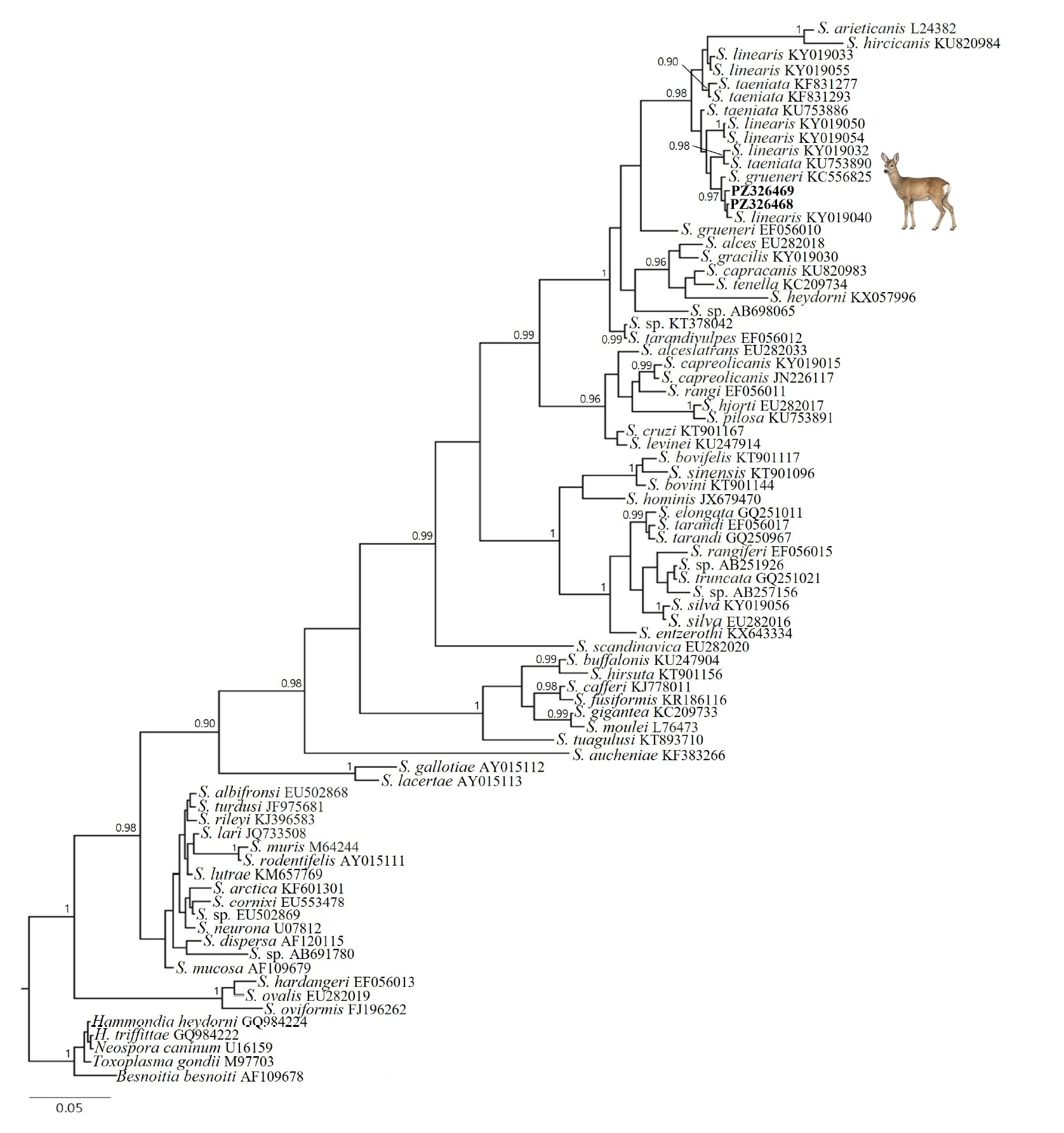
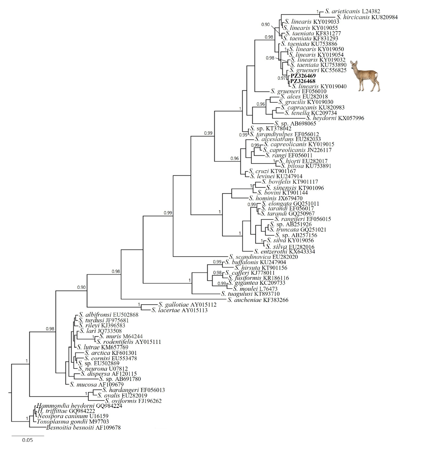
cox1 gene was performed on these samples; however, no specific amplification products were obtained. Two PCR products yielded high-quality sequences and were deposited in GenBank under accession numbers PZ326468 and PZ326469, corresponding to cases 17 and 18, respectively. The 2 sequences showed high similarity, differing by a single nucleotide within an overlapping 568-bp region, corresponding to 99.82% identity. BLAST analysis revealed that both sequences shared ≥99% identity with previously reported
Sarcocystis linearis sequences and were also highly similar to closely related species, including
S. taeniata and
S. grueneri. Phylogenetic analysis based on the SSU rRNA gene placed the sequences within a clade of ungulate-associated
Sarcocystis spp., including
S. linearis,
S. taeniata, and related taxa (
Fig. 2). Within this clade, sequences assigned to different species were intermingled rather than forming distinct species-specific clades.
In the present study,
Sarcocystis infection was identified in wild Korean roe deer from Jeju Island based on histopathological and molecular findings. Intramuscular sarcocysts were consistently observed within cardiac and skeletal muscle fibers, without any associated inflammatory response, indicating a subclinical infection pattern typical of intermediate hosts [
1].
Molecular analysis targeting the 18S rRNA gene revealed that the obtained sequences were highly similar to
S. linearis and closely related species, including
S. taeniata and
S. grueneri. However, species-level identification could not be achieved. This limitation is consistent with previous studies demonstrating that the SSU rRNA gene is highly conserved among closely related
Sarcocystis species, particularly those infecting cervids, and therefore lacks sufficient discriminatory power for precise species differentiation [
6].
In addition, the use of FFPE tissues may have further limited molecular resolution in this study. DNA extracted from FFPE samples is often fragmented and chemically modified, which can reduce amplification efficiency and restrict the length and quality of obtainable sequences [
17]. This limitation may have contributed to the restricted number of sequences obtained and the difficulty in achieving species-level identification.
The phylogenetic analysis further supported this limitation, as sequences assigned to different Sarcocystis species were intermingled within the same clade rather than forming distinct species-specific groupings. This pattern highlights the difficulty of resolving taxonomic relationships within this group using SSU rRNA markers alone and underscores the need for additional genetic loci, such as mitochondrial loci (e.g., cox1), to improve species-level resolution.
Cervids are well-recognized intermediate hosts of diverse
Sarcocystis species, and multiple species have been reported in roe deer in Europe [
6,
7]. However, information on
Sarcocystis infection in wildlife in Korea remains limited. In particular, data on wild Korean roe deer are lacking despite their ecological importance. Previous studies in Korea have reported
Sarcocystis infections in various domestic and wild animals, but investigations in wildlife remain scarce [
7-
10,
18,
19]. Therefore, the present study provides baseline data on the occurrence and molecular characteristics of
Sarcocystis infection in this host species in Korea.
From an ecological perspective, the absence of inflammatory response observed in the present study suggests a well-adapted host–parasite relationship in wild roe deer. Such subclinical infections are characteristic of intermediate hosts and may facilitate the maintenance of
Sarcocystis life cycles in natural ecosystems without causing overt disease [
1]. Given the high population density of roe deer on Jeju Island, this host species may play an important role in sustaining parasite transmission dynamics in the region. However, completion of the
Sarcocystis life cycle also depends on the presence of suitable definitive hosts. Although the definitive host of the
Sarcocystis species detected in this study could not be determined, domestic dogs, free-ranging dogs, and other carnivorous mammals present on Jeju Island may contribute to the maintenance of transmission cycles in the local ecosystem.
Although the number of sequences obtained in this study was limited, the combined histopathological and molecular findings provide preliminary evidence of Sarcocystis infection in wild roe deer. However, species-level identification was not possible based on the available 18S rRNA sequences alone. Future studies incorporating a larger number of samples and higher-resolution genetic markers will be necessary to further clarify species diversity and host–parasite relationships in this system.
Notes
-
Author contributions
Conceptualization: Jung JY. Data curation: Jang J, Yun Y. Formal analysis: Nam DY, Kim S. Investigation: Nam DY. Methodology: Nam DY, Jung JY. Resources: Jang J, Yun Y. Software: Kim S. Supervision: Jung JY. Validation: Kim JH. Writing – original draft: Nam DY. Writing – review & editing: Kim S, Kim JH, Jung JY.
-
Conflict of interest
The authors have no conflicts of interest to declare.
-
Funding
This work was supported by the National Institute of Wildlife Disease Control and Prevention as a “Special graduate school support project for wildlife disease specialists.”
Supplementary information
Fig. 1.Histopathologic findings. (A) Low-power view of multiple intramuscular sarcocysts distributed among myocardial fibers. Hematoxylin and eosin, scale bar=100 µm. (B) Longitudinal section of a sarcocyst within skeletal muscle. The cysts are located within the sarcoplasm of muscle fibers and contain numerous bradyzoites. No inflammatory response is present. Hematoxylin and eosin, scale bar=50 µm.
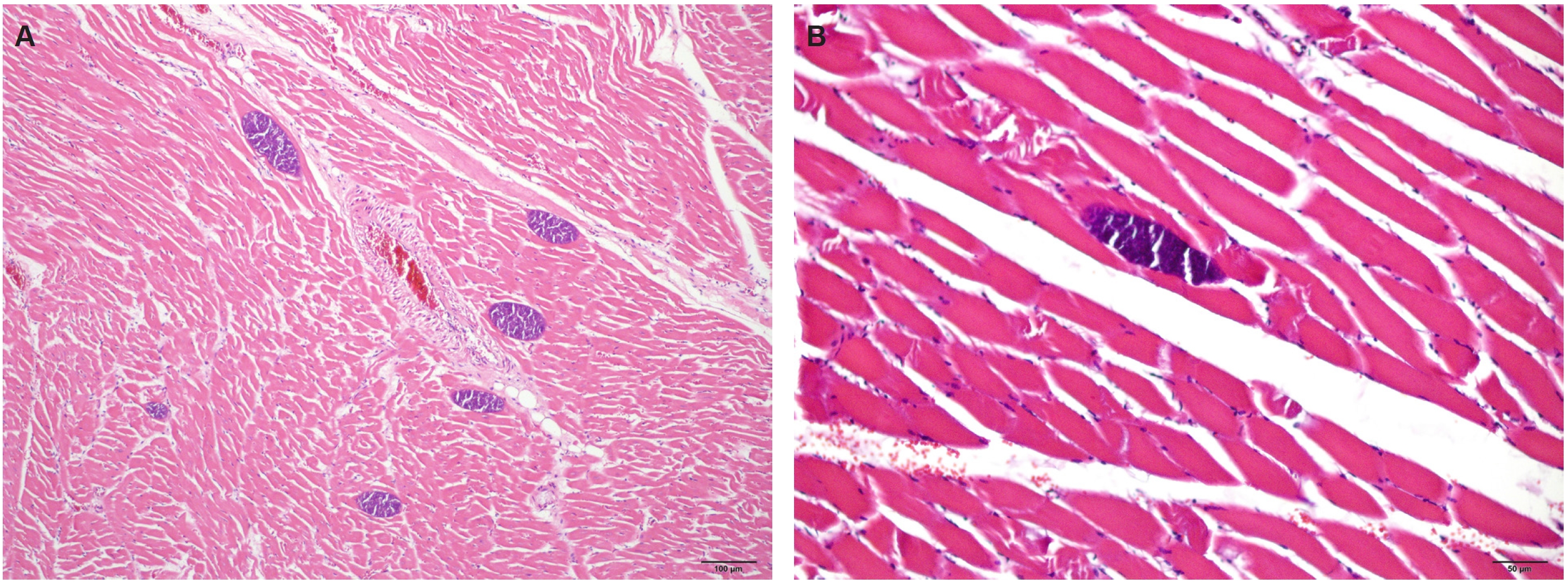
Fig. 2.Phylogenetic relationships within the genus Sarcocystis inferred from the small subunit rRNA dataset (639 bp) constructed under the HKY+I+G substitution model using Bayesian inference. The 2 newly generated sequences from the present study are shown in bold. Tree topology is based on the Bayesian all-compatible consensus tree. Posterior probabilities are shown at nodes only for values ≥0.90.
References
- 1. Otranto D, Wall R. Veterinary protozoology. In: Otranto D, Wall R, Taylor MA, editors. Veterinary parasitology. Wiley-Blackwell; 2024. p. 138-215.
- 2. Dubey JP, Calero-Bernal R, Rosenthal BM, Speer CA, Fayer R. Sarcocystosis of animals and humans. CRC Press; 2015.
- 3. Pérez-Creo A, Panadero R, López C, et al. Prevalence and identity of Sarcocystis spp. in roe deer (Capreolus capreolus) in Spain: a morphological study. Res Vet Sci 2013;95:1036-40. https://doi.org/10.1016/j.rvsc.2013.08.003
- 4. Reissig EC, Helman E, Moré G. Sarcocystis spp. infection in South American deer huemul (Hippocamelus bisulcus) and pudu (Pudu puda) from Patagonian National Parks, Argentina. Parasitol Res 2020;119:3915-22. https://doi.org/10.1007/s00436-020-06889-9
- 5. Gjerde B, Luzón M, Alunda JM, de la Fuente C. Morphological and molecular characteristics of six Sarcocystis spp. from red deer (Cervus elaphus) in Spain, including Sarcocystis cervicanis and three new species. Parasitol Res 2017;116:2795-811. https://doi.org/10.1007/s00436-017-5590-z
- 6. Gjerde B, Giacomelli S, Bianchi A, et al. Morphological and molecular characterization of four Sarcocystis spp., including Sarcocystis linearis n. sp., from roe deer (Capreolus capreolus) in Italy. Parasitol Res 2017;116:1317-38. https://doi.org/10.1007/s00436-017-5410-5
- 7. Moon MH. Sarcocystis infection and identification of Sarcocystis species in pigs in Korea. Korean J Vet Res 1989;29:325-31.
- 8. Kang SS, Yi YJ, Cui XS, et al. Sarcocystis infestation in farmed elk (Cervus canadensis). J Vet Clin 1999;16:529-32.
- 9. Hong EJ, Sim C, Chae JS, et al. Ultrastructural and molecular identification of Sarcocystis tenella (Protozoa, Apicomplexa) in naturally infected Korean native goats. Vet Med 2016;61:374-81. https://doi.org/10.17221/93/2015-vetmed
- 10. Choi TI, Hong EJ, Ryu SY, et al. Detection and identification of Sarcocystis cruzi (Protozoa: Apicomplexa) by molecular and ultrastructural studies in naturally infected Korean cattle (Bos taurus coreanae) from Daejeon, Korea. Korean J Parasitol 2018;56:121-7. https://doi.org/10.3347/kjp.2018.56.2.121
- 11. Moré G, Schares S, Maksimov A, et al. Development of a multiplex real time PCR to differentiate Sarcocystis spp. affecting cattle. Vet Parasitol 2013;197:85-94. https://doi.org/10.1016/j.vetpar.2013.04.024
- 12. Gjerde B. Phylogenetic relationships among Sarcocystis species in cervids, cattle and sheep inferred from the mitochondrial cytochrome c oxidase subunit I gene. Int J Parasitol 2013;43:579-91. https://doi.org/10.1016/j.ijpara.2013.02.004
- 13. Kearse M, Moir R, Wilson A, et al. Geneious Basic: an integrated and extendable desktop software platform for the organization and analysis of sequence data. Bioinformatics 2012;28:1647-9. https://doi.org/10.1093/bioinformatics/bts199
- 14. Baker CS, Dalebout ML, Lavery S, Ross HA. www.DNA-surveillance: applied molecular taxonomy for species conservation and discovery. Trends Ecol Evol 2003;18:271-2. https://doi.org/10.1016/S0169-5347(03)00101-0
- 15. Larkin MA, Blackshields G, Brown NP, et al. Clustal W and Clustal X version 2.0. Bioinformatics 2007;23:2947-8. https://doi.org/10.1093/bioinformatics/btm404
- 16. Ronquist F, Teslenko M, van der Mark P, et al. MrBayes 3.2: efficient Bayesian phylogenetic inference and model choice across a large model space. Syst Biol 2012;61:539-42. https://doi.org/10.1093/sysbio/sys029
- 17. Srinivasan M, Sedmak D, Jewell S. Effect of fixatives and tissue processing on the content and integrity of nucleic acids. Am J Pathol 2002;161:1961-71. https://doi.org/10.1016/S0002-9440(10)64472-0
- 18. Kim TH, Han JH, Chang SN, et al. Detection of sarcocystic infection in a wild rodent (Apodemus agrarius chejuensis) captured on Jeju island. Lab Anim Res 2011;27:357-9. https://doi.org/10.5625/lar.2011.27.4.357
- 19. Kim HW, Kim HC, Ryu SY, et al. Molecular identification of Sarcocystis grueneri in wild Korean water deer (Hydropotes inermis argyropus). Korean J Parasitol 2018;56:129-34. https://doi.org/10.3347/kjp.2018.56.2.129