Abstract
Intestinal helminth infections have become uncommon in Korea but still occur sporadically, either as residual native infections or as imported infections among immigrants. We retrospectively reviewed 4 cases of intestinal helminth infections incidentally identified during colonoscopy at a single academic hospital between January 2019 and March 2026. One Cambodian immigrant had hookworm infection (species-level differentiation between Ancylostoma duodenale and Necator americanus was not possible). Of the 3 Korean patients, 2 had histopathologically confirmed Trichuris trichiura infection, and in 1, T. trichiura infection was suspected endoscopically, but the retrieved organism was not submitted for histopathologic examination. All patients were asymptomatic or had only nonspecific symptoms, and none showed peripheral eosinophilia. Colonoscopy allowed direct visualization and removal of each organism, demonstrating mucosal embedding in T. trichiura infection and firm mucosal attachment of the hookworm in the rectum. Three patients received a single 400-mg oral dose of albendazole; the case without histopathologic confirmation was managed without anthelmintic therapy. Recognition of characteristic endoscopic morphology can guide diagnosis even when histopathologic confirmation is unavailable, and management should be individualized according to diagnostic certainty and clinical context.
-
Key words: Trichuris trichiura, hookworm, colonoscopy, helminthiasis, incidental findings, Republic of Korea
Introduction
Soil-transmitted helminth infections caused by
Trichuris trichiura and the hookworms
Ancylostoma duodenale and
Necator americanus remain a major global health burden, especially in tropical and subtropical regions [
1-
3]. In Korea, the prevalence of intestinal helminth infections has declined markedly over the past several decades following improvements in sanitation, widespread anthelmintic use, and national parasite control programs [
4-
6].
Despite this decline, sporadic cases continue to occur, either as residual infections in the native population or as imported infections among immigrants and travelers from endemic regions [
4-
7]. As international migration increases, clinicians in low-prevalence settings may encounter parasitic infections that are no longer routinely seen [
7].
With the widespread use of colonoscopy for colorectal cancer screening and for evaluation of gastrointestinal symptoms, intestinal helminth infections may be detected incidentally during endoscopy [
8-
10]. Because affected patients are often asymptomatic or report only nonspecific symptoms, helminth infection is rarely suspected before the procedure. In this setting, colonoscopy offers a unique opportunity for direct visualization, endoscopic removal, and subsequent diagnostic evaluation.
Endoscopic appearance can guide the diagnosis:
T. trichiura typically presents as a thin, whip-like worm with its anterior portion embedded in the colonic mucosa and its posterior portion freely mobile within the lumen, most often in the cecum or ascending colon [
8-
10]. Hookworms are small, cylindrical nematodes that usually inhabit the small intestine but may rarely be encountered during lower endoscopy, appearing as motile or mucosa-attached worms within the intestinal lumen [
2,
7]. Because these findings are now uncommon in routine endoscopy, parasites may be overlooked or mistaken for mucus strands, food residue, or other luminal debris, leading to missed diagnosis or inconsistent management.
We report 4 cases of intestinal helminth infections incidentally detected during colonoscopy at a single academic hospital: 1 Cambodian immigrant with hookworm infection in whom species-level differentiation between A. duodenale and N. americanus was not possible, 2 Korean patients with histopathologically confirmed T. trichiura infection, and 1 Korean patient with endoscopically suspected T. trichiura infection but without histopathologic examination of the retrieved organism. Rather than presenting these findings as entirely novel entities, this series highlights several practical issues that remain insufficiently addressed in low-prevalence settings: recognition of characteristic endoscopic morphology when pre-procedural suspicion is low, interpretation of unusual parasite location such as rectal hookworm, diagnostic uncertainty when specimen confirmation is unavailable, and individualized decisions regarding additional evaluation and anthelmintic therapy after endoscopic removal.
Case Report
Between January 1, 2019 and March 31, 2026, we retrospectively identified 4 patients in whom intestinal helminth infections were incidentally detected during colonoscopy at our institution. Demographic, clinical, endoscopic, pathologic, parasitologic, treatment, and follow-up data were collected from the electronic medical records. The diagnosis was based on colonoscopic morphology and on subsequent specimen evaluation when available. The demographic characteristics, colonoscopic findings, specimen evaluations, treatments, and clinical outcomes of the 4 patients are summarized in
Table 1.
We conducted this study in compliance with the principles of the Declaration of Helsinki. The study protocol was reviewed and approved by the Institutional Review Board of Seoul Metropolitan Government–Seoul National University Boramae Medical Center (IRB No. 20-2026-33), and the requirement for informed consent was waived because of the retrospective design.
Case 1
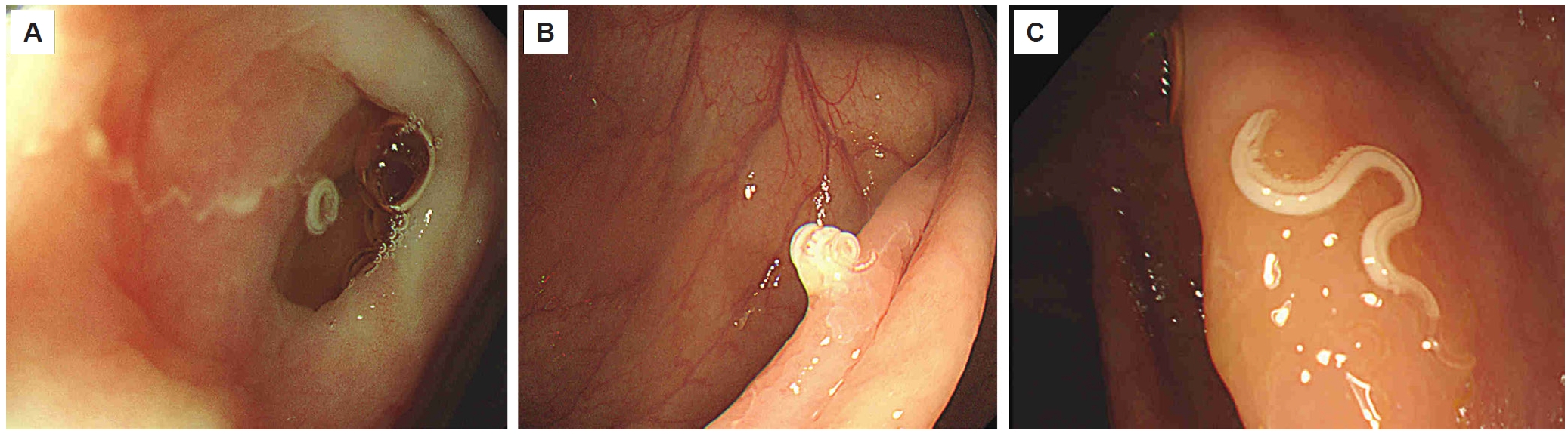
A 57-year-old Cambodian man presented to our institution with anal bleeding and a protruding anal mass. His past medical history included hypertension and a recent total thymectomy for thymoma. Colonoscopy was performed for suspected hemorrhoidal disease. Pre-colonoscopy laboratory tests were unremarkable. Hemoglobin level and eosinophil count were within the reference ranges, and inflammatory markers were not elevated. Colonoscopy was performed after standard bowel preparation. A slender, translucent, slightly reddish, cylindrical worm approximately 12 mm long was incidentally observed in the rectum, firmly attached to the mucosa with its posterior portion trailing into the lumen (
Fig. 1A,
B). No mucosal inflammation, ulceration, or bleeding was seen.
Because parasitic infection had not been suspected before the procedure, the worm was grasped and removed with endoscopic forceps for identification and referred to a reference parasitology laboratory (Green Cross Laboratories). On morphologic examination, the retrieved organism was slender and cylindrical, approximately 12 mm in length, with the anterior portion curved in a characteristic hook shape. The uterus in the lower body was filled with multiple ova, identifying the worm as an adult female, and numerous erythrocytes were visible within the body of the worm, consistent with active blood feeding. These features supported the diagnosis of adult female hookworm and differed from T. trichiura (whip-like body with a thin anterior portion embedded in the colonic mucosa and a characteristic stichosoma on histopathology), from Enterobius vermicularis (of comparable length but far more slender, with a sharply attenuated pointed tail, no buccal capsule, and no evidence of blood feeding), and from larger luminal nematodes such as Ascaris lumbricoides. However, species-level differentiation between A. duodenale and N. americanus was not possible because the buccal capsule, the anatomical structure used to distinguish these 2 species, was damaged during retrieval.
The patient had emigrated from Cambodia to Korea approximately 1 year before presentation to join his adult children, who live and work in Korea, and to undergo a comprehensive health screening examination. A thymoma was identified incidentally during that screening. Before emigrating, he reported possible exposure to agricultural fields in Cambodia where human and animal feces were used as fertilizer. He usually wore shoes but occasionally walked barefoot during rainy weather, and vegetables were typically washed before consumption. Based on this exposure history, percutaneous infection through contaminated soil in Cambodia was considered the most plausible route, although oral acquisition could not be excluded because the hookworm species was not determined.
After endoscopic removal he received a single 400-mg oral dose of albendazole. Small-bowel evaluation, including capsule endoscopy or enteroscopy, was not performed because the patient had no anemia, eosinophilia, abdominal symptoms, or other evidence suggesting residual small-bowel infection. Treatment was well tolerated, and a follow-up stool examination performed 1 month after treatment was negative for parasites. He remained asymptomatic during follow-up without evidence of recurrence.
Case 2
A 53-year-old Korean man underwent colonoscopy for evaluation of a positive fecal occult blood test. Pre-procedure laboratory tests, including the eosinophil count, were within normal limits. Colonoscopy showed a whitish, coiled, whip-like organism at the appendiceal orifice. The anterior portion appeared embedded in the mucosa while the posterior portion was freely mobile within the lumen (
Fig. 2A). The organism was removed endoscopically.
Based on the whitish, coiled, whip-like morphology, location at the appendiceal orifice, and partial mucosal embedding with a freely mobile posterior portion, the finding was considered endoscopically suspected T. trichiura infection. However, the retrieved organism was not submitted for histopathologic or parasitologic examination; therefore, the diagnosis remained morphology-based and lacked laboratory confirmation.
Because the visible worm had been completely removed and histopathologic confirmation was not pursued, anthelmintic therapy was not given, and the patient was observed without further treatment.
Case 3
A 58-year-old Korean woman underwent screening colonoscopy. Pre-procedure laboratory tests, including the eosinophil count, were within normal limits. Colonoscopy showed a thin, whip-like worm attached to the cecal mucosa, with the anterior portion embedded in the mucosa and the posterior portion freely mobile within the lumen (
Fig. 2B). The worm was removed endoscopically. Histopathologic examination showed a grayish-white, whip-shaped nematode with the characteristic stichosoma (the elongated anterior esophagus formed by a chain of stichocytes) and a uterus filled with ova, confirming an adult female
T. trichiura. After endoscopic removal she received a single 400-mg oral dose of albendazole and had no reported complications.
A 66-year-old asymptomatic Korean woman underwent screening colonoscopy. Pre-procedure laboratory tests, including the eosinophil count, were within normal limits. Colonoscopy showed a single, curved worm in the ascending colon with partial embedding in the mucosa (
Fig. 2C). The worm was removed endoscopically. Histopathologic examination again demonstrated the characteristic stichosoma and an ova-filled uterus, confirming an adult female
T. trichiura. After endoscopic removal she received a single 400-mg oral dose of albendazole and recovered without complications.
Discussion
This case series illustrates the continued occurrence of intestinal helminth infections in a setting where such infections have become uncommon. National parasite-control programs and improved sanitation have markedly reduced soil-transmitted helminthiasis in Korea, yet sporadic infections still occur in native residents, and imported infections may be encountered with increasing population movement [
4-
7]. In our 4 patients, identified over a 7-year period at a single center, 3 Korean patients had confirmed or suspected
T. trichiura infection, whereas the single hookworm infection occurred in a Cambodian immigrant. Although the small number of cases precludes formal epidemiologic inference, this distribution is compatible with residual low-level whipworm infection among Korean residents and exposure-related hookworm infection in a patient from an endemic region [
2,
3,
7], and supports consideration of intestinal helminth infections in both native and immigrant patients even where these infections are now uncommon. The value of the present series is therefore not in documenting colonoscopic detection itself, which has been described previously, but in integrating endoscopic morphology, epidemiologic context, diagnostic limitations, and actual management decisions into a practical framework for clinicians who encounter unexpected helminthic findings during routine colonoscopy.
The presumed route of infection differed according to the organism and epidemiologic context. In Case 1, hookworm infection was most plausibly related to exposure in Cambodia, where the patient reported agricultural contact with soil fertilized with human or animal feces and occasional barefoot walking during rainy weather. Because species-level identification was not possible, the exact transmission route could not be determined; however, percutaneous penetration by larvae in contaminated soil was considered the most likely route, while oral acquisition could not be completely excluded [
2,
7]. In Cases 2–4,
T. trichiura infection was presumed to have occurred through fecal–oral ingestion of embryonated eggs from contaminated soil, food, or water [
1,
3,
7]. No definite individual exposure source was identified in these Korean patients, and the infections may represent residual low-level native transmission or remote exposure in a low-prevalence setting rather than recent clinically apparent acquisition [
4-
7].
The hookworm case underscores the importance of integrating epidemiologic context with morphology. Although species-level differentiation between
A. duodenale and
N. americanus was not possible because the anterior structures, including the buccal capsule, were damaged during retrieval, the diagnosis was supported by compatible morphology together with the exposure history described above [
2,
7]. The organism was slender and cylindrical, approximately 12 mm in length, with a characteristic hook-shaped curvature of the anterior portion, features that are compatible with adult hookworm. Reference-laboratory examination further confirmed a female adult worm on the basis of multiple ova within the lower-body uterus, and demonstrated numerous erythrocytes within the worm body, consistent with active blood feeding. This morphology differed from
T. trichiura, which characteristically has a whip-like body with mucosal embedding of the thin anterior portion in the cecum or ascending colon and shows the pathognomonic stichosoma on histopathology, from
E. vermicularis, which is of comparable length but far more slender, has a sharply attenuated pointed tail, lacks a buccal capsule, and does not feed on blood, and from
A. lumbricoides, which is usually larger and more robust. Notably, the patient presented for an unrelated indication and had no peripheral eosinophilia or other suggestive laboratory abnormality, reinforcing that a low index of suspicion based on routine tests alone may overlook the diagnosis.
All 4 infections were detected incidentally during colonoscopy. With the expansion of colorectal cancer screening, colonoscopy is increasingly performed in asymptomatic individuals, and helminthic findings may be encountered unexpectedly [
8-
10]. Three patients underwent colonoscopy for screening or a positive fecal occult blood test, and one for anal bleeding and an anal mass; none had symptoms clearly attributable to helminth infection, so clinical suspicion was low before the procedure. Colonoscopy nonetheless offers a distinct advantage in this setting, permitting direct visualization of the organism, immediate endoscopic removal, and subsequent parasitologic or histopathologic evaluation within a single procedure.
The endoscopic findings were useful for diagnosis and were consistent with previous reports:
T. trichiura is classically recognized as a thin, whip-like worm with its anterior portion embedded in the colonic mucosa—most often in the cecum or ascending colon—and its posterior portion freely mobile within the lumen [
8-
10]; this appearance was seen in the 2 histopathologically confirmed cases and in the endoscopically suspected case at the appendiceal orifice. The hookworm, in contrast, appeared as a slender, translucent, slightly reddish, cylindrical worm firmly attached to the rectal mucosa, with its posterior portion trailing into the lumen. Familiarity with these features is important, as intestinal helminth infections are now rarely seen at endoscopy and may be mistaken for mucus strands, food residue, or other luminal debris, with the potential for misdiagnosis [
8-
10].
Because adult hookworms typically attach within the small intestine, an attached hookworm in the rectum is unusual [
2]. In Case 1, small-bowel evaluation by capsule endoscopy, enteroscopy, or other modalities was not performed. We infer that the worm was most likely dislodged from its usual small-intestinal site by bowel preparation and transported distally to the rectum, where it was visualized attached to the mucosa at colonoscopy; whether this represented an ectopic attachment at the rectal site or an incidental encounter during distal transit could not be determined. This interpretation is supported by the absence of abdominal symptoms, anemia, eosinophilia, or endoscopic evidence of mucosal injury, as well as by the negative follow-up stool examination after albendazole treatment. Nevertheless, because the small intestine was not directly evaluated, residual small-bowel infection could not be completely excluded.
Case 2 highlights a missed opportunity for diagnostic confirmation and empirical treatment, as well as a broader reality of low-prevalence settings. The endoscopic appearance—a coiled, whip-like worm with partial mucosal embedding at the appendiceal orifice and a freely mobile posterior portion—was highly suggestive of
T. trichiura, yet the retrieved organism was not submitted for parasitologic or histopathologic examination and no anthelmintic therapy was given; the finding was therefore left without laboratory confirmation and without targeted treatment. Because histopathologic or parasitologic confirmation was not obtained, this case should be regarded as endoscopically suspected rather than laboratory-confirmed
T. trichiura infection. Given the typical endoscopic appearance and the favorable safety profile of short-course albendazole, empirical therapy could reasonably have been offered. This sequence reflects a practical reality: helminthic findings at colonoscopy have become uncommon, and many endoscopists understandably feel unprepared to commit to both specimen submission and empirical treatment when parasitic infection is not high on the pre-procedural differential. The practical lesson is that when a colonoscopist visualizes a parasite with characteristic endoscopic features, the morphology itself should prompt an accurate working diagnosis, deliberate retrieval with care to preserve diagnostic structures, formal submission for parasitologic or histopathologic examination, and timely anthelmintic therapy. Detailed case series—including the present one, which combines histopathologically confirmed and morphology-only presentations together with the actual management decisions taken—may help build the practical experience that endoscopists in low-prevalence settings can draw on when they encounter similar unexpected findings [
8-
10].
None of the 4 patients had peripheral eosinophilia. Although this small series cannot define the frequency of eosinophilia in intestinal helminthiasis, the finding is consistent with prior data indicating that eosinophilia may be absent in light, localized, or incidentally detected intestinal parasitic infections [
11]. A normal eosinophil count therefore should not be used to exclude helminth infection, particularly when a suspicious organism is seen at endoscopy.
Management of incidentally detected intestinal helminth infections should be individualized according to the suspected organism, diagnostic certainty, endoscopic burden, symptoms, laboratory findings, and epidemiologic risk [
1-
3,
7]. When a worm is unexpectedly encountered during colonoscopy, the first practical step is careful retrieval with minimal damage to diagnostic structures, followed by formal submission for parasitologic or histopathologic examination whenever possible [
7]. Additional evaluation may include a complete blood count with eosinophil count and assessment for anemia or iron deficiency, especially in suspected hookworm infection [
2,
7]. Stool ova-and-parasite examination is useful for confirming ongoing infection, documenting clearance after treatment, and estimating infection burden when eggs are detected; however, a single negative stool examination does not necessarily exclude light infection [
7]. Repeated stool examinations or quantitative egg assessment may be considered when multiple worms are seen, symptoms are present, eosinophilia or anemia persists, exposure risk is high, or treatment response is uncertain. Small-bowel evaluation, including capsule endoscopy, is not routinely required for a solitary colonic
T. trichiura infection after complete endoscopic removal. However, it may be considered when hookworm infection is suspected or confirmed, because adult hookworms usually inhabit the small intestine [
2,
7]. It may also be considered in patients with unexplained iron-deficiency anemia, obscure gastrointestinal bleeding, persistent abdominal symptoms, persistent eosinophilia, positive stool examination, multiple parasites, or concern for residual small-bowel infection [
2,
7]. In the present series, systematic stool ova-and-parasite examination and egg-burden quantification were not performed in all patients; only the patient with hookworm underwent follow-up stool examination, which was negative. No patient underwent small-bowel capsule endoscopy, reflecting the absence of persistent symptoms, eosinophilia, anemia, or recurrent parasitologic evidence after management.
Endoscopic removal of a visible worm does not necessarily eradicate infection, because additional adult worms or eggs may remain undetected. Therefore, anthelmintic therapy is generally appropriate after confirmed or strongly suspected soil-transmitted helminth infection, even when the visible organism has been removed, particularly when the patient has symptoms, multiple worms, eosinophilia, anemia, positive stool examination, relevant endemic exposure, or high diagnostic certainty based on characteristic morphology [
1-
3,
7].
Current evidence further informs the choice of anthelmintic regimen for incidentally detected intestinal helminth infections. For hookworm, single-dose albendazole 400 mg remains highly effective, with a cure rate of 79.5% in a network meta-analysis of randomized trials [
12] and 97.2% in a recent prospective cohort [
13]. For
T. trichiura, however, single-dose albendazole has limited and apparently declining efficacy: in the same network meta-analysis the albendazole cure rate fell from 38.6% in 1995 to 16.4% in 2015, with a parallel fall in the egg-reduction rate from 72.6% to 43.4% [
12], and a 2024 cohort study reported a cure rate of only 49.5%, with reinfection in more than half of the children who had been cured [
13]. Accordingly, multi-day regimens—typically albendazole 400 mg daily for 3 consecutive days—or co-administration of albendazole with ivermectin are now considered preferable for trichuriasis [
14], and the albendazole–ivermectin combination is endorsed by the World Health Organization for preventive chemotherapy against
T. trichiura [
15], with a recent randomized superiority trial reporting that the combination achieved a higher cure rate (31.3% versus 12.3% with albendazole alone) and a substantially higher egg-reduction rate (91.4% versus 52.7%) [
15]. For confirmed or strongly suspected
T. trichiura infection, treatment is generally advised even in asymptomatic patients to prevent chronic morbidity, including iron-deficiency anemia and occult colitis, and to limit ongoing fecal–oral transmission [
3,
14]. Applied to our series, this evidence supports offering empirical anthelmintic therapy when endoscopic morphology is highly characteristic, as in Case 2, and would favor either a 3-day albendazole regimen or an albendazole–ivermectin combination over the single 400-mg dose used historically in our 2 histopathologically confirmed trichuriasis cases (Cases 3 and 4). In Korea, ivermectin has no approved indication for trichuriasis, and its use for this indication would therefore be off-label; a multi-day albendazole regimen (400 mg daily for 3 consecutive days) is the most implementable, regulatory-compliant option for endoscopists locally.
Beyond the parasites encountered in our cases, several other intestinal parasites have been incidentally identified at colonoscopy in low-prevalence settings, and an appreciation of their management is helpful for endoscopists who may face similar unexpected findings. In a Korean single-center retrospective review of 24 colonoscopy-detected parasitic infections—predominantly
T. trichiura and
A. lumbricoides, with single cases of
E. vermicularis and
Anisakis sp. infection—many patients were asymptomatic and had negative stool examinations, supporting colonoscopy itself as a useful diagnostic modality when conventional testing is unrevealing [
16].
Individual case reports document
Ascaris lumbricoides retrieved at screening colonoscopy followed by single-dose albendazole [
17], hookworm visualized at the terminal ileum during colonoscopy and treated with a benzimidazole [
18], and fish-borne diphyllobothriid cestodes—in East Asia predominantly
Dibothriocephalus nihonkaiensis (formerly
Diphyllobothrium nihonkaiense), historically misidentified as
Diphyllobothrium latum (now
Dibothriocephalus latus)—extracted at colonoscopy with confirmatory praziquantel therapy to ensure scolex expulsion [
19]. By contrast, asymptomatic colonic anisakiasis has been managed effectively by endoscopic forceps removal alone, without additional pharmacotherapy [
20]. These reports collectively suggest that management is organism- and context-specific: most soil-transmitted helminth infections are managed by endoscopic removal of the visible organism together with a benzimidazole (with multi-day or combination regimens preferred for
T. trichiura); fish tapeworms warrant praziquantel after endoscopic recovery; and biopsy-diagnosed
Strongyloides stercoralis or
Schistosoma spp. infection in patients with relevant endemic exposure should generally be treated in view of the autoinfection and hyperinfection risk of
Strongyloides and the chronic morbidity of schistosomiasis [
7]. The unifying principle, regardless of organism, is careful retrieval of the visible parasite, accurate identification, epidemiologic assessment, and individualized treatment.
This study has several limitations. It included only 4 patients from a single center and was retrospective, with the attendant potential for selection and ascertainment bias. Diagnosis rested on endoscopic morphology and specimen evaluation; stool ova-and-parasite examinations and egg-burden quantification were not systematically performed, so infection intensity could not be graded and the retrieved organism in Case 2 was not submitted for histopathologic examination, leaving that case without laboratory parasitologic confirmation. Species-level identification of the hookworm was precluded by damage to the anterior structures, and molecular methods were not applied. Finally, follow-up was limited and post-treatment parasitologic testing was obtained in only 1 patient—the hookworm case, in whom a stool examination 1 month after treatment was negative—so cure, recurrence, and reinfection could not be systematically assessed in the remaining patients. In addition, because the multi-day and combination regimens now favored for trichuriasis were adopted after the index procedures, retrospective reconsideration of anthelmintic re-treatment for Cases 3 and 4 was not undertaken.
Despite these limitations, this case series shows that intestinal helminth infections may still be encountered incidentally during colonoscopy in low-prevalence settings such as Korea, in both Korean residents and immigrants from endemic regions. Recognition of characteristic endoscopic features—mucosal embedding by T. trichiura and small cylindrical worms attached to or motile within the intestinal lumen in hookworm infection—can guide diagnosis when histopathologic confirmation is limited or unavailable, and a normal peripheral eosinophil count should not be used to exclude infection. By enabling direct visualization, endoscopic removal, and subsequent specimen evaluation within a single procedure, colonoscopy can establish the diagnosis and guide cautious, individualized management even when parasitic infection is not suspected before the procedure, and greater awareness among endoscopists should support its timely recognition in routine practice.
Notes
-
Data availability
The data supporting the findings of this case series are available from the corresponding author upon reasonable request, subject to institutional data-sharing policies.
-
Author contributions
Conceptualization: Shin R. Data curation: Na J, Kim KW, Choi JS. Formal analysis: Na J, Kim KW, Shin R. Investigation: Na J, Kim KW, Kim B, Kim JH, Kang HW, Kim JW, Choi JS, Heo SC, Shin R. Supervision: Shin R. Writing – original draft: Na J, Kim KW. Writing – review & editing: Kim B, Kim JH, Kang HW, Kim JW, Choi JS, Heo SC, Shin R.
-
Conflict of interest
The authors have no conflicts of interest to declare.
-
Acknowledgments
The authors thank the colonoscopy unit and pathology staff of Seoul Metropolitan Government–Seoul National University Boramae Medical Center for their assistance. The authors also acknowledge the use of Claude Code (Anthropic; Claude Opus 4.7) and OpenAI Codex CLI (GPT-5.5) during preparation of this manuscript for English-language refinement and formatting to the journal’s style; the authors reviewed and verified all output and take full responsibility for the content of the manuscript.
Fig. 1.Colonoscopic findings of hookworm infection (species not determined) in Case 1. (A) Colonoscopic image showing a slender, slightly reddish, cylindrical hookworm firmly attached to the rectal mucosa. (B) Close-up view demonstrating the elongated morphology of the hookworm. On reference-laboratory examination the retrieved worm measured approximately 12 mm in length. All images were obtained using standard white-light colonoscopy without optical magnification. The requirement for informed consent for the publication of clinical images was waived because of the retrospective design of the study.
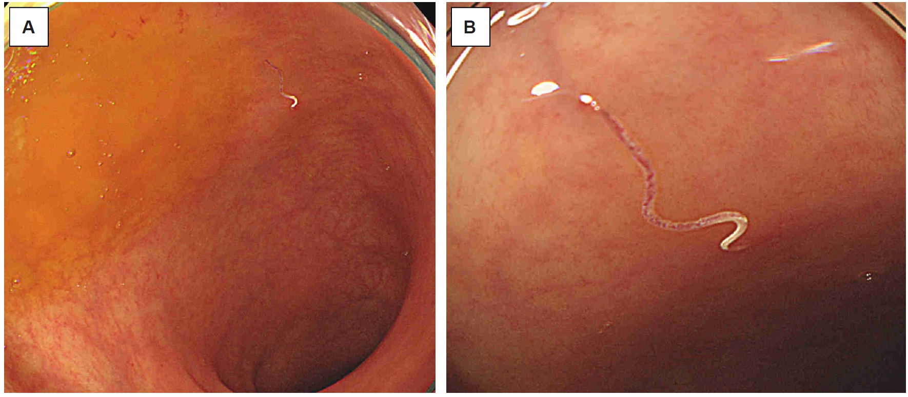
Fig. 2.Colonoscopic findings of suspected and confirmed Trichuris trichiura infection in Cases 2–4. (A) Case 2: endoscopic image showing a whitish, coiled, whip-like organism at the appendiceal orifice, with the anterior portion embedded in the mucosa and the posterior portion freely mobile within the lumen. The retrieved organism was not submitted for histopathologic or parasitologic examination; an accurate size measurement is therefore not available. (B) Case 3: endoscopic image showing histopathologically confirmed T. trichiura attached to the cecal mucosa in a coiled configuration, with the anterior portion embedded and the posterior portion freely mobile. The coiled morphology in situ precluded precise length measurement; the worm straightened and appeared to stretch during endoscopic retrieval, consistent with the elongated body of an adult T. trichiura. (C) Case 4: endoscopic image showing histopathologically confirmed T. trichiura in the ascending colon, with a wave-like curved configuration approximately 30 mm in length and partial mucosal embedding. All images were obtained using standard white-light colonoscopy without optical magnification. The requirement for informed consent for the publication of clinical images was waived because of the retrospective design of the study.
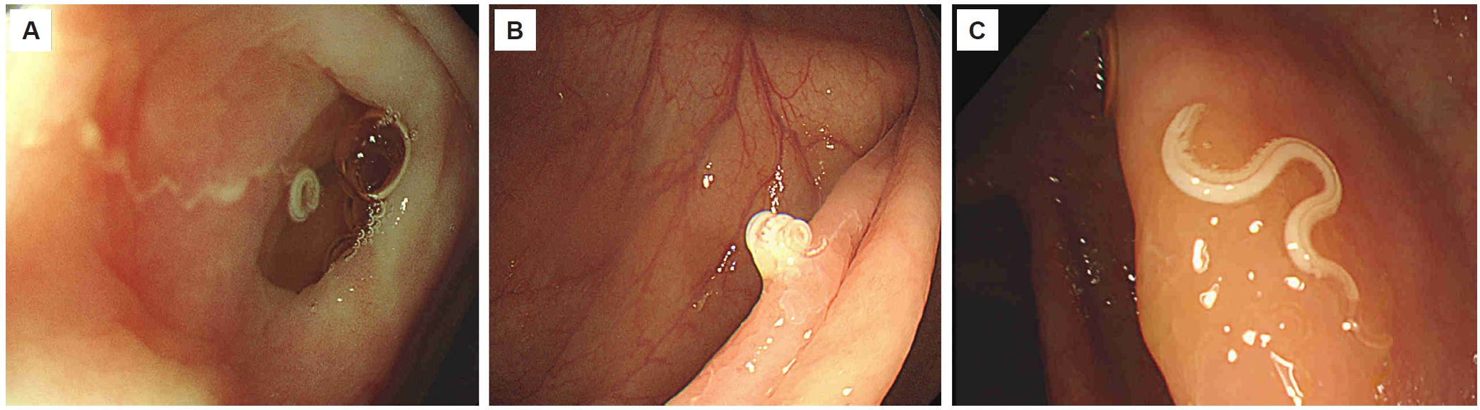
Table 1.Summary of patients with incidentally detected intestinal helminth infections during colonoscopy
Table 1.
|
Case |
Age (year)/sex/nationality |
Indication/symptoms |
Location/endoscopic findings |
Specimen evaluation/diagnosis |
Peripheral eosinophil count |
Stool examination |
Presumed route of infection |
Treatment |
Outcome/follow-up |
|
1 |
57/male/Cambodian |
Anal bleeding, protruding anal mass; no symptoms attributable to helminth infection |
Rectum; slender, translucent, slightly reddish worm firmly attached to rectal mucosa |
Hookworm infection supported by morphology; species undetermined because anterior structures were damaged |
Within reference range |
Follow-up stool examination at 1 month: negative |
Contaminated soil exposure in Cambodia; percutaneous route most likely, oral acquisition not excluded |
ER + ALB |
Stool examination negative at 1 month; asymptomatic without documented recurrence |
|
2 |
53/male/Korean |
Positive fecal occult blood test; no symptoms attributable to helminth infection |
Appendiceal orifice; whitish, coiled, whip-like organism with anterior embedding and posterior mobility |
Endoscopically suspected Trichuris trichiura infection; no histopathologic or parasitologic confirmation |
Within reference range |
Not performed |
Presumed fecal–oral ingestion of embryonated T. trichiura eggs; source not identified |
ER only |
Observed without anthelmintic therapy; formal follow-up not available |
|
3 |
58/female/Korean |
Screening colonoscopy; asymptomatic |
Cecum; thin, whip-like worm with anterior embedding and freely mobile posterior portion |
Histopathologically confirmed T. trichiura infection |
Within reference range |
Not performed |
Presumed fecal–oral ingestion of embryonated T. trichiura eggs; source not identified |
ER + ALB |
No recurrence documented in available records; formal follow-up not available |
|
4 |
66/female/Korean |
Screening colonoscopy; asymptomatic |
Ascending colon; single worm approximately 30 mm long with a wave-like curved shape and partial mucosal embedding |
Histopathologically confirmed T. trichiura infection |
Within reference range |
Not performed |
Presumed fecal–oral ingestion of embryonated T. trichiura eggs; source not identified |
ER + ALB |
No recurrence documented in available records; formal follow-up not available |
References
- 1. Jourdan PM, Lamberton PHL, Fenwick A, Addiss DG. Soil-transmitted helminth infections. Lancet 2018;391:252-65. https://doi.org/10.1016/S0140-6736(17)31930-X
- 2. Loukas A, Hotez PJ, Diemert D, et al. Hookworm infection. Nat Rev Dis Primers 2016;2:16088. https://doi.org/10.1038/nrdp.2016.88
- 3. Else KJ, Keiser J, Holland CV, et al. Whipworm and roundworm infections. Nat Rev Dis Primers 2020;6:44. https://doi.org/10.1038/s41572-020-0171-3
- 4. Hong ST, Yong TS. Review of successful control of parasitic infections in Korea. Infect Chemother 2020;52:427-40. https://doi.org/10.3947/ic.2020.52.3.427
- 5. Shin H, Hong S, Lee YH, et al. Intestinal helminth infections among health examinees: 10-year (2011-2020) nationwide observations in Korea. Ann Clin Microbiol 2024;27:41-8. https://doi.org/10.5145/ACM.2024.27.2.2
- 6. Lee MR, Ju JW, Baek SO, Lee YJ, Lee HI. Infection status of intestinal helminths in 2022. Public Health Wkly Rep 2023;16:1131-40. https://doi.org/10.56786/PHWR.2023.16.32.2
- 7. Vanhooren M, Stoefs A, Van Den Broucke S, et al. Intestinal helminthic infections: a narrative review to guide the hepatogastroenterologist. Acta Gastroenterol Belg 2023;86:460-73. https://doi.org/10.51821/86.3.11895
- 8. Ok KS, Kim YS, Song JH, et al. Trichuris trichiura infection diagnosed by colonoscopy: case reports and review of literature. Korean J Parasitol 2009;47:275-80. https://doi.org/10.3347/kjp.2009.47.3.275
- 9. Wang XJ, Eaton JE. Whipworm found during a colonoscopy. ACG Case Rep J 2019;6:e00259. https://doi.org/10.14309/crj.0000000000000259
- 10. Bathobakae L, Wilkinson T, Yasin S, et al. An unpleasant souvenir: whipworm as an incidental finding during a screening colonoscopy. J Investig Med High Impact Case Rep 2024;12:23247096231224328. https://doi.org/10.1177/23247096231224328
- 11. Webster JL, Stauffer WM, Mitchell T, et al. Cross-sectional assessment of the association of eosinophilia with intestinal parasitic infection in U.S.-bound refugees in Thailand: prevalent, age dependent, but of limited clinical utility. Am J Trop Med Hyg 2022;106:1552-9. https://doi.org/10.4269/ajtmh.21-0853
- 12. Moser W, Schindler C, Keiser J. Efficacy of recommended drugs against soil transmitted helminths: systematic review and network meta-analysis. BMJ 2017;358:j4307. https://doi.org/10.1136/bmj.j4307
- 13. Gebreyesus TD, Makonnen E, Tadele T, et al. Reduced efficacy of single-dose albendazole against Ascaris lumbricoides, and Trichuris trichiura, and high reinfection rate after cure among school children in southern Ethiopia: a prospective cohort study. Infect Dis Poverty 2024;13:8. https://doi.org/10.1186/s40249-024-01176-6
- 14. Chai JY, Jung BK, Hong SJ. Albendazole and mebendazole as anti-parasitic and anti-cancer agents: an update. Korean J Parasitol 2021;59:189-225. https://doi.org/10.3347/kjp.2021.59.3.189
- 15. Palmeirim MS, Hürlimann E, Beinamaryo P, et al. Efficacy and safety of albendazole alone versus albendazole in combination with ivermectin for the treatment of Trichuris trichiura infections: an open-label, randomized controlled superiority trial in south-western Uganda. PLoS Negl Trop Dis 2024;18:e0012687. https://doi.org/10.1371/journal.pntd.0012687
- 16. Do KR, Cho YS, Kim HK, et al. Intestinal helminthic infections diagnosed by colonoscopy in a regional hospital during 2001-2008. Korean J Parasitol 2010;48:75-8. https://doi.org/10.3347/kjp.2010.48.1.75
- 17. Shah J, Shahidullah A. Ascaris lumbricoides: a startling discovery during screening colonoscopy. Case Rep Gastroenterol 2018;12:224-9. https://doi.org/10.1159/000489486
- 18. Alavinejad P, Sina AH, Nikfam M. Hookworm diagnosed during colonoscopy and terminal ileum intubation. Endoscopy 2021;53:E33-4. https://doi.org/10.1055/a-1173-8265
- 19. Adachi E, Nagai H, Saito M, Osawa A. Colonoscopy-based diagnosis of Dibothriocephalus nihonkaiensis infection protruding into the ascending colon. Am J Trop Med Hyg 2025;113:833-5. https://doi.org/10.4269/ajtmh.25-0254
- 20. Omori M, Shibahara K, Murakami T, et al. Incidental detection of asymptomatic colonic anisakiasis during surveillance colonoscopy: a case report and literature review. Clin Case Rep 2026;14:e72593. https://doi.org/10.1002/ccr3.72593


 , Kwang Woo Kim2,†