Abstract
Three anopheline mosquitoes in Korea were studied for their abilities as vectors for Plasmodium vivax. The female mosquitoes of Anopheles lesteri, An. pullus and An. sinensis were allowed to suck malaria patient blood until fully fed, and they were then bred for 2 weeks to develop from malaria parasites to sporozoites. The result from the above confirmed the sporozoites in one An. lesteri of one individual and five An. sinensis of six individuals. We also confirmed that An. sinensis was the main vector to transmit malaria and An. lesteri as well as An. sinensis were able to carry Korean malaria parasites. Therefore, we propose that diversified study is needed to manage malaria projects.
-
Key words: Anopheles lesteri, Anopheles sinensis, Anopheles pullus, Plasmodium vivax, vector, Korea
INTRODUCTION
Several anopheline mosquitoes have been recognized as major vectors of Plasmodium vivax. There are 6 Anopheline mosquitoes distributed in Korea, including Anopheles (Anopheles) lindesayi japonicus Yamada, An. (An.) koreicus Yamada & Watanabe, An. (An.) lesteri Baisas & Hu, An. (An.) pullus Yamada, An. (An.) sinensis Wiedemann, and An. (An.) sineroides Yamada.
Among them,
An. sinensis is distributed in Japan, southern parts of China including Korean Peninsula (
WHO, 1989) and is the primary vector in Korea (
Ree et al., 1967;
Hong, 1977;
Lee et al., 2000;
Lee et al., 2001).
An. pullus, named as
An. yatsushiroensis, is also distributed in the Korean Peninsula, Japan, and mid-northern parts of China (
Tanaka et al., 1979) and is another malaria vector by which sporozoites from wild female mosquitoes and oocysts from experimental infection are confirmed in Korea (
Hong, 1977).
An. lesteri was known as malaria vector in China (
WHO, 1989) and suggested as the malaria vector at outbreak period of malaria in Japan (
Otsuru, 1949;
Kamimura, 1968). Shim et al. (
1997) showed
An. lesteri to have low population density in Korea compared with
An. sinensis. However, it inhabits in domestic area up to now and has a possibility to be a vector which transmits
P. vivax in Korea. Therefore, an evaluation of the vector competence of Anopheline mosquitoes could be important in explaining the distribution and epidemic of malaria in Korea.
In this study, anopheline mosquitoes were fed blood of malaria patient who was confirmed as malaria patient through the blood smear, and then we have observed sporozoites in salivary glands at internal of mosquito for regular hours.
MATERIALS AND METHODS
Malaria patient who had experienced the human bait collection at Dongjung-ri (N 38°04'08", E 126°57'15"), Yeoncheon-gun, Gyeonggi-do, as the malaria prevalent area from June to September in 2000 and had never been bitten by mosquitoes in 2001, first developed symptoms of malaria with a chill at 6:00 AM 27
th on May in 2001, and showed a slight fever intermittently thereafter, revealing the symptom of typical longterm incubation period malaria in the Korean Peninsula (
Hall and Loomis, 1952;
Hankey et al., 1953;
Arnold et al., 1954;
Garnham et al., 1975;
WHO, 1977).
Approximately 28 hours after the patient revealed the first symptom, malaria protozoa with ring form was confirmed in the red blood cell by blood smear method and tropozoites and gametocytes were confirmed after about 48 hours. When malaria protozoa with ring form was detected in the blood of the patient, the individuals which did not suck the blood in cow shed were collected by aspirator at Ogum-ri, [Tanhyeon-myeon, Paju-si, Gyeonggi-do as the northeast part of Korea (N 37°49'20", E 126°42'20")] from 21:00 PM to 22:00 PM and were left overnight at the insectarium (26±1℃, RH 80±5%) for preparing the blood sucking. After past 48 hours since the first showing of symptom, we could find tropozoites and gametocytes, so that prepared mosquitoes could be induced to suck blood at patient's arm.
To propagate malaria protozoa, the blooded mosquitoes were kept at 26±1℃ and RH80±5% in insectarium with a supply of 5% sucrose solution for 2 weeks. In order to examine whether the sporozoites were in internal of mosquitoes or not, the mosquitoes were dissected under an anatomical microscope to extract salivary glands and covered with cover a glass, so that we could confirm motility of sporozoites under an optics microscope (×1000) and took a photograph with dyeing of PROTOCOL™ HEMA 3R.
To confirm the species of mosquitoes, each individual blood sucked mosquitoes were placed in the paper cup covered with gauze and a wet towel for digestion for one day. At first, deck of eggs was observed after oviposition of eggs with an addition of a quarter distilled water and was then continuously bred for production of adult, and finally we confirmed the species of mosquitoes.
RESULTS
The malaria patient who had past 48 hours from the first showing of symptom was exposed to prepared mosquitoes and then collected the blood from patient. As shown in
Table 1, parasitemia was investigated by blood smear on slide glass.
In
Table 2, the blood sucked mosquitoes, which were exposed to the arm of malaria patient were shown from 17 individuals of
An. lesteri, 16 individuals of
An. pullus, and 35 individuals of
An. sinensis. However, one individual of
An. lesteri and 6 individuals of
An. sinensis survived for 14 days at 26±1℃ and RH80±5% in insectarium, but all
An. pullus died.
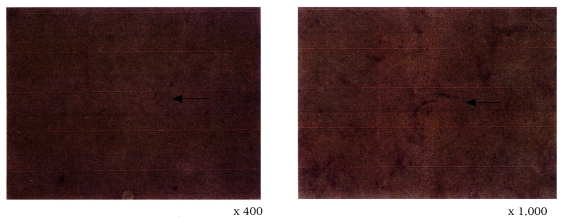
We could confirm the motility of sporozoites in salivary glands of the survival mosquitoes through anatomical microscope (×400). The result on sporozoites in salivary glands with dye confirmed one
An. lesteri of one individual and 5
An. sinensis of 6 individuals (
Fig. 1). The above result confirmed for the first time that
An. lesteri was able to carry of
P. vivax in Korea.
DISCUSSION
An. sinensis and
An. pullus (=
An. yatsushiroensis, this species was reported as a synomym of
An. pullus by
Shin & Hong, 2001) have been known as vectors of
P. vivax in Korea.
An. sinensis was confirmed as vector by natural infectious confirmation method with collected mosquitoes (
Ree et al., 1967;
Hong, 1977), polymerase chain reaction (
Lee et al., 2000), and by artificial infection with ELISA (
Lee et al., 2001).
An. pullus was confirmed as a vector through confirmation of oocysts in mid-gut external wall and sporozoites in salivary gland by natural infection and artificial blood-sucking method (
Hong, 1977).
An. lesteri, confirmed as a vector of malaria in Korea in this study, has already been known as major or potential vector of malaria in China and Japan. However, it was reported that
An. lesteri had the low population density in Korea, consequently it was not recognized as a important malaria vector. In the morphologic features,
An. lesteri is similar to female adult of
An. sinensis and it is difficult to identify without the observation of patch at the part of coax in mid leg. Also, in the shape of wing,
An. lesteri, which has no pale fringe spot at the end part of wing vein 5.2, is within the variation category of wing shape of
An. sinensis and
An. pullus (
Otsuru and Ohmori, 1960;
Kanda and Oguma, 1976;
Shin and Hong, 2001). It is, therefore, difficult to identify each species, particularly in the case of partially damaged scale in body and wing.
Due to possible mistake in identifications, an ecological characteristic and population of An. lesteri in Korean remain unclear.
Generally, it was reported that the population density of
An. lesteri in Korea was low however, our study did not shown the low density in time of collection (
Table 2). Further studies on seasonal prevalences and geographical distributions are needed to determine ecology of
An. lesteri in Korea. Furthermore, the rate of
An. lesteri as a malaria vector in Korea has to be confirmed through investigations of natural infection rates and susceptibilities in
P. vivax. Moreover, other Korean anopheline mosquitoes which are not established as malaria vector need to be investigated similar to this study.
We suggest that a management counterplan could be established to eliminate the malaria early through reanalysis of epidemiological characteristics about present malaria patients, based on entomological approach such as this study.
Notes
-
This study was supported by a grant of the Korea Health 21 R & D Project, Ministry of Health and Welfare, Republic of Korea (HMP-99-M-04-0002).
References
- 1. Arnold J, Alving AS, Hockwald RS, Clayman CB, Dern RJ, Beutler E. Natural history of Korean vivax malaria after deliberate inoculation of human volunteers. J Lab Clin Med 1954;44:723-726.
- 2. Garnham PCC, Bray RS, Bruce-Chwatt LJ, et al. A strain of Plasmodium vivax characterized by prolonged incubation: morphological and biological characteristics. Bull World Health Organ 1975;52:21-32.
- 3. Hall WH, Loomis GW. Vivax malaria in veterans of the Korean War. A preliminary report of 25 cases. New Engl J Med 1952;246:90-93.
- 4. Hankey DD, Jones R, Coatney RS, et al. Korean vivax malaria. I. Natural history and response to chloroquine. Am J Trop Med Hyg 1953;2:958-969.
- 5. Hong HK. Ecological studies of important mosquitoes in Korea. 1977, Dongkook University Graduates School. pp 1-6 Ph.D. Theses.
- 6. Kamimura K. The distribution and habit of medically important mosquitoes of Japan. Jpn J Sanit Zool 1968;19:15-34.
- 7. Kanda T, Oguma Y. Morphological variations of Anopheles sinensis Wiedemann, 1828 and A. lesteri Baisas and Hu, 1936 and frequency of clasper movements of the males of several Anopheles species during induced copulation. Jap J Sanit Zool 1976;27:325-331.
- 8. Lee HW, Cho SH, Shin EH, et al. Experimental infection of Anopheles sinensis with Korean isolates of Plasmodium vivax. Korean J Parasitol 2001;39:177-183.
- 9. Lee WJ, Lee HW, Shin EH, et al. Vector determination of tertian malaria (Plasmodium vivax) by polymerase chain reaction. Korean J Parasitol 2000;39:77-83.
- 10. Otsuru M. A new species of Anopheles hyrcanus in Japan. Fukoka Igaku Zassi 1949;40:139-148.
- 11. Otsuru M, Ohmori Y. Malaria studies in Japan after World War II, Part II. The research for Anopheles sinensis sibling species group. Japan J Exp Med 1960;31(1):33-65.
- 12. Ree HI, Hong HK, Paik YH. Study on natural infections of Plasmodium vivax in Anopheles sinensis in Korea. Korean J Parasitol 1967;5:3-4.
- 13. Shim JC, Shin EH, Yang DS, Lee WK. Seasonal prevalence and feeding time of mosquitoes (Dipter: Culicidae) at outbreak regions of domestic malaria (P. vivax) in Korea. Korean J Entomol 1997;27:265-277.
- 14. Shin EH, Hong HK. A new synomym of Anopheles (Anopheles) pullus Yamada, 1937 - A. (A.) yatsushiroensis Miyazaki, 1951. Korean J Entomol 2001;31:1-5.
- 15. Tanaka K, Mizusawa K, Saugstad ES. A revision of the adult and larval mosquitoes of Japan (including the Ryukyu Archipelageo and the Ogasawara island) and Korea (Diptera: Culicidae). Cont. Amer Entomol Inst. 1979, pp 79-81.
- 16. WHO. WHO/MAL/77.895. The course of infection caused by the North Korean strain of Plasmodium vivax. 1977.
- 17. WHO. WHO/VBC/89/967. Geographical distribution of arthropodborne diseases and their principal vector. 1989.
Fig. 1Sporozoites of Plasmodium vivax, Korean strain

Table 1.The parasitemia of Plasmodium vivax in blood of the malaria patient who was exposed to mosquitoes to for infection
Table 1.
|
Sex/age |
No. of parasitesa)
|
Total |
|
Trophozoites |
Schizonts |
Gametocytes
|
|
male |
female |
|
male/38 |
920 |
ND |
NDb)
|
40 |
960 |
Table 2.Results of dissection of the mosquitoes 14 days after feeding the blood of a volunteer infected with Plasmodium vivax
Table 2.
|
Species |
No. of fed mosquitoes |
No. of survived mosquitoes |
No. of infected mosquitoesa)
|
|
An. lesteri
|
17 |
1 |
1 |
|
An. pullus
|
16 |
0 |
0 |
|
An. sinensis
|
35 |
6 |
5 |

