Abstract
A case of human infection with Heterophyes nocens (Heterophyidae) was incidentally found in a biopsy specimen of the Meckel's diverticulum at the upper part of the small intestine. The patient was a 58-year-old man living in a rural area of Talsong-gun, Kyongsangbuk-do. He had gastrointestinal symptoms such as epigastric pain, indigestion, and abdominal discomfort for 3 months, and severe diarrhea, abdominal pain, and vomiting for about 1 month before hospitalization. Endoscopy of the upper part of the small intestine revealed a Meckel's diverticulum, and it was excised and histopathologically examined. Three adult flukes were incidentally found sectioned in the mucosa, and they were identified as H. nocens. The patient had a history of eating raw mullets at a fish market in Pusan 6 months ago, and the mullets were presumed to be the source of infection. This case brings a considerable interest in that specific diagnosis of heterophyid infections could be done by sectional morphology of the worms.
-
Key words: Heterophyes nocens, heterophyid, case report, human, small intestine, Meckel's diverticulum
INTRODUCTION
Almost 20 species of minute intestinal flukes have been reported from human infections in Korea (
Chai and Lee, 1990). Among them, it is agreed that heterophyids (Heterophyidae) are the most important species from epidemiological points of view. Especially,
Heterophyes nocens (
Chai et al., 1994,
1997,
1998) and three species of
Metagonimus,
M. yokogawai (
Chai and Lee, 1990),
M. takahashii (
Ahn and Ryang, 1988), and
M. miyatai (
Chai et al., 1993), are the major species showing considerable degrees of endemicity in various localities.
Heterophyes nocens was first described by Onji and Nishio (
1916) and now is known to be distributed in southeast Asian countries, including Korea and Japan. In Korea, Seo et al. (
1980) first identified the presence of
H. nocens metacercariae in
Mugil cephalus. The first human case was subsequently reported from a 52-year-old man living in Okku-gun, Chollabuk-do (
Seo et al., 1981a). Since then, 13 cases were sporadically reported (
Chai et al., 1984,
1985;
Sohn et al., 1989), and finally, several endemic foci were discovered in Shinan-gun, Muan-gun, Puan-gun, Chollanam-do, and Sachon-gun, Kyongsangnam-do (
Chai et al., 1994,
1997,
1998).
The diagnosis of
H. nocens infection in Korea has usually been based upon discovery of eggs in fecal samples followed by recovery of adult flukes after anthelmintic treatment and purgation (
Chai et al., 1998). The worm recovery is needed because of great similarities in the morphology of eggs among different species of heterophyids in fecal smears. Alternatively, intestinal excision and histopathological observation could be helpful for a specific diagnosis (
Africa et al., 1940;
Seo, 1979). However, cases of
H. nocens infection diagnosed in biopsy specimens have seldom been reported in the literature.
We histopathologically examined intestinal tissues of a 58-year-old man admitted to Yeungnam University Hospital with clinical complaints of severe diarrhea, abdominal pain, and vomiting, and diagnosed as the Meckel's diverticulum by endoscopy. Sections of H. nocens worms were incidentally found in the mucosa of the small intestine of the patient for which the present paper mainly focuses on.
CASE RECORD
The patient was a 58-year-old male farmer, living in a rural area of Talsong-gun, Kyongsangnam-do. He visited Yeungnam University Hospital with severe abdominal pain, diarrhea, and vomiting on September, 1998. He had a history of eating raw mullets several times in a fish market located in Pusan 6 months ago, and raw fresh-water fish caught from a small stream in Talsong-gun. He often drank alcohol, but didn't have any history of special diseases nor treatments. He experienced mild gastrointestinal symptoms such as indigestion for about 3 months and abruptly developed severe symptoms such as abdominal pain, diarrhea, and vomiting about 1 month before visiting the hospital.
Laboratory examinations revealed that the total number of white blood cells (WBCs), hematocrit, erythrocyte sedimentation rate, and leucocyte differential counts were within normal limits, but the levels of hemoglobin (12.8 g/dL) and platelets (141,000/mm2) were lower than normal values. In liver function tests, the total protein, albumin, aspartate aminotransferase, and alanine aminotransferase were within normal values, but the level of gamma glutamyl transferase (205 unit/L) was higher than normal. The blood glucose level was increased to 191 mg/dl, and the urine glucose level was triple positive (+++). Hepatitis B antigens and antibodies were all negative, except for HBc IgG antibody. The fecal occult blood was also positive.
In fecal examinations, heterophyid and Trichuris trichiura eggs were found by formalin-ether sedimentation technique. By Stoll's egg counting technique, the EPG (eggs per gram of feces) of heterophyid eggs was 400, and that of T. trichiura was below 100. The size of heterophyid eggs was 28.2 × 15.4 µm. The skin tests for Clonorchis sinensis and Paragonimus westermani showed negative reactions.
PARASITOLOGICAL DESCRIPTION
The specimen was excised from the Meckel's diverticulum at the upper part of the small intestine. It was fixed in 10% formalin and a paraffin block was made. The block was sectioned serially at 6 µm thickness and stained with hematoxylin and eosin. Three adult flukes (worms A, B and C) were sectioned which were identified as H. nocens Onji and Nishio, 1916 (Heterophyidae).
The worms A and B were sliced vertically with a little inclination from the central line (worm A;
Figs. 1-2) or from the oral sucker to the posterior part of the genital sucker (worm C). The worm C was sectioned horizontally so that the protruded genital sucker was easily recognized (
Figs. 3-4). The maximum length and width of the worm A were 512.0 µm and 210.8 µm, respectively, and the maximum length of the worm C was 358.4 µm. The maximum thickness of the worm C, on the other hand, was 180.7 µm (
Fig. 3). The shape of the worm A looked like a long gourd dipper, and the narrow anterior part became wider and extended down to the posterior part (
Fig. 1).
Small tegumental spines were distributed densely over the anterior 1/3 of the body at the interval of about 1.2 µm (worm B). The average length of each spine was 5.4 µm with the maximum thickness of 1.6 µm. The oral sucker was round, 87.0 µm in diameter (worm B), and consisted of strong muscles. The ventral sucker was also round, 89.6 µm in diameter (worm C), and located on the midline of the body.
The genital sucker was well sectioned, and its characteristic structures were clearly seen (worm A). In another section, it was ventrally protruded to show its typical lateral view (worm C). It was round in shape, 133.1 µm in diameter, and located at the anterior 1/3 level of the body (
Fig. 1). Sixty-two small chitinous rodlets were arranged circularly on the gonotyl (
Fig. 2). The gonotyl was typically hammer-shaped when it is protruded (worm C). One of the chitinous rodlets, 4.6 µm long, was seen to be protruded outwards (
Figs. 3-4).
The ejaculatory duct was 18.0 µm in diameter, and cross-sectioned just posterior to the genital sucker (
Fig. 4). The seminal vesicle with sperms was located postero-dorsally to the genital sucker and was 56.3 µm in diameter (
Fig. 4). Two sections of seminal receptacles were seen posteriorly to the seminal vesicle at 84.5 × 6.8 µm and 64.0 × 43.5 µm in size, respectively (
Fig. 3). The ovary was round and was located in front of two testes. Two testes were measured 140.8 × 87.0 µm in size (
Fig. 1) and were located a bit askew at the lower part of the body. The diameter of the intestinal cecum was 38.4 µm (worm C). Round vitelline follicles were scattered over posterior sides of the worm (worm B). In the uterus, many eggs were seen, and the eggs were measured 28.1 × 15.4 µm in average size (n=15).
DISCUSSION
Although taxonomic problems were raised after
H. nocens Onji and Nishio was first described in 1916 (
Cort and Yokogawa, 1921;
Faust and Nishigori, 1926;
Witenberg, 1929), Asada (
1934) compromised the situation by designating
H. nocens as a sub-species of
H. heterophyes. However, Chai and Lee (
1990) acknowledged
H. nocens as a distinct species because of a consistent feature in the number of chitinous rodlets on the gonotyl;
H. heterophyes having 70-85 rodlets (
Chai et al., 1986) and
H. nocens 52-64 rodlets (
Chai et al., 1984).
In this study, however, it was fortunate that the genital sucker of the worm A was sectioned in a very good plane to show complete orientation and arrangement of the rodlets on the gonotyl. The number of rodlets in the worm A was counted as 62 which was consistent with
H. nocens (
Chai et al., 1984). In worms B and C, the genital suckers were not well sectioned, and the number of rodlets was difficult to count, but they were diagnosed also as
H. nocens based on the presence of the genital suckers and other morphological characteristics.
Several mature eggs were seen in the uterus of 3 worms. They were operculated, ellipsoid to oval in shape, and measured 28.1 × 15.4 µm in average size. All of them agreed well with
H. nocens (
Chai et al., 1984). The arrangement of other genital organs such as the ejaculatory duct, seminal vesicle, ovary, seminal receptacle, and testes were all consistent with the descriptions of
H. nocens (
Chai et al., 1984).
Although the present case was diagnosed as H. nocens infection, the symptoms complained by the patient may have not been necessarily correlated with the fluke infection. The patient had a Meckel's diverticulum at the upper part of the small intestine, hence, occurrence of various gastrointestinal symptoms was possible. Therefore, it is likely that H. nocens infection is an incidental finding in this patient. A supporting evidence for this is that the EPG of heterophyid eggs in the feces of the patient was not high, only 400, which means that this patient is not heavily infected with H. nocens to cause significant clinical manifestations.
In clinicopathological aspects, the pathogenicity as well as symptoms caused by
H. nocens infection in humans are yet unclear and have to be further documented. For example, two cases reported by Chai et al. (
1984) suffered from epigastric discomfort and indigestion, but they were co-infected with
C. sinensis and other intestinal parasites. Eight cases infected with
H. nocens (
Chai et al., 1985) complained of gastrointestinal troubles, but they were also co-infected with other kinds of intestinal parasites. Erratic parasitism in the heart, brain, and spinal cord by eggs and/or adults of heterophyid flukes were reported (
Africa et al., 1940). In
H. nocens infection, however, such possibility has not yet been elucidated.
According to the past history of the present case, the source of infection is presumed to be raw mullets. In Korea, mullets,
Mugil cephalus, and gobies,
Acanthogobius flavimanus, caught from various localities were reported to have metacercariae of
H. nocens (
Seo et al., 1980,
1981b).
There are, however, difficulties when diagnosing human
H. nocens infection; the low count of daily eggs produced by the worms (
Chai and Lee, 1990), relatively low worm burdens in most infected cases (
Chai et al., 1994,
1997,
1998), and morphological similarities of eggs to other heterophyids or
C. sinensis (
Chai and Lee, 1990). The present case brings a considerable interest in that a specific diagnosis was made based on sectional morphology of the worms in a mucosal biopsy specimen.
References
- 1. Africa CM, Leon W, Garcia EY. Visceral complications in intestinal heterophyidiasis of man. Acta Medica Philippina, Monographic Series 1940;1:1-132.
- 2. Ahn YK, Ryang YS. Epidemiological studies on Metagonimus infection along the Hongcheon River, Kangwon Province. Korean J Parasitol 1988;26:207-213. (in Korean).
- 3. Asada J. On the Metagonimus and its related species. Clin Med 1934;22:43-56. (in Japanese).
- 4. Chai JY, Hong SJ, Sohn WM, Lee SH, Seo BS. Further cases of human Heterophyes heterophyes nocens infection in Korea. Seoul J Med 1985;26:197-200.
- 5. Chai JY, Huh S, Yu JR, et al. An epidemiological study of metagonimiasis along the upper reaches of the Namhan River. Korean J Parasitol 1993;31:99-108.
- 6. Chai JY, Kim IM, Seo M, Guk SM, Kim JL, Sohn WM, Lee SH. A new endemic focus of Heterophyes nocens, Pygidiopsis summa, and other intestinal flukes in a coastal area of Muan-gun, Chollanam-do. Korean J Parasitol 1997;35:233-238.
- 7. Chai JY, Lee SH. Intestinal trematodes of humans in Korea: Metagonimus, heterophyids and echinostomes. Korean J Parasitol 1990;28(suppl.):103-122.
- 8. Chai JY, Nam HK, Kook J, Lee SH. The first discovery of an endemic focus of Heterophyes nocens (Heterophyidae) infection in Korea. Korean J Parasitol 1994;32:157-161.
- 9. Chai JY, Seo BS, Lee SH. Studies on intestinal trematodes in Korea XI. Two cases of human infection by Heterophyes heterophyes nocens. Korean J Parasitol 1984;22:37-42.
- 10. Chai JY, Seo BS, Lee SH, Hong SJ, Sohn WM. Human infections by Heterophyes heterophyes and H. dispar imported from Saudi Arabia. Korean J Parasitol 1986;24:82-88.
- 11. Chai JY, Song TE, Han ET, Guk SM, Park YK, Choi MH, Lee SH. Two endemic foci of heterophyids and other intestinal fluke infections in southern and western coastal areas in Korea. Korean J Parasitol 1998;36:155-161.
- 12. Cort WW, Yokogawa S. A new human trematode from Japan. J Parasitol 1921;8:66-69.
- 13. Faust EC, Nishigori M. The life cycles of two new species of Heterophyidae, parasitic in mammals and birds. J Parasitol 1926;13:91-128.
- 14. Onji Y, Nishio T. On the trematodes whose intermediate host is brackish water fishes. J Chiba Coll Med Prof 1916;82 & 81:229-249. (in Japanese).
- 15. Seo BS. Biology and clinical aspects of Heterophyidae. Human Science 1979;3:784-791. (in Korean).
- 16. Seo BS, Cho SY, Chai JY, Hong ST. Studies on the intestinal trematodes in Korea II. Identification of the metacercariae of Heterophyes heterophyes nocens in mullets of three southern coastal areas. Seoul J Med 1980;21:30-38.
- 17. Seo BS, Hong ST, Chai JY. Studies on intestinal trematodes in Korea III. Natural human infections of Pygidiopsis summa and Heterophyes heterophyes nocens. Seoul J Med 1981a;22:228-235.
- 18. Seo BS, Hong ST, Chai JY, Cho SY. Studies on intestinal trematodes in Korea IV. Geographical distribution of Pygidiopsis and Heterophyes metacercariae. Seoul J Med 1981b;22:236-242.
- 19. Sohn WM, Chai JY, Lee SH. Two cases of natural human infections by Heterophyes nocens and the infection status of heterophyid metacercariae in mullets from Samcheonpo, Kyongnam Province. Inje Med J 1989;10:443-452. (in Korean).
- 20. Witenberg Y. Studies on the trematode-family Heterophyidae. Ann Trop Med Parasitol 1929;23:131-268.
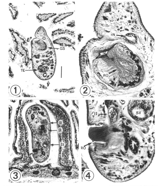
Figs. 1-4
Figs. 1-2 Micrographs showing a longitudinally sectioned worm (worm A) in the small intestine of the patient. Fig. 1. A round genital sucker is located at the anterior 1/3 of the body. H & E stain. ×100. Fig. 2. Along the inner side of the genital sucker, there were 62 chitinous rodlets on the gonotyl arranged ovally. H & E stain. ×450.
Figs. 3-4. Micrographs showing a horizontally sectioned worm (worm C) in the small intestine of the patient. Fig. 3. The gonotyl (arrowhead) is protruded ventrally. The seminal receptacle and seminal vesicle are sectioned well. H & E stain. ×100. Fig. 4. A rodlet is seen on the gonotyl. H & E stain. ×450. ED; ejaculatory duct, GS; genital sucker, IN; intestine, RO; rodlet, SR; seminal receptacle, SV; seminal vesicle. TE; testis.