Abstract
The present study surveyed the prevalence of natural infection of the sheep esphagus muscle with sarcocysts of Sarcocystis ovicanis and examined induction of protective immunity using UV-attenuated sporocysts. The overall prevalence of natural infection of the sheep was 95%. Infectivity of the collected sarcocysts was confirmed by shedding of sporulated oocysts after feeding infected esophageal tissues to dogs. To induce protective immunity, lambs were immunized 3 times (once a week) with 1.5 × 10⁴ sporocysts exposed to UV-light for 30 min (UV-30 group) or 60 (UV-60 group) min and then challenged with 1.5 × 10⁴ normal sporocysts at the 3rd week post the 1st vaccination. These lambs showed high survival and less clinical signs of sarcocystosis than normal infected lambs. The attenuated sporocysts produced abnormal cysts; small in size and detached from the muscle fiber. These abnormalities were more obvious in UV-60 group than UV-30 group. Also, the IFN-γ level and lymphocyte percentage were increased while the total leukocyte count was decreased in the UV-60 group compared with other groups. The high level of IFN-γ may be an evidence for the induction of Th1 responses which may have protective effect against a challenge infection.
-
Key words: Sarcocystis ovicanis, UV-attenuated sporocysts, protective immunity, IFN-γ
INTRODUCTION
Sarcocystis infections of sheep are common throughout the world [
1]. Of the 4 known species of
Sarcocystis that infected sheep (
Sarcocystis ovicanis,
S. tenella,
S. medusiformis, and
S. gigantea), only
S. ovicanis (
S. arieticanis) and
S. tenella are pathogenic. Dogs (
Canis) act as the definitive host for the pathogenic species whereas felids serve as the definitive host for the non-pathogenic species [
1].
Sarcocystis infection can cause anorexia, fever, decreased weight gain, anemia, and death in experimentally infected lambs, and has been associated with abortions in ewes. Neurologic signs including encephalomyelitis, muscle weakness, hind limb paresis, and ataxia have been seen in naturally infected sheep. Moreover, condemnation of cattle and sheep containing macroscopic cysts was a serious economic problem [
2-
5].
The control of
Sarcocystis relied on preventing contamination of pasture and water with feces of dogs, foxes, and cats or by controlling access of young susceptible stocks to the contaminated land [
6]. Some studies revealed that amprolium and salinomycin are somewhat effective in controlling clinical
Sarcocystis resulting from infection with
S. ovicanis as they reduced the number of deaths and severity of the infection signs in lambs [
7,
8]. Relatively little has been known about the immunity induced by infection with
Sarcocystis species; however, some studies referred to the protective immunity and cell-mediated mechanisms [
6].
The present study was carried out to address the prevalence of natural infection of domestic sheep (Ovis ammon aries) at Beni-Suef Governorate, Egypt, with S. ovicanis and to induce protective immunity against S. ovicanis using UV-attenuated sporocysts.
MATERIALS AND METHODS
The natural infection
About 120 sheep esophageal samples of different ages were collected from different localities at Beni-Suef Governorate, Egypt, during 2007. The natural infection with Sarcocystis was detected by fresh tissue squash preparations.
Experimental infection of the final host
For experimental infection of the final host, 14 young dogs (2-3 months old) were used. All dogs were coccidia-free at the time of the experiment as neither sporocysts nor oocysts were detected in their feces by daily examination for 3 wk. Then, dogs were fed on segments of the naturally infected sheep esophagus. Their feces were examined daily for
Sarcocystis oocysts and sporocysts by the flotation technique [
9].
The sporulated oocysts and sporocysts were concentrated by centrifugation, washed several times with distilled water to remove the salt, then suspended in 2.5% potassium dichromate solution and stored at 4℃. Two dogs were kept non-infected as a negative control.
UV-attenuation of sporocysts
Sporocysts were washed with phosphate-buffered saline solution (PBS) (pH 7.2) several times and the number of sporocysts per ml was determined by Neubauer hemocytometer. A UV Lamp, VL6-LC (ETS Viber-Louramat, Marne La Vallaee, Cedex, France), of output at 254 nm was used for the sporocysts attenuation. A 15-ml sample of PBS containing 1.5 × 10⁴ oocysts or sporocysts per ml was poured into a clear glass dish. Then, this dish was placed at the center of a closed carton box of 27 cm in length, 18 cm in width, and 17 cm in height. The UV lamp was adjusted above this box through an opening in the center of the box roof. The cyst suspension was exposed to UV light for either 30 or 60 min.
Experimental infection and vaccination of the intermediate host
Total 12 lambs of 2 months old were divided equally into 4 groups. The first group was kept as a non-infected control group. The second group (normal infected group) was inoculated with 1.5 × 10⁴ normal oocysts or sporocysts per lamb. The lambs of the second group were sacrificed at the 5th week post-infection (PI). Each lamb of the third group was immunized with 1.5 × 10⁴ oocysts or sporocysts exposed to UV light for 30 min once a week for a period of 3 wk (UV-30 group). Lambs of the fourth group were immunized each with 1.5 × 10⁴ sporocysts exposed to UV light for 60 min once a week for a period of 3 wk (UV-60 group). The third and the fourth groups were challenged at the 3rd week post the 1st vaccination (PV) with 1.5 × 10⁴ normal sporocysts and were sacrificed at the 8th week PV (5 wk post-challenge infection).
Organ collection
From the sacrificed lambs, tissue samples from the esophagus, liver, and kidney were fixed in 10% buffered formalin. Fixed tissues were processed for histological studies, sectioned, and stained with hematoxylin and eosin (H-E).
Blood sampling and serum preparation
EDTA-treated blood samples and non-EDTA-treated blood samples were taken before and after infection each week from the subjugular vein. Sera were collected from the clotted blood samples after centrifugation at 400 g for 15 min at 4℃, then divided into aliquots and stored at -80℃ until used.
Total and differential leukocyte count
The total WBC count was carried out in EDTA-treated blood samples by using Turk's solution and Neubauer hemocytometer. Differential leukocyte counts were obtained in Giemsa-stained blood smears according to the method of Dacie and Lewis [
10].
The IFN-γ level in sera was determined using sandwich ELISA. Measurement of IFN-γ was performed using commercially available reagents and ELISA kits purchased from Koma Biotech Inc. (Seoul, Korea) according to manufacturer's directions. IFN-γ concentration was determined using a standard curve obtained from the known concentration of cytokine standard included in each assay plate according to manufacture instructions.
Statistical analysis
The data were analyzed using 1-way analysis of variance (ANOVA) followed by LSD analysis to compare various groups with each other [
11]. Results were expressed as mean ± SD, and values of
P > 0.05 were considered statistically insignificant while those of
P < 0.05 and
P < 0.01 were considered statistically significant and highly significant, respectively. F-probability for each variable expressed the general effects between groups.
RESULTS
During the present study, a total of 120 sheep esophagus were examined for the infection with Sarcocystis. The prevalence of infection was 95% (114/120).
Light microscopy
Mature and immature microscopic sarcocysts were observed in the examined sheep esophagus samples (
Figs. 1, 2). The mature sarcocysts measured 700 (670-860) µm in length and 188 (170-200) µm in width in the longitudinal section (
Fig. 1), while it was measured 75 (22-118) µm in the cross section (
Fig. 2). The cysts located within muscle fibers and surrounded by striated primary cyst wall which was about 10 (9-11) µm in thickness (
Figs. 1, 2). The interior of the mature cyst was divided into many chamber-like compartments by septa. The ground substance was found directly underneath the cyst wall and extended inside the cyst to form numerous septa (
Fig. 1).
The infected dogs shed sporulated oocysts and sporocysts in their feces at days 9-11 PI. The number of sporocysts shed by dogs gradually increased till reaching the maximum at days 16-17 PI, then it decreased to the minimum at days 21-58 PI. No sporogonic stages were found in the control dogs.
Experimental infection of the intermediate host with normal or attenuated sporocysts
All lambs infected with normal sporocysts showed clinical manifestations of acute sarcocystosis after day 21 PI. These signs included reduced food intake, weakness, fever, anorexia, severe weight loss, nervousness, hypersalivation, lameness, and hair loss on the extremities, erosions and ulcerations in the oral cavity and esophagus, and severe laminitis. Such lambs were expected to die at week 5 PI; therefore, we sacrificed them at that time. No cysts were observed in the normal infected group at week 5 PI.
On the other hand, lambs of UV-30 group showed clinical symptoms of acute sarcocystosis at week 3 post-challenge infection, but these symptoms were less in severity when compared with the normal infected group and they survived healthy until sacrificed at week 5 post-challenge infection (week 8 PV). Several young cysts were found in the skeletal muscles of these lambs. These cysts were small in size detached from the muscle fibers (
Fig. 3). Similarly, lambs of UV-60 group survived healthy without any clinical signs of sarcocystosis until sacrificed at week 5 post-challenge. In such lambs cysts were also small in size and detached from the muscles fibers (
Figs. 4, 5). The abnormalities and the deformation of cysts in this group were higher than those in the UV-30 group.
As shown in
Fig. 6, the total leukocyte count in the non-infected control group was around the normal level along the experiment, while in the normal infected group it reached its maximum value at week 4 PI and then it slightly decreased at week 5 PI. In the UV-30 group and the UV-60 group, the count was nearly around the normal level at week 1 PV and reached its maximumat week 3 PV, and then it made a plateau line until week 7 PV and finally decreased around the normal level at week 8 PV. The difference in both groups was in the 2nd week as the count increased in the UV-30 group and decreased in the UV-60 group. The total leukocyte count of UV-60 group decreased significantly (
P < 0.01) than those of the other groups except at weeks 1 and 2 PV. The total leukocyte count of UV-30 group was not significantly different (
P > 0.05) from that of the normal infected group at weeks 3-5 PV, and also there was no significant difference (
P > 0.05) with UV-60 group at weeks 1 and 8 PV. The total leukocyte count of the normal infected group was significantly higher than that of UV-30 group (
P < 0.01) at week 1 PI and all other groups at week 2 PI, and became not different (
P > 0.05) with UV-30 group at weeks 3-5 PI.
As shown in
Table 1, the lymphocyte percentage of UV-60 group was significantly higher (
P < 0.01) than those of all other groups at weeks 1-4 PV. Also, the lymphocyte percentage of UV-30 group was significantly higher (
P < 0.01) than those of non-infected and normal infected groups only at weeks 2 and 5 PV. At weeks 5 and 6 PV, the lymphocyte percentage of UV-30 group was significantly higher (
P < 0.01) than that of UV-60 group and not significantly different at week 7 PV. However, at week 8 PV, the lymphocyte percentage of UV-60 group significantly increased than those of UV-30 and non-infected groups.
The neutrophil percentage of UV-60 group was significantly lower than those of all other groups at weeks 1-4 and week 8 PV. However, the neutrophil percentage of UV-30 group significantly decreased than those of the other groups during weeks 5 and 6 PV. At week 7 PV, the neutrophil percentage of UV-60 group was not significantly different from that of UV-30, but significantly increased than that of the non-infected group (
Table 1).
The monocyte percentage of UV-30 group was the highest during week 1 PV, while at weeks 2 and 3 PV there was a significance decrease in all groups when compared with the non-infected group. At weeks 4 and 5 PV, the monocyte percentage of normal infected and UV-60 groups significantly decreased than those of non-infected and UV-30 groups, while at week 6 PV there was no significant difference among the different groups. At weeks 7 and 8 PV, the monocyte percentages of non-infected group was significantly higher than that of UV-60 group and significantly lower than that of UV-30 group (
Table 1).
The eosinophil percentage showed little significant differences between groups at weeks 2, 4, 5, and 7 PV, and no significant differences in the other weeks; however, all values were within the normal range (
Table 1).
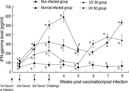
Fig. 7 showed IFN-γ concentrations of different groups compared within the same week. In the control, non-infected group, IFN-γ was in a plateau manner and recorded the lowest level in all weeks along the experiment. However, the IFN-γ level of the normal infected group increased from week 1 to 3 PI, and slightly decreased till week 5 PI. In UV-30 group, the level of IFN-γ increased from week 1 to 3 PV; then it decreased at weeks 4 and 5, and re-increased at week 6 and reached its maximum at week 7 PV. On the other hand, the IFN-γ level of UV-60 group increased from week 1 PV until reached the maximum level at week 4, and then decreased sharply at weeks 5 and 6 and re-increased at weeks 7 and 8 PV.
In weeks 1 and 2 PV, there was no significant difference in the IFN-γ levels among the all groups (P > 0.05). The IFN-γ level of UV-60 group was significantly higher than those of all the other groups (P < 0.01) except for UV-30 group at weeks 6 and 7 PV. The IFN-γ level of UV-30 group was significantly lower than those of UV-60 group and the infected group (P < 0.01) along the experiment except at weeks 6 and 7 PV.
DISCUSSION
The present study showed that the prevalence of natural infection of the sheep esophagus muscles with sarcocysts was 95%, and lambs showed lower prevalences than old sheep. Similar results were reported by Ford [
12] and Bashtar et al. [
13]. They found that the prevalence were 100% in sheep in South Australia and 88-98% in sheep at Aswan province, Egypt.
Each species of
Sarcocystis has a characteristic primary cyst wall structure distinguishing it from all other species occurring in the same host [
14,
15]. In the present study, the cyst wall was thick and striated as same as that of S. ovicanis described by Heydron et al. [
16]. No secondary cyst wall was observed in our study which was in agreement with Bashtar et al. [
17], and this supported the suggestion that the tissue surrounding the parasitized cells showed no significant morphological reactions to the parasite [
18]. The components of sarcocysts were separated by fine branches of the ground substances that occur in the cysts of
Sarcocystis and
Frenkelia [
19]. Similar results were recoded in the present study. The measurements of the present sarcocysts had the same range of sarcocysts reported by Bashtar et al. [
13].
In lambs of UV-30 group and UV-60 group, cysts were found in the skeletal muscles and appeared detached from the muscle fibers and smaller in size than normal cysts. The abnormalities and the deformation of sarcocysts in the UV-60 group were higher than in UV-30 group which may have been due to vaccination by UV-exposed sporocysts but not from a challenge infection. This assumption was confirmed by the result of the normal infected group as no cyst was formed in such group at week 5 PI. This result was also supported by the study of Bashtar et al. [
13] and Dubey et al. [
20] as they found that the beginning of the cyst formation, only metrocytes within the young cyst, were found at day 35 PI and the mature cysts were found at day 60 PI. The abnormal cysts that were formed in UV-30 and UV-60 groups might have been due to the effects of UV radiation on sporocysts inoculated to these groups. These cysts could be attributed to harmful effects of UV-light on the living organisms resulting from the initiation of photochemical absorption by molecules of biological significance.
The major affected molecules by UV were the nucleic acids (DNA and RNA), proteins, and other molecules [
21,
22]. The nucleotide bases represent the chromophores (absorbing centers) within nucleic acids. Among the nucleic acid bases, pyrimidines turned out to be about 10 times as sensitive to UV-light as purines [
23]. The major photoproducts following the absorption of UV-photons by DNA are pyrimidine derivatives [
24]. These photoproducts are cyclobutyl-type dimers, such as pyrimidines dimers, pyrimidines adducts (spore phtoproducts), pyrimidines hydrates, and DNA-protein cross links [
22]. Consequently, UV-light can damage the DNA molecule, which holds the genetic information in cells, and the RNA molecule, which was important in translating genetic information. Both of these molecules absorb energy in the specific UV wave length (maximum 254-265 nm) and build molecular bridges that destroy their biological functions. The cells have some ability to repair the damage by photo-reactivation, post-replication, and excision repair [
25,
26]. However, certain damage is more serious, permanent, cannot be repaired, and thus can affect the survival and development of the organism [
27].
It was found that the exposure to radiation-attenuated infective stages remains one of the best methods for eliciting consistently high levels of protective immunity against many protozoan parasites [
28]. Therefore, the present study was designed to evaluate vaccination against
Sarcocystis infection by using UV-attenuated (oocysts) sporocysts. The choice of UV-light was due that UV-light attenuation was cheaper and more convenient to perform in comparison with γ- or X-ray irradiation [
29]. The data of the present study revealed that lambs infected with normal sporocysts showed clinical manifestations of acute sarcocystosis at week 5 PI, and lambs were expected to die from acute sarcocystosis at this week. However, lambs of vaccinated groups survived healthy until sacrificed at week 5 post-challenge infection, which may be due that the vaccinated lambs developed protective immunity against a challenge infection.
The UV-60 group showed a highly significant serum level of IFN-γ (a Th1 associated cytokine) than the other groups. This indicated that immunized lambs with 60 min attenuated sporocysts stimulated production of high levels of IFN-γ than other groups that was an indication for a predominant Th1 response. Such Th1 responses may have a protective effect against the challenge infection. Other studies proved that CD8
+ T cells and IFN-γ production by Th1 cells (a subset of CD4
+ T cells) protect against chronic
Toxoplasma infection and against challenge infections in previously immunized mice [
30].
The present study showed that the total leukocyte count of UV-60 group was around the normal level at weeks 1 and 2 PV, but decreased below the normal level from week 3 to 7, then back to the normal level at week 8 PV. This significant decrease might be due to the infection which stimulated migration of leukocytes from the peripheral blood toward the tissue where the parasite was found or to the regional lymphoid tissues, and hence the total leukocyte count dropped below the normal level. The percentage of the lymphocytes which is responsible for mediating immunity against the parasite increased especially from week 1 to 4 PV in UV-60 group. Our data showed a correlation between IFN-γ level and lymphocyte percentage in UV-60 and UV-30 groups. This might be due to the elevated level of IFN-γ stimulates Th1 response that produced a high level of IL-2 and IFN-γ. IL-2 enhanced mitogenesis of T cells that led to an increase in the number of lymphocytes. Also, IFN-γ induced a predominated cell-mediated immune response which was the most effective mechanism in combating intracellular parasites. On the other hand, the percentage of monocytes in the blood of UV-60 decreased to below the normal level due to the monocytes that were drawn from the blood toward the infected tissue and transformed into macrophages to attack the parasite which found mainly in the tissues.
In conclusion, the present study addressed the prevalence of S. ovicanis in sheep, Ovis ammon aries, at Beni-Suef Governorate, Egypt. Also, the study induced protective immunity against S. ovicanis using UV-attenuated sporocysts which accompanied with a high level of IFN-γ
References
Fig. 1-5Fig. 1. Longitudinal section through a normal mature cyst (C) of Sarcocystis ovicanis in the sheep muscle (M) surrounded by a cyst wall (arrows). Scale bar = 10 µm. Fig. 2. Cross section through a normal cyst in the sheep muscle showing the thick striated cyst wall and merozoites. Fig. 3-5. Scale bar = 10 µm. Sections through abnormal cysts (C) detached from the surrounding muscles of experimentally challenged lambs after infection with attenuated sporocysts. Scale bar = 10 µm.
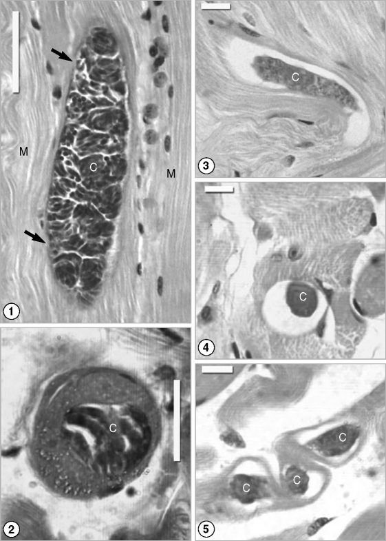
Fig. 6Total leukocyte counts of different groups. Blood was collected weekly and diluted at 1 : 20 in Turk's solution. Leukocytes were count under × 40 light microscope using improved Neubauer hemocytometer. Data are expressed as mean ± SD of 3 lambs per each group. Data of the different groups are compared in the same week. Values not sharing common superscripts denote significant differences.
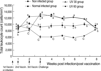
Fig. 7INF-γ concentrations of different groups. Blood was collected weekly and IFN-γ level was measured in serum by sandwich ELISA. Data are expressed as mean ± SD of 3 lambs per each group. Data of different groups are compared in the same week. Values not sharing common superscripts denote significant differences.

Table 1.Differential leukocyte counts of non-infected and infected lambs with normal or attenuated sporocysts of Sarcocystis ovicanis
Table 1.
|
|
Groups |
Non-infected group |
Normal infected group |
UV-30 group |
UV-60 group |
F-probability |
F-value |
|
Weeks |
|
|
Lymphocyte % |
1 |
52 ± 2b
|
54 ± 0.5b
|
53 ± 1b
|
61 ± 1a
|
P < 0.01 |
27 |
|
2 |
51 ± 3c
|
50 ± 1c
|
56 ± 0.5b
|
64 ± 1.5a
|
P < 0.01 |
34 |
|
3 |
50 ± 1.5b
|
48 ± 1.5b
|
49 ± 2.6b
|
84 ± 0.5a
|
P < 0.01 |
317 |
|
4 |
52 ± 3b
|
49 ± 1b
|
49 ± 1b
|
63 ± 2a
|
P < 0.01 |
33.9 |
|
5 |
50 ± 1.5c
|
53 ± 1.5b,c
|
61 ± 2.5a
|
54 ± 1.5b
|
P < 0.01 |
18.2 |
|
6 |
51 ± 1.7b
|
ND |
60 ± 1a
|
53 ± 1.5b
|
P < 0.01 |
30.9 |
|
7 |
53 ± 2a
|
ND |
50 ± 0.5a
|
50 ± 1.5a
|
P > 0.05 |
2 |
|
8 |
51 ± 2b
|
ND |
52 ± 0.5b
|
71 ± 1a
|
P < 0.01 |
188 |
|
Neutrophil % |
1 |
39 ± 3a
|
39 ± 1a
|
38 ± 1a
|
34 ± 1b
|
P < 0.05 |
5.2 |
|
2 |
44 ± 0.5a
|
45 ± 1.7a
|
40 ± 1b
|
33 ± 1.5c
|
P < 0.01 |
56 |
|
3 |
43 ± 2.6b
|
48 ± 0.5a
|
49 ± 2.8a
|
14 ± 1c
|
P < 0.01 |
215.6 |
|
4 |
42 ± 2b
|
49 ± 1a
|
46 ± 1.7a
|
33 ± 1.5c
|
P < 0.01 |
52.4 |
|
5 |
42 ± 0.5a
|
43 ± 1.5a
|
31 ± 3.5b
|
42 ± 0.5a
|
P < 0.01 |
24.2 |
|
6 |
43 ± 3a
|
ND |
32 ± 2.5b
|
42 ± 1a
|
P < 0.01 |
14.7 |
|
7 |
39 ± 2b
|
ND |
41 ± 0.5a,b
|
44 ± 2.6a
|
P < 0.05 |
5.2 |
|
8 |
42 ± 3a
|
ND |
38 ± 1.7b
|
26 ± 1.5c
|
P < 0.01 |
39.5 |
|
Monocyte % |
1 |
6 ± 0.5b
|
4 ± 0.5c
|
8 ± 0.5a
|
3 ± 0c
|
P < 0.01 |
62.5 |
|
2 |
4 ± 0.5a
|
3 ± 0.5b
|
2 ± 0.5b
|
2 ± 0.5b
|
P < 0.01 |
16.25 |
|
3 |
4 ± 0.5a
|
3 ± 1b
|
1 ± 0.5b
|
1 ± 0.5b
|
P < 0.01 |
12.7 |
|
4 |
4 ± 1.5a
|
1 ± 0b
|
4 ± 0.5a
|
2 ± 1b
|
P < 0.01 |
9.3 |
|
5 |
6 ± 1a
|
3 ± 0c
|
4 ± 0.5b
|
3 ± 0.5c
|
P < 0.01 |
21.3 |
|
6 |
5 ± 2a
|
ND |
6 ± 0.5a
|
4 ± 0.5a
|
P > 0.05 |
2.4 |
|
7 |
6 ± 0.5b
|
ND |
8 ± 0a
|
4 ± 0.5c
|
P < 0.01 |
37.9 |
|
8 |
5 ± 0.5b
|
ND |
8 ± 1a
|
2 ± 0c
|
P < 0.01 |
64.7 |
|
Eosinophil % |
1 |
3 ± 0.5a
|
3 ± 0.5a
|
2 ± 0.5a
|
2 ± 0.5a
|
P > 0.05 |
3.33 |
|
2 |
1 ± 0.5b
|
2 ± 0a
|
2 ± 0a
|
1 ± 0.5b
|
P < 0.01 |
16.6 |
|
3 |
3 ± 1a
|
1 ± 0a
|
1 ± 0.5a
|
1 ± 0.5a
|
P > 0.05 |
2.6 |
|
4 |
2 ± 0.5a
|
1 ± 0.5b
|
1 ± 0.5b
|
2 ± 0.5a,b
|
P < 0.05 |
6 |
|
5 |
2 ± 0.5a
|
1 ± 0b
|
2 ± 0.5a
|
1 ± 0b
|
P < 0.01 |
9.2 |
|
6 |
1 ± 0.5a
|
ND |
2 ± 0.5a
|
1 ± 0.5a
|
P > 0.05 |
4.3 |
|
7 |
2 ± 0.5a
|
ND |
1 ± 0.5b
|
2 ± 0.5a
|
P < 0.05 |
6 |
|
8 |
2 ± 1a
|
ND |
2 ± 0.5a
|
1 ± 0.5a
|
P > 0.05 |
4.2 |