Abstract
Primary renal echinococcosis, a rare disease involving the kidney, accounts for 2-3% of human echinococcosis. A 64-year-old female patient from Uzbekistan presented with complaints of left flank pain. A CT scan revealed a cystic mass in the upper to midpole of the left kidney. We regarded this lesion as a renal malignancy and hand-assisted laparoscopic radical nephrectomy was performed to remove the renal mass. The mass consisted of a large unilocular cyst and multiple smaller cysts without any grossly visible renal tissue. The final pathologic diagnosis was a renal hydatid cyst. For patients from endemic areas, hydatid cyst should be included in the differential diagnosis. Here, we present a case of renal hydatid cyst in a female patient who relocated from Uzbekistan to Korea.
-
Key words: Echinococcosis, kidney, nephrectomy
INTRODUCTION
Hydatid cyst of the kidney is caused by the larval stage of
Echinococcus granulosus [
1]. Endemic areas are located in the Middle East Asia, South America, Australia, New Zealand, and Alaska, where people raise sheep and cattle. Hydatid disease is a major health problem in many developing countries [
2]. Because of an expanding economy, Korea has become a labor importing country. The number of foreign nationals has significantly increased in Korea in recent years. In 1988, the total number of legally registered foreign nationals was approximately 6,500; this has increased to over 1.2 million people according to 2010 estimates. Even though Korea is not an endemic area for hydatid disease, the number of people from such endemic areas is increasing continuously.
Humans are an intermediate host of
E. granulosus through ingestion of water or vegetables contaminated by its eggs [
3]. Usually, it takes 3-6 years for the hydatid cyst to grow up to the size of a hen's egg, so there is an adequate delay for clinical manifestations to appear in a new local if people relocate from an endemic area.
Echinococcus, usually called the flat worm, is classified as a member of the Taeniidae family. It is approximately 5 mm long and the adult resides in the bowel lumen of the final host or lumen of the dog. The adult worm lives in the proximal jejunum of the definitive host and attaches to the mucosa using hooklets. Its eggs are excreted in the host's feces and when ingested by an appropriate intermediate host, such as sheep, cattle, or humans, the embryos break out from the eggs, then penetrate the intestinal mucosa and enter the systemic circulation [
4]. Once the parasitic embryo passes through the intestinal wall, it can reach the portal venous or the lymphatic system. The liver plays an important defensive role and is the most commonly involved site (75%), subsequently there is pulmonary involvement (15%), which acts as the second site of involvement for hydatid cysts. Other systemic dissemination may occur at almost any anatomical location in the human body. However, primary renal hydatid cysts are very rare [
5,
6]. From a 10-year case review of 144 renal hydatid cysts in Tunis, one of the most affected endemic areas, patients ranged in age from 8 to 65 years, with a mean age of 35 years. Women were more affected than men (87 vs 57) because of prolonged exposure to domestic animals at home [
3]. It is assumed that the cysts pass through the portal system into the liver and retroperitoneal lymphatics to reach the kidneys. The most frequent clinical manifestations are abdominal pain and hydaturia as a result of rupture of the cyst into the collecting system of the patient, typically passing collapsed daughter cyst-like material in the urine [
7]. Fever, lumbar mass, and hematuria are also major complaints [
8].
The differential diagnosis of renal hydatid cysts is difficult even in endemic areas. Radiologic studies are suggestive, but usually inconclusive, because the usual findings of complicated cysts in renal echinococcosis can mimic renal malignancies or a benign ureteropelvic junction obstruction. Here, we report a rare case of a renal hydatid cyst in a female patient from Uzbekistan that was preoperatively misinterpreted as a renal cell carcinoma.
CASE RECORD
A 64-year-old female patient presented with complaints of a 2 month history of left flank pain. The nature was slow onset dull, aching pain. There was no history of fever, hematuria, or pyuria. Upon admission, vital signs were normal and the patient was stable. The patient's medical history was unremarkable. Previously, she was living in a rural area of Uzbekistan and working as a housekeeper. She had no history of past radiation exposure or renal stones. Physical examination revealed mild costovertebral angle tenderness, but there was no palpable left flank mass. Routine hematology and biochemical tests revealed a mild leukocytosis (13,200 mm3), but there was no eosinophilia (5%). Urinalysis and urine cytologic examination were unremarkable.
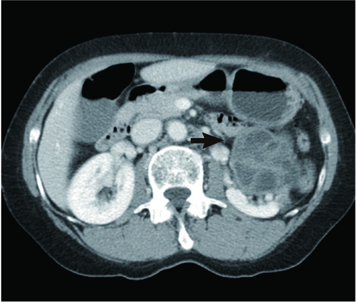
A computed tomography scan of the abdomen and pelvis demonstrated a 6.2-cm left renal cystic lesion in the upper to midpole of the kidney with soft tissue infiltration to anterior perirenal fascia. The cyst had irregular wall thickening and heterogeneous attenuation (
Fig. 1). The right kidney and ureter were normal and there was no ascites or lymphadenopathy. Other organs were without definite abnormalities.
This lesion was thought to represent a renal malignancy, leading to a decision to perform a nephrectomy. A 7-cm supraumbilical midline incision was made for a hand port and a camera was placed through a 10-mm umbilical port. A 10-mm working port for introduction of the operating instrument was made in the midclavicular line 4 fingers above the level of the umbilicus.
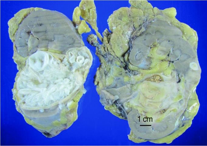
The surgical approach to the kidney was prepared, and the perinephric space was entered by incision into Gerota fascia on the lateral aspect of the kidney. The renal artery was divided into 2 branches and 1 vein and the major vessels were clamped with a hemolock. For specimen removal, a lap sac was utilized. The specimen was extracted and sent to the pathology department. A drain was brought out through a separate stab incision. The kidney specimen measured 12.2×7×5.2 cm in size. The entire mass was a large unilocular cyst with multiple smaller associated cysts without any grossly apparent renal parenchyma (
Fig. 2).
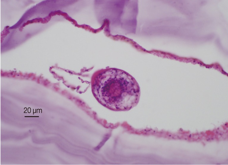
At ×40 magnification, the specimen demonstrated atrophic granulomatous inflammation and fibrosis of the kidney. The outermost pericystic layer was composed of fibro-collagenous tissue along with an eosinophilic infiltrate and the innermost germinal layer was apparently comprised of degenerated brood capsules (
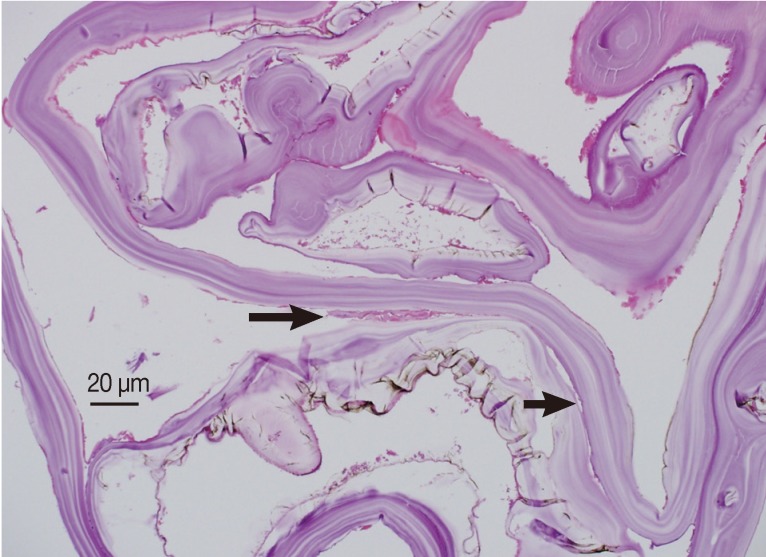
Fig. 3). At ×400 magnification, multiple protoscolex-like structures were found inside of the cyst (
Fig. 4). Following nephrectomy, the intermittent pain and tenderness resolved.
DISCUSSION
Preoperative diagnosis of hydatid cysts could result from various investigations, including intravenous pyelography (IVP), ultrasonography, and serology tests. Computerized scans are indicated when a clarification of the differential diagnosis of solid tumors is required. Usually, renal hydatid cysts present as a single large cyst with smaller daughter cysts of varying sizes. In our case, ultrasonography and CT scan showed a typical daughter cyst within the 6 cm sized mother cyst.
Serology is generally useful in the diagnosis of hydatid disease. However, the sensitivity of serological tests is influenced by the site and maturation of the hydatid cysts. Hydatid cysts in human lungs, spleen, or kidney tend to be associated with lower serum antibody levels [
9]. Eosinophilia is reported in 25-50% of hydatid disease cases and may occur in other parasitic diseases [
10].
Generally, surgery is the treatment of choice for renal hydatid cysts. Renal sparing or total nephrectomy is available irrespective of the surgical method, so laparoscopic removals of renal hydatid cysts have been reported [
8]. If preoperative diagnosis of a hydatid cyst is made, the area around the cyst can be carefully isolated by gauze packs and initial cyst aspiration and replacement of the cystic liquid with scolicide can be performed. A second technique, the so called partial cysto-pericystectomy is also an available option: the cyst, including the hydatid membrane and the daughter cysts, are opened and removal of the laminated membrane with scolicide-soaked swabs is performed. The margin of the remnant pericyst tissue is then sutured by running absorbable sutures [
5]. As a general rule, nephrectomy must be reserved for a non-functioning kidney as a renal hydatid cyst is a benign infection. So, in localized cases, focal or partial resection would be better option if a preoperative diagnosis is possible. In cases of a suspected renal mass, complete removal of a definite lesion is the first priority in non-endemic areas, such as Korea. Even though the patient was from Uzbekistan, we did not consider a hydatid cyst as a diagnostic option.
In a Tunisian study of 175 renal masses, hydatid cysts were found in 32% of cases, malignant tumors in 36.5%, simple cysts in 27%, and benign tumors in 4.5%. If the possibility of a hydatid cyst were considered in the present case, a different approach could have been considered [
11]. Hydatid cysts at unusual sites, such as the kidney, especially in non-endemic areas, can lead to difficulties in the diagnosis and management of this unfamiliar parasite, possibly leading to complications, such as an acute surgical emergency or a chronic illness leading to morbidity. Further, inadequate or over treatment may happen even in a controlled situation.
Although hydatid cysts of the kidney are relatively rare, this disease must be considered in people with renal cystic masses from endemic countries. A multimodality approach, which includes clinical history, hematology, serology, ultrasonography, IVP, or CT, with histo-pathological confirmation is required for optimal management. The diagnosis of a renal hydatid cyst is based primarily on imaging findings. In Korea, it is uneasy to differentiate a renal hydatid cyst from other renal diseases, because a hydatid cyst is not a familiar disease to Korean clinicians. Actually, there was 1 renal hydatid cyst case reported in Korea. In this case, CT scan shows the simple cyst lesion with a daughter cyst and slight enhancement of the cystic wall [
12]. So, laparoscopic cyst marsupialization and local excision was possible differently from our case.
Korea is becoming more multi-ethnic because of various economic and socio-political factors. Therefore, clinicians in Korea may encounter a greater number of echinococcosis patients. A total of 33 echinococcosis cases have been reported in the literature based on the diagnosis of the parasite; these patients included 25 Koreans, 7 Uzbeks, and 1 Mongolian [
9,
13]. With the exclusion of 2 Korean patients in whom the origin of infection is unclear, the remaining 31 patients are considered as imported echinococcosis. The areas of acquisition were primarily the Middle East and other endemic regions [
9,
13]. Furthermore, more Koreans travel or work abroad, and imported cases are increasing steadily. Finally, for an enlarged kidney with a renal mass, especially in someone from an endemic area, hydatid cyst should be included in the differential diagnosis, even though Korea is not an endemic area.
2011-0020128
Soonchunhyang University
Notes
-
We have no conflict of interest related to this study.
ACKNOWLEDGMENTS
This work was supported by a National Research Foundation of Korea (NRF) grant funded by the Korea Government (MSIP) (No. 2011-0020128) and by the Soonchunhyang University Research Fund.
References
- 1. Bhandarwar AH, Tayade MB, Makki K, Mhaske S. Primary renal hydatid cyst. Bombay Hosp J 2009;51:401-404.
- 2. Vuitton DA. The WHO informal working group on echinococcosis. Coordinating Board of the WHO-IWGE. Parassitologia 1997;39:349-353.
- 3. Fazeli F, Narouie B, Firoozabadi MD, Afshar M, Naghavi A, Ghasemirad M. Isolated hydatid cyst of kidney. Urology 2009;73:999-1001.
- 4. Sayilir K, Iskender G, Ogan C, Arik AI, Pak I. A case of isolated renal hydatid disease. Int J Infect Dis 2009;13:110-112.
- 5. Horchani A, Nouira Y, Kbaier I, Attyaoui F, Zribi AS. Hydatid cyst of the kidney. A report of 147 controlled cases. Eur Urol 2000;38:461-467.
- 6. Kumar SA, Shetty A, Vijaya C, Geethamani V. Isolated primary renal echinococcosis: a rare entity. Int Urol Nephrol 2013;45:613-616.
- 7. Sountoulides P, Zachos I, Efremidis S, Pantazakos A, Podima-tas T. Nephrectomy for benign disease? A case of isolated renal echinococcosis. Int J Urol 2006;13:174-176.
- 8. Mongha R, Naraya S, Kundu AK. Primary hydatid cyst of kidney and ureter with gross hydatiduria. A case report and evaluation of radiological features. Indian J Urol 2008;24:116-117.
- 9. Byun SJ, Moon KC, Suh KS, Han JK, Chai JY. An imported case of echinococcosis of the liver in a Korean who traveled to western and central Europe. Korean J Parasitol 2010;48:161-165.
- 10. Afsar H, Yagci F, Meto S, Aybasti N. Hydatic disease of the kidney: evaluation and features of diagnostic procedures. J Urol 1994;151:567-570.
- 11. Wani R, Wani I, Malik A, Parray FQ, Wani AA, Dar AM. Hydatid disease at unusual sites. Int J Case Rep Images 2012;3:1-6.
- 12. Jeon SH, Kim TH, Lee HL. Laparoscopic treatment of isolated renal hydatid cyst. Korean J Urol 2007;48:555-557.
- 13. Ahn KS, Hong ST, Kang YN, Kwon JH, Kim MJ, Park TJ, Kim YH, Lim TJ, Kang KJ. An imported case of cystic echinococcosis in the liver. Korean J Parasitol 2012;50:357-360.
Fig. 1A cystic renal mass (arrow) at the left kidney upper pole, 6.2 cm in size with multiseptation, showing irregular wall thickening, heterogeneous attenuation, and soft tissue infiltration.

Fig. 2A large unilocular cyst in the upper kidney with multiple smaller associated cysts without any grossly apparent renal parenchyma.

Fig. 3Microscopic findings of the cystic region in the kidney. At ×40 magnification, atrophic granulomatous inflammation (long arrow) and fibrosis (short arrow) are seen (H&E stain).

Fig. 4Microscopic findings inside the cyst showing multiple protoscolex-like structures (H&E stain. ×400).


