Abstract
Toxoplasma gondii, a common protozoan parasite, poses significant public health risks due to its potential to cause toxoplasmosis in humans and can be contracted from pigs, which are considered its critical intermediate host. The aim of this study is to evaluate the prevalence of T. gondii in slaughtered pigs for human consumption, emphasizing the zoonotic implications and the need for improved biosecurity and monitoring practices in pig farming. A total of 1,526 pig samples (1,051 whole blood samples and 384 lung tissue samples from the local slaughterhouse and 91 aborted fetus samples from local farms) were collected throughout the whole country of Korea in 2020. Among them, 6 (0.4%) were found to be infected with T. gondii by nested PCR. When compared by sample type, the prevalence of T. gondii was significantly higher in the aborted fetus samples (2.2%, 2/91) than in the blood (0.3%, 3/1,051) and lung tissue samples (0.3%, 1/384). The B1 gene sequence of T. gondii was similar (97.9–99.8%) to that of the other T. gondii isolates. This study represents the first molecular genotyping survey of T. gondii in the lung tissue of fattening pigs and aborted fetuses in Korea. Our findings indicated the importance of adopting preventive measures including the implementation of rigorous farm hygiene protocols and the promotion of public awareness about the risks of consuming undercooked pork. By addressing the gaps in current control strategies and encouraging the One Health approach, this study contributes to the development of more effective strategies to mitigate the transmission of T. gondii from pigs to humans, ultimately safeguarding public health.
-
Key words: Toxoplasma gondii, pig, molecular epidemiology, genotyping, Korea
Introduction
Toxoplasmosis, a zoonosis with a global distribution, is estimated to infect approximately one-third of the world’s human population [
1].
Toxoplasma gondii has consequently emerged as the most significant protozoan foodborne pathogen, which is mostly contracted through meat consumption [
2]. A primary mode of human transmission involves the ingestion of raw or undercooked meat from various animals, particularly pigs. Pigs can contract
T. gondii by consuming food or water contaminated with sporulated oocysts or ingesting cysts in the tissues of infected animals (e.g., rodents, birds, and other pigs) or through congenital transmission [
3].
Effective management and monitoring of
T. gondii infection are important to implement in public health programs. In Korea, surveillance programs for
T. gondii infection in pigs at slaughter are limited, and the suitability of
Toxoplasma-infected meat for human consumption remains to be unmonitored. In contrast, the European Food Safety Authority has recognized
T. gondii as a significant biological hazard. It advocates for the inclusion of
T. gondii in the revised meat inspection regulations, which include not only pigs but also other animals, such as sheep, goats, farmed deer, and farmed wild boar [
4].
Numerous seroprevalence studies on
T. gondii in pigs (1.8–38.3%) were conducted in Korea between 2007 and 2018 [
5–
8]. Despite this, only a few studies have focused on the molecular surveillance of
T. gondii within the country’s pig population. Given the limited information on the molecular detection of
T. gondii in pigs designated for human consumption, the present study aims to genetically identify the infectious strains of
T. gondii as well as gather epidemiological data concerning
T. gondii in both intensively raised fattening pigs and aborted fetuses throughout Korea’s pig farming regions. Through molecular analysis, this study identified and characterized the genotypes of
T. gondii that are prevalent on these farms. Furthermore, this study presents our efforts to molecularly delineate the strains of
T. gondii circulating in Korea’s domestic pig population.
Materials and Methods
Ethics statement
This study, which was conducted in 2020, was beyond the purview of the Institutional Animal Care and Use Committee (IACUC) at Kyungpook National University (KNU) because the IACUC at KNU only evaluates studies that involve laboratory animals maintained in indoor facilities and does not regulate research involving outdoor animals. Clinically healthy pigs were slaughtered for pig meat at the local slaughterhouse, and the blood and lung tissue samples were collected at that time. The aborted fetus samples were submitted to KNU for diagnostic investigation.
Sample size determination and collection
In 2020, 11,078,032 pigs from a total of 6,078 farms were reared in Korea [
9]. The sample size was determined using the following formula with an expected disease prevalence of 10%, accepted absolute error of 5%, and confidence level of 95% with a simple random sampling design [
10]:
where n=required sample size, pexp=expected prevalence, and d=desired absolute precision.
Based on the formula, a minimum of 138 samples was required; however, 1,526 pig samples (1,051 whole blood samples and 384 lung tissue samples from the local slaughterhouse from clinically healthy pigs and 91 aborted fetus samples from local farms) were selected throughout the entire country in 2020. The regions were classified as the northern, central, and southern regions and Jeju Island. In the northern region, the samples were collected from Gyeonggi and Gangwon Province. In the central region, collection was performed in Chungcheong Province. The samples in the southern region were collected from Gyeongsang and Jeolla Province. Finally, the remaining samples were collected from Jeju Island. Data on the regions, sample types, seasons, and farm sizes were recorded for subsequent analysis.
DNA extraction and PCR detection
Genomic DNA was extracted using the DNeasy Blood & Tissue Kit (Qiagen, Hilden, Germany) in accordance with the manufacturer’s instructions. Nested PCR (nPCR) was performed using the AccuPower HotStart PCR Premix Kit (Bioneer, Daejeon, Korea). nPCR was performed to detect
T. gondii by amplifying the B1 gene with external (Tg1 and Tg2) and internal (Tg3 and Tg4) primers that generated a 531-bp amplicon, as previously described [
11]. A sample of
T. gondii isolated from cats in Korea [
12] was used as the positive control, whereas a sample lacking a DNA template was used as the negative control.
The amplicons from the infected animals were purified using the QIAquick Gel Extraction Kit (Qiagen), ligated into the pGEM-T Easy vector (Promega, Madison, WI, USA), transformed into Escherichia coli DH5α-competent cells (Thermo Fisher Scientific, Wilmington, DE, USA), and incubated at 37°C overnight. A plasmid DNA extraction was performed using a plasmid miniprep kit (Qiagen) in accordance with the manufacturer’s instructions.
Sequencing and phylogenetic analyses
Recombinant clones were selected and sent to Macrogen, Daejeon, Korea, for nucleotide sequencing. The sequences obtained in this study were aligned and analyzed using the CLUSTAL Omega multiple sequence alignment program Omega (v. 1.2.1 (Bioweb, Ferndale, WA, USA)), and the alignment was corrected using BioEdit (v. 7.2.5 (BioEdit Company, Manchester, UK)). Moreover, a phylogenetic analysis was subsequently performed using MEGA v. 7.0 (Mega software solutions, Madhurawadha, India) based on the maximum likelihood method with the Kimura 2-parameter distance model. The aligned sequences were analyzed using a similarity matrix. The stability of the trees obtained was then estimated via bootstrap analysis with 1,000 replicates.
Statistical analysis
The GraphPad Prism software (v. 5.04; GraphPad, La Jolla, CA, USA) was used for statistical analyses. The differences among the groups were analyzed using a chi-square test. A P-value of <0.05 was considered statistically significant. A 95% confidence interval (CI) was calculated for all estimates.
Results
Prevalence
Of the 1,526 pigs, 6 (0.4%; 95% CI, 0.1–0.7) tested positive for the
T. gondii B1 gene (
Table 1). With regard to region,
T. gondii was the most prevalent on Jeju Island (1.2%; 1/85; 95% CI, 0–3.5), followed by the southern region (0.6%; 3/484; 95% CI, 0–1.3) and central region (0.3%; 2/752; 95% CI, 0–0.6). However, none were detected in the northern region. When compared by sample type, the prevalence of
T. gondii was significantly higher in the aborted fetus samples (2.2%; 2/91; 95% CI, 0–5.2;
P=0.0179) than in the blood (0.3%; 3/1,051; 95% CI, 0–0.6) and lung tissue samples (0.3%; 1/384; 95% CI, 0–0.8). With regard to season,
T. gondii was prevalent during the summer (0.7%; 2/280; 95% CI, 0–1.7), followed by the other seasons, such as autumn (0.4%; 1/246; 95% CI, 0–1.2), winter (0.3%; 1/350; 95% CI, 0–0.8), and spring (0.2%; 1/650; 95% CI, 0–0.5). In terms of farm size,
T. gondii was prevalent on farms with fewer than 500 pigs (1.2%; 2/252; 95% CI, 0–2.5), followed by those with more than 500 but less than 2,000 pigs (0.4%; 2/562; 95% CI, 0–0.8) and with more than 2,000 pigs (0.1%; 1/712; 95% CI, 0–0.4).
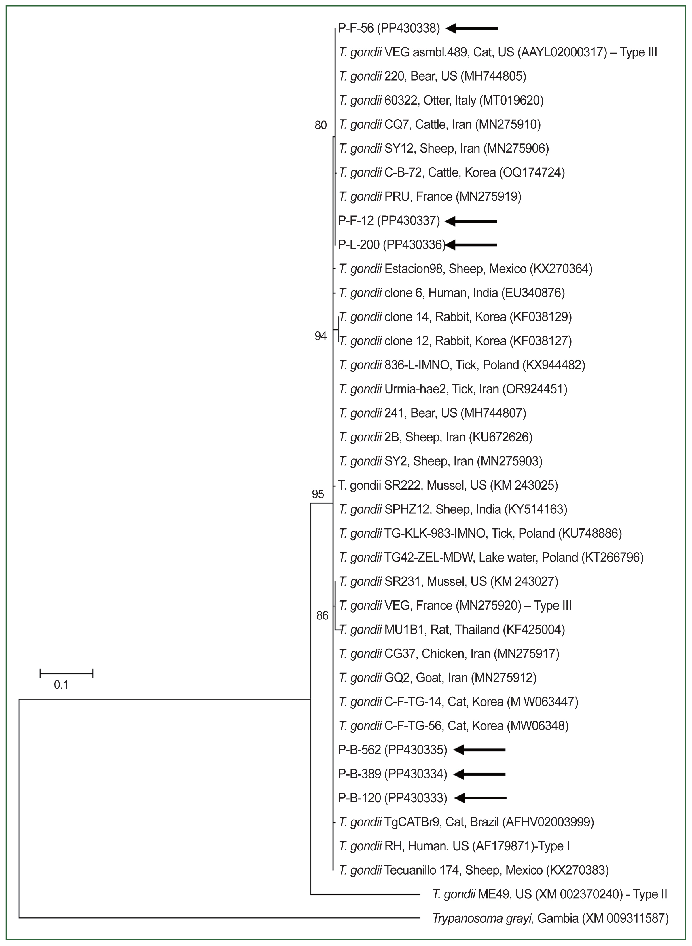
The phylogenetic analysis revealed that the B1 gene nucleotide sequences of
T. gondii identified in this study clustered with previously identified
T. gondii sequences (
Fig. 1). The 6 sequences of the
T. gondii B1 gene in this study shared a 99.4–100% identity. Moreover, they were 97.9–99.8% identical to the B1 gene sequences of previously reported
T. gondii isolates. All sequences used in the phylogenetic analysis were submitted to the GenBank database (accession numbers: PP430333–PP430338).
Discussion
In this study, we provided epidemiological and molecular insights into
T. gondii in slaughtered pigs and aborted fetuses in Korea. Our findings revealed a low prevalence of
T. gondii infections on swine farms. While the prevalence in pigs decreased, the seroprevalence among Korean residents reportedly increased in recent years [
13]; this is mainly attributed to the increased consumption of local or imported pork and other meats susceptible to
T. gondii infection [
2]. Moreover, asymptomatic animals that have
T. gondii cysts in their muscles may enter the fresh pork market and thus represent a significant foodborne toxoplasmosis risk and public health concern. In Poland,
T. gondii was found in retail raw meat products including sausages (45.1% of positives), smoked meats (27.4%), ham (8.0%), and minced meat (19.5%) [
14]. The results of these studies indicate that raw meat products could be a critical source of
T. gondii infection for humans. Therefore, the government should implement health education measures that encourage proper hygiene as well as proper cooking temperatures and methods to deactivate
T. gondii cysts in meats to prevent human infections caused by contaminated meat [
15].
In Korea, studies on
T. gondii infection in pigs have included various investigations. On Jeju Island in 2002, dead sows (100%, 2/2) and aborted fetuses from the same litter (60%, 3/5) tested positive for
T. gondii via histopathology and immunohistochemistry, and sows that had aborted or normal sows (41.2%, 7/17) were evaluated using the latex agglutination test [
16]. In Gyeonggi Province in 2007, the latex agglutination test (22.8%, 118/516) and PCR test (57.6%, 68/118) were used [
7]. In the eastern areas of Gyeongbuk Province in 2008, the prevalence of
T. gondii infection was evaluated via ELISA (16.8%, 62/368) [
5], and in Gyeongnam Province in 2009, ELISA (38.3%, 115/300) and PCR (0%) were used [
6]. Finally, in 2018, local (9.1%, 53/583) and imported pork (1.8%, 7/386) from Korean retail meat markets were tested for
T. gondii using ELISA [
8]. These findings suggest that
T. gondii is widely distributed in pigs from several farms in Korea, reflecting a cumulative probability of exposure to
T. gondii and the lifelong persistence of antibodies.
With regard to sample type,
T. gondii showed a significant prevalence in aborted fetuses. Moreover,
T. gondii infections in pigs are often asymptomatic; however, several instances of clinical disease and mortality were recorded.
Toxoplasma gondii infection has been linked to reproductive failure in sows, manifesting as abortion, fetal mummification, stillbirth, and neonatal death [
17]. This presents significant clinical and economic implications for the agribusiness sector, making it a crucial area for research. Infections in aborted fetuses have also been reported in Korea [
16] and Switzerland [
17]. A previous study reported elevated abortion rates (up to 44%) and unusually high sow mortality rates (up to 19%) that were primarily attributed to toxoplasmosis in Korea [
16]. Moreover, the process of
T. gondii detection in the lung tissues in this study is consistent with that in studies focusing on piglets and fattening pigs in China [
18] as well as aborted fetuses in Korea [
16].
With regard to seasonal prevalence,
T. gondii was more commonly isolated during the summer than during other seasons. This finding is consistent with those of other studies [
5,
19]. The warmer temperatures and increased humidity that are typical during the summer season create conditions that favor the survival and proliferation of
T. gondii oocysts in environments including pig farms [
19]. Furthermore, the heightened interactions between farm environments and potential intermediate hosts, such as rodents, during the summer months could facilitate the spread of
T. gondii. These factors, even in predominantly indoor pig farming systems, can lead to higher infection rates during the summer, emphasizing the critical need for stringent biosecurity and hygiene practices throughout the year.
With regard to farm size, small farms have shown a higher prevalence of
T. gondii than large farms. This finding is consistent with those of other studies [
6,
20]. Small, family-run farms have been found to have an increased risk of
T. gondii infections, which is likely due to lower hygienic standards than those found in larger, intensive operations [
21]. Therefore, it is important to implement preventive and control measures for swine toxoplasmosis in farms to prevent pigs from coming into contact with sources of
T. gondii infection, such as cat feces and infected rodents [
22]. By adopting appropriate hygiene and preventive strategies, the risk of infection can be further minimized.
In a prior investigation,
Toxoplasma strains were categorized into 3 genotypes according to their virulence levels in outbred mice: Type I (highly virulent), Type II (moderately virulent), and Type III (non-virulent) [
23]. Type II is identified as the most common genotype in humans [
24]. Further research has revealed various
T. gondii types in pigs. For instance, all 3 genotypes have been distributed in Poland as follows: Type III (49%), Type II (17.3%), Type I (10.2%), and mixed genotypes (23.5%) [
14]. Moreover, molecular identification enhances our understanding of the risks associated with meat consumption because the impact of
T. gondii infection is influenced by host factors and the specific parasite lineages involved, leading to different clinical outcomes for each genotype [
25]. In this study, 6 pigs were confirmed to be positive for
T. gondii, while the analysis of the B1 gene, Type I/III was identified. However, additional phylogenetic studies are necessary to distinguish between Types I and III.
In the present study, we identified a relatively low T. gondii infection rate (0.4%) in pigs. This low rate may be due to diminished exposure to common sources of T. gondii infection, which includes enhancements in pig farming infrastructure and strong regulations. To our knowledge, this study represents the first molecular genotyping survey of T. gondii in the lung tissues of fattening pigs and aborted fetuses in Korea. Toxoplasmosis in pigs can cause substantial economic losses for pig farmers. Thus, it is crucial to educate pig farmers on proper hygiene practices to cultivate healthy and disease-free pigs, thereby mitigating economic losses and preventing foodborne zoonotic diseases. Furthermore, from a public health perspective, the establishment of regulatory measures and control strategies for foodborne toxoplasmosis in Korea is essential.
Notes
-
Author contributions
Conceptualization: Kwak D
Formal analysis: Seo MG
Investigation: Kwak D
Project administration: Seo MG
Supervision: Seo MG
Writing – original draft: Kwak D
Writing – review & editing: Seo MG
-
The authors declare no conflict of interest related to this study.
Acknowledgements
We would like to thank the pig farm owners or managers who supplied pig blood and lung tissue specimens for our research.
Fig. 1The phylogenetic tree of Toxoplasma gondii on the basis of the B1 gene sequences. The tree was constructed using the maximum likelihood method. The black arrows represent the sequences analyzed in this study. Trypanosoma grayi was used as the outgroup. The GenBank accession numbers of other sequences are presented with their names. The numbers on the branches represent the mean bootstrap support based on 1,000 replicates. The scale and scale bar indicate the phylogenetic distance.

Table 1Prevalence of Toxoplasma gondii in Korean pigs in 2020
Table 1
|
Group |
|
No. tested |
No. positive (%) |
95% CIa
|
χ2 (dfb) (P-valuec) |
|
Regiond
|
Northern |
205 |
0 |
0 |
3.086 (3) 0.3785 |
|
Central |
752 |
2 (0.3) |
0–0.6 |
|
|
Southern |
484 |
3 (0.6) |
0–1.3 |
|
|
Jeju Island |
85 |
1 (1.2) |
0–3.5 |
|
|
|
Sample type |
Blood |
1,051 |
3 (0.3) |
0–0.6 |
8.051 (2) 0.0179 |
|
Lung tissue |
384 |
1 (0.3) |
0–0.8 |
|
|
Aborted fetus |
91 |
2 (2.2) |
0–5.2 |
|
|
|
Season |
Spring |
650 |
1 (0.2) |
0–0.5 |
1.949 (3) 0.5831 |
|
Summer |
280 |
2 (0.7) |
0–1.7 |
|
|
Autumn |
246 |
1 (0.4) |
0–1.2 |
|
|
Winter |
350 |
1 (0.3) |
0–0.8 |
|
|
|
Farm size |
<500 |
252 |
3 (1.2) |
0–2.5 |
5.271 (2) 0.0717 |
|
500–2,000 |
562 |
2 (0.4) |
0–0.8 |
|
|
>2,000 |
712 |
1 (0.1) |
0–0.4 |
|
|
|
Total |
|
1,526 |
6 (0.4) |
0.1–0.7 |
|
References
- 1. Hill DE, Chirukandoth S, Dubey JP. Biology and epidemiology of Toxoplasma gondii in man and animals. Anim Health Res Rev 2005;6(1):41-61.
https://doi.org/10.1079/ahr2005100
- 2. Djurković-Djaković O, Bobić B, Nikolić A, Klun I, Dupouy-Camet J. Pork as a source of human parasitic infection. Clin Microbiol Infect 2013;19(7):586-594.
https://doi.org/10.1111/1469-0691.12162
- 3. García-Bocanegra I, Dubey JP, Simon-Grifé M, Cabezón O, Casal J, et al. Seroprevalence and risk factors associated with Toxoplasma gondii infection in pig farms from Catalonia, north-eastern Spain. Res Vet Sci 2010;89(1):85-87.
https://doi.org/10.1016/j.rvsc.2010.01.017
- 4. EFSA. Surveillance and monitoring of Toxoplasma in humans, food and animals1Scientific Opinion of the Panel on Biological Hazards. EFSA Journal 2007;583:1-64.
https://doi.org/10.2903/j.efsa.2007.583
- 5. Seo MG, Jang YS, Lee EM, Park NC, Kwak D. Prevalence of antibodies to Toxoplasma gondii in cattle and pigs reared in eastern areas of Gyeongbuk province. Korean J Vet Serv 2009;32(2):131-137. (in Korean).
- 6. Kim EG, Park HJ, Son BG, Jung MH, Heo JH, et al. Prevalence of Toxoplasma gondii infection from domestic pigs in Gyeongnam province. Korean J Vet Serv 2010;33(4):345-351. (in Korean).
- 7. Shim HS, Choi GM, Jeon OS, Lee SJ, Woo JT, et al. Investigation of swine toxoplasmosis by latex agglutination and polymerase chain reaction(PCR). Korean J Vet Serv 2008;31(1):87-91. (in Korean).
- 8. Yoo WG, Kim SM, Won EJ, Lee JY, Dai F, et al. Tissue fluid enzyme-linked immunosorbant assay for piglets experimentally infected with Toxoplasma gondii and survey on local and imported pork in Korean retail meat markets. Korean J Parasitol 2018;56(5):437-446.
https://doi.org/10.3347/kjp.2018.56.5.437
- 9. KSIS. Number of farms in households and animals in heads by type. Korean Statistical Information Service; 2017.
- 10. Thrusfield MV, Christley R. Veterinary epidemiology. Fourth ed. Wiley; Hoboken, USA. 2018.
- 11. Grigg ME, Boothroyd JC. Rapid identification of virulent type I strains of the protozoan pathogen Toxoplasma gondii by PCR-restriction fragment length polymorphism analysis at the B1 gene. J Clin Microbiol 2001;39(1):398-400.
https://doi.org/10.1128/jcm.39.1.398-400.2001
- 12. Kwak D, Seo MG. Genetic analysis of zoonotic gastrointestinal protozoa and microsporidia in shelter cats in South Korea. Pathogens 2020;9(11):894.
https://doi.org/10.3390/pathogens9110894
- 13. Jung J, Lee J, Chang YK, Ahn SK, Park SH, et al. Seroprevalence of Toxoplasma gondii assayed using rapid diagnostic tests among residents in three counties adjacent to the demilitarized zone, Korea. Korean J Parasitol 2021;59(1):9-14.
https://doi.org/10.3347/kjp.2021.59.1.9
- 14. Sroka J, Bilska-Zając E, Wójcik-Fatla A, Zając V, Dutkiewicz J, et al. Detection and molecular characteristics of Toxoplasma gondii DNA in retail raw meat products in Poland. Foodborne Pathog Dis 2019;16(3):195-204.
https://doi.org/10.1089/fpd.2018.2537
- 15. Jones JL, Dubey JP. Foodborne toxoplasmosis. Clin Infect Dis 2012;55(6):845-851.
https://doi.org/10.1093/cid/cis508
- 16. Kim JH, Kang KI, Kang WC, Sohn HJ, Jean YH, et al. Porcine abortion outbreak associated with Toxoplasma gondii in Jeju Island, Korea. J Vet Sci 2009;10(2):147-151.
https://doi.org/10.4142/jvs.2009.10.2.147
- 17. Basso W, Handke M, Sydler T, Borel N, Grimm F, et al. Involvement of Toxoplasma gondii in reproductive disorders in Swiss pig farms. Parasitol Int 2015;64(2):157-160.
https://doi.org/10.1016/j.parint.2014.11.017
- 18. Wang H, Zhang L, Ren Q, Yu F, Yang Y. Diagnosis of swine toxoplasmosis by PCR and genotyping of Toxoplasma gondii from pigs in Henan, Central China. BMC Vet Res 2017;13(1):152.
https://doi.org/10.1186/s12917-017-1079-3
- 19. Xu Y, Li RC, Liu GH, Cong W, Zhang XX, et al. Seroprevalence of Toxoplasma gondii infection in sows in Hunan province, China. ScientificWorldJournal 2014;2014:347908.
https://doi.org/10.1155/2014/347908
- 20. Sroka J, Karamon J, Wójcik-Fatla A, Piotrowska W, Dutkiewicz J, et al.
Toxoplasma gondii infection in slaughtered pigs and cattle in Poland: seroprevalence, molecular detection and characterization of parasites in meat. Parasit Vectors 2020;13(1):223.
https://doi.org/10.1186/s13071-020-04106-1
- 21. Papatsiros VG, Athanasiou LV, Stougiou D, Papadopoulos E, Maragkakis GG, et al. Cross-sectional serosurvey and risk factors associated with the presence of Toxoplasma gondii antibodies in pigs in Greece. Vector Borne Zoonotic Dis 2016;16(2):48-53.
https://doi.org/10.1089/vbz.2015.1845
- 22. Ortega-Pacheco A, Acosta-Viana KY, Guzman-Marin E, Uitzil-Álvarez B, Rodríguez-Buenfil JC, et al. Infection dynamic of Toxoplasma gondii in two fattening pig farms exposed to high and low cat density in an endemic region. Vet Parasitol 2011;175(3–4):367-371.
https://doi.org/10.1016/j.vetpar.2010.10.018
- 23. Howe DK, Sibley LD.
Toxoplasma gondii comprises three clonal lineages: correlation of parasite genotype with human disease. J Infect Dis 1995;172(6):1561-1566.
https://doi.org/10.1093/infdis/172.6.1561
- 24. Su C, Shwab EK, Zhou P, Zhu XQ, Dubey JP. Moving towards an integrated approach to molecular detection and identification of Toxoplasma gondii
. Parasitology 2010;137(1):1-11.
https://doi.org/10.1017/s0031182009991065
- 25. Maubon D, Ajzenberg D, Brenier-Pinchart MP, Dardé ML, Pelloux H. What are the respective host and parasite contributions to toxoplasmosis? Trends Parasitol 2008;24(7):299-303.
https://doi.org/10.1016/j.pt.2008.03.012