Abstract
IgA vasculitis (IgAV) and Strongyloides stercoralis infection may both present with abdominal pain and purpura; however, glucocorticoid, the treatment for IgAV, is a major risk factor for S. stercoralis hyperinfection syndrome and disseminated strongyloidiasis. To date, only 2 cases of IgAV with concurrent S. stercoralis infection have been reported globally, both in children. We report a 72-year-old man presenting with abdominal pain and purpura. Diagnosed with IgAV during this admission, he received glucocorticoid therapy. However, the patient’s condition did not improve as expected. Subsequently, S. stercoralis was detected in the patient's sputum and stool. We report the first adult case of IgAV with concurrent S. stercoralis infection, highlighting the importance of screening patients with IgAV for strongyloidiasis before initiating immunosuppressive therapy.
-
Key words: IgA vasculitis, Strongyloides stercoralis, severe strongyloidiasis, immunosuppressive therapy
Introduction
IgA vasculitis (IgAV) is a systemic small-vessel vasculitis characterized by a clinical tetrad that includes palpable purpura, arthralgia and/or arthritis, abdominal pain, and renal disease [
1]. Pathogens are known to induce IgAV. However, the relationship between IgAV and parasites has rarely been reported [
2].
Strongyloides stercoralis is a soil-transmitted helminth widely distributed in tropical and subtropical regions worldwide. Its clinical manifestations can involve the skin, respiratory system, and digestive system. Immunosuppressive conditions can cause life-threatening hyperinfection syndrome and disseminated strongyloidiasis [
3]. Given the similar clinical manifestations of IgAV and strongyloidiasis, and the use of glucocorticoids in IgAV [
4], we report the first case of IgAV complicated by
S. stercoralis infection in an adult, highlighting the importance of screening for strongyloidiasis in patients with IgAV.
Case Report
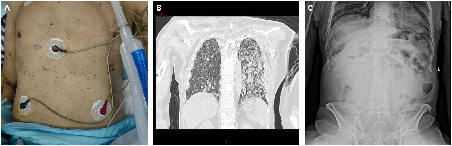
A 72-year-old man was admitted to the People’s Hospital of Guangxi Zhuang Autonomous Region presenting with a 10-day history of upper mid‑abdominal pain and nausea. He had a medical history of coronary heart disease, hypertension, and diabetes mellitus. Two months earlier, the patient had been evaluated at another hospital for a hepatic hilar mass, which was suspected to be caused by IgG4-related cholangitis. Treatment was initiated with oral methylprednisolone (glucocorticoid) 32 mg daily, with tapering by 4 mg every 2 weeks. He was admitted to the gastroenterology department for abdominal pain evaluation. Physical examination revealed extensive non-blanching purpura and epigastric tenderness without significant rebound (
Fig. 1A). Laboratory tests showed elevated inflammatory markers (C-reactive protein, 32.13 mg/L; normal range, 0–10 mg/L), hyperglycemia (blood glucose, 10.44 mmol/L; normal range, 3.9–6.1 mmol/L), and impaired cardiac function (high-sensitivity cardiac troponin, 39.13 ng/L; normal range, 0–14 ng/L); coagulation, liver and renal function, tumor markers, and autoimmune antibodies were all unremarkable. Fecal microscopy was not performed due to the patient's inability to defecate. Computed tomography imaging confirmed right lung inflammatory changes (
Fig. 1B), and abdominal radiography revealed incomplete bowel obstruction (
Fig. 1C). The presentation of typical palpable purpura with abdominal pain met the EULAR/PRINTO/PRES criteria for IgAV. Admission laboratory studies ruled out thrombotic thrombocytopenic purpura and other hemorrhagic rash disorders, as well as antineutrophil cytoplasmic antibody-associated vasculitis. Clinical and laboratory findings substantiated the diagnosis of IgAV, and methylprednisolone was initiated; concomitantly, right-sided pneumonia was treated with piperacillin-tazobactam, and incomplete bowel obstruction was managed by nasogastric decompression and therapeutic enemas.
On hospital day 7, the patient developed acute respiratory failure and was managed with endotracheal intubation and mechanical ventilation. Later the same day,
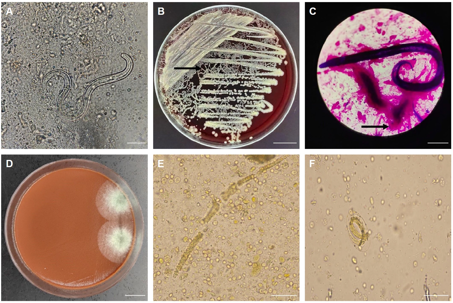
S. stercoralis were identified in sputum under microscopic examination (
Fig. 2A). Given the limited availability of ivermectin in China, albendazole was administered for the treatment of
S. stercoralis infection. Blood agar culture of sputum showed bacterial colonies arranged in numerous serpiginous tracts, suggestive of multiple live, motile parasites (
Fig. 2B). Gram staining of the smear prepared from the bacterial colony revealed the presence of Gram positive bacilli and
S. stercoralis under the microscope (
Fig. 2C). Bacteria were identified as
Corynebacterium striatum by MALDI TOF mass spectrometry (
Supplementary Fig. S1). The metagenomic next-generation sequencing of tracheal aspirate showed consistency with above identification (
Supplementary Table S1). On hospital day 9, the family obtained ivermectin from overseas. Ivermectin was subsequently administered to treat
S. stercoralis infection. On hospital day 11,
Aspergillus fumigatus was isolated from tracheal aspirate cultures (
Fig. 2D). On hospital day 13, stool examination demonstrated significant quantities of lifeless
S. stercoralis (
Fig. 2E) and Strongyloides-like eggs (
Fig. 2F). Unfortunately, the patient’s condition failed to improve. On hospital day 15, gastrointestinal bleeding was indicated by dark red gastric aspirate and melena. Emergency bedside endoscopy revealed diffuse bleeding of the rectal mucosa. On hospital day 16, the patient’s family chose automatic discharge due to the poor prognosis and multiple comorbidities. Regrettably, the patient died following discharge. Written informed consent for publication was obtained from the patient.
Discussion
S. stercoralis is a soil-transmitted helminth found worldwide. It is mainly prevalent in tropical and subtropical areas. The parasite has a complex life history. It parasitizes the human small intestine and reproduces by parthenogenesis. In the intestine, eggs hatch into non-infective rhabditiform larvae. Most of these larvae leave the host through feces and enter the environment. Some can develop directly into infectious filariform larvae in the intestine, leading to autoinfection. Autoinfection enables
S. stercoralis to persist within the host, potentially resulting in lifelong infection [
5]. A case report showed that an 83-year-old man had been infected with
S. stercoralis for 75 years [
6]. In this case, the patient reported soil exposure over a decade ago that likely led to chronic
S. stercoralis infection, which remained latent until worsening comorbidities and long-term corticosteroid use triggered hyperinfection.
IgAV is a multifactorial leukocytoclastic vasculitis characterized by IgA-dominant immune complex deposition within or around small vessels. It exhibits 4 clinical manifestations: non-thrombocytopenic palpable purpura, arthralgia or arthritis, abdominal pain, and renal impairment [
1]. A variety of factors, such as infections, medications, genetic susceptibility, malignancies, and monoclonal gammopathy, have been implicated in the pathogenesis of IgAV [
7]. IgAV with parasitic infection is extremely rare. Currently, only 2 cases of IgAV with
S. stercoralis infection have been described, both in children [
8,
9]. This is the first adult case of IgAV with strongyloidiasis, providing further evidence for the role of
S. stercoralis in the pathogenesis of IgAV. In addition, the clinical manifestations of strongyloidiasis are similar to those of IgAV. The glucocorticoid used to treat IgAV may induce
S. stercoralis hyperinfection syndrome and disseminated strongyloidiasis. Thus, clinicians must perform
S. stercoralis screening before starting immunosuppressive treatment for IgAV.
The diagnosis of strongyloidiasis is challenging. To date, there is neither a recognized ‘gold standard’ nor a standardized testing process. The best diagnosis approach often varies according to the patient's disease status. Current diagnostic methods include microscopic examination, immunological detection, and molecular technology. The sensitivity of microscopic examination is limited, and as shown in this case, severe strongyloidiasis can be manifested as intestinal obstruction and constipation; the patient’s stool cannot be obtained for microscopic examination. Immune diagnosis is of great value for chronic strongyloidiasis, but false negatives may occur in patients with acute infection, early infection, or who are immunocompromised. In addition, the antigen of
S. stercoralis is prone to cross-reaction with other helminth antigens. Generally speaking, molecular techniques have high sensitivity and specificity, but the cost is high [
10]. A comprehensive understanding of the diagnostic method is very important for clinicians because missing
S. stercoralis may lead to serious consequences.
There are several shortcomings in this case. First, the patient comes from the epidemic area of S. stercoralis, and ivermectin is not available locally; there is a possibility of exposure. However, the possibility of strongyloidiasis was not considered before immunosuppressive treatment was started. Second, the fecal sample could not be sent in time after an enema and gastrointestinal decompression. Third, given the known association between severe strongyloidiasis and HTLV-1 infection, HTLV-1 testing was not performed. In summary, the lack of awareness of strongyloidiasis led to diagnostic and therapeutic delays.
Notes
-
Author contributions
Conceptualization: Zhao L, Hu L. Data curation: Liang Y. Investigation: Liang Y. Resources: Zhao L, Hu L, Chen Y. Supervision: Chen Y. Writing – original draft: Hu L, Chen Y. Writing – review & editing: Zhao L.
-
Conflict of interest
The authors have no conflicts of interest to declare.
Supplementary information
Fig. 1.Cutaneous manifestations and radiological features. (A) Diffuse non-blanching purpuric macules and petechiae involved the trunk and bilateral thighs. (B) Multiple small patchy and cord-like opacities with indistinct margins scattered in all lobes of the right lung, indicating pulmonary inflammation. (C) Mild bowel dilatation with short air-fluid levels and residual intra-abdominal bowel gas, suggesting incomplete bowel obstruction. Written informed consent for publication was obtained from the patient.
Fig. 2.Pathogenic microscopic and culture findings. (A) Filariform larva of Strongyloides stercoralis recovered from clinical sputum specimen, characterized by slender somatic form and a lengthy esophagus devoid of posterior esophageal bulb. Scale bar=50 µm. (B) Sputum cultured on blood agar exhibited extensive serpiginous bacterial trails (arrow), signifying active larval migration. Scale bar=1 cm. (C) Gram staining of bacterial colony smear revealed Gram-positive bacilli (arrow) and S. stercoralis. Scale bar=50 µm. (D) Tracheal aspirate cultured on chocolate agar showed Aspergillus fumigatus with smoky green, velvety colonies. Scale bar=1 cm. (E) Stool smear revealed numerous non-viable larvae of S. stercoralis with degenerated internal structures. Scale bar=30 µm. (F) Stool smear revealed numerous S. stercoralis-like eggs that were oval, thin-shelled, and larvated. Scale bar=30 µm.
References
- 1. Xu L, Li Y, Wu X. IgA vasculitis update: epidemiology, pathogenesis, and biomarkers. Front Immunol 2022;13:921864. https://doi.org/10.3389/fimmu.2022.921864
- 2. Mastroianni A, Greco S, Vangeli V, et al. Lambliasis-associated Schönlein-Henoch purpura in an Italian traveller: first case report in Italy. Int Marit Health 2023;74:89-91. https://doi.org/10.5603/IMH.2023.0013
- 3. Buonfrate D, Bradbury RS, Watts MR, Bisoffi Z. Human strongyloidiasis: complexities and pathways forward. Clin Microbiol Rev 2023;36:e0003323. https://doi.org/10.1128/cmr.00033-23
- 4. Floege J, Barratt J, Cook HT, et al. Executive summary of the KDIGO 2025 Clinical Practice Guideline for the Management of Immunoglobulin A Nephropathy (IgAN) and Immunoglobulin A Vasculitis (IgAV). Kidney Int 2025;108:548-54. https://doi.org/10.1016/j.kint.2025.04.003
- 5. Lo NC, Addiss DG, Buonfrate D, et al. Review of the WHO guideline on preventive chemotherapy for public health control of strongyloidiasis. Lancet Infect Dis 2025;25:e146-52. https://doi.org/10.1016/S1473-3099(24)00595-4
- 6. Prendki V, Fenaux P, Durand R, Thellier M, Bouchaud O. Strongyloidiasis in man 75 years after initial exposure. Emerg Infect Dis 2011;17:931-2. https://doi.org/10.3201/eid1705.100490
- 7. Stichert VR, Santoso B, Winters BG, et al. Updates on the diagnosis, prognosis, and management of IgA vasculitis in adults: a narrative review. J Am Acad Dermatol 2026;S0190-9622(26):00170-2. https://doi.org/10.1016/j.jaad.2026.02.007
- 8. Janković S, Nikolić M, Simović A, Vujić A. Henoch-Schönlein purpura associated with Strongyloides stercoralis infection. Vojnosanit Pregl 2016;73:491-5. https://doi.org/10.2298/vsp140821028j
- 9. Konstantopoulou A, Tsoliakos I, Berikopoulou MM, et al. A case-based review of IgA vasculitis complicated with gastrointestinal infections: insights from a norovirus-associated case in an adolescent. Rheumatol Int 2025;45:114. https://doi.org/10.1007/s00296-025-05876-6
- 10. Gordon CA, Utzinger J, Muhi S, et al. Strongyloidiasis. Nat Rev Dis Primers 2024;10:6. https://doi.org/10.1038/s41572-023-00490-x
Citations
Citations to this article as recorded by