Abstract
Ticks are important vectors of numerous zoonotic pathogens, yet integrated molecular surveillance of wildlife hosts and their parasitizing ticks remains limited in Korea. In this study, we investigated the occurrence of major tick-borne bacterial pathogens in wildlife and their associated ticks. Blood samples were collected from Korean water deer (Hydropotes inermis), raccoon dogs (Nyctereutes procyonoides), and wild boars (Sus scrofa), and a total of 246 ticks were obtained from these 34 animals. Molecular analyses identified multiple bacterial pathogens, including Anaplasma phagocytophilum, Ehrlichia canis, Bartonella schoenbuchensis, Rickettsia raoultii, Rickettsia monacensis, and Candidatus Rickettsia longicornii. Notably, E. canis was detected for the first time in blood samples from raccoon dogs and wild boars in Korea, and B. schoenbuchensis was identified in ticks for the first time in the country. In addition, R. monacensis exhibited a remarkably high minimum infection rate in Ixodes nipponensis, and all positive ticks were collected from raccoon dogs. Strikingly, Ca. R. longicornii was detected at a very high prevalence (94.1%) in wildlife blood samples, suggesting extensive circulation among wildlife hosts in the study area. These findings indicate that wildlife and their parasitic ticks may serve as important reservoirs of diverse tick-borne bacteria in Korea and highlight the importance of continuous molecular surveillance within a One Health framework.
-
Key words: Tick-borne diseases, ticks, wildlife, One Health
Introduction
Ticks are hematophagous ectoparasites that parasitize a wide range of vertebrate hosts and serve as important vectors for numerous zoonotic pathogens. The transmission of tick-borne pathogens is influenced not only by complex interactions among vectors, pathogens, and hosts, but also by environmental and ecological factors as well as human behavior [
1]. Rickettsial diseases are caused by obligate intracellular bacteria transmitted by arthropod vectors and have re-emerged as significant threats to both human and animal health worldwide. Bacteria belonging to the order Rickettsiales, including
Anaplasma,
Ehrlichia, and
Rickettsia, are transmitted to humans, livestock, companion animals, and wildlife through the bites of infected ticks [
2].
Anaplasma phagocytophilum, the causative agent of human granulocytic anaplasmosis, has been detected in various tick species in Korea [
3]. Ehrlichiosis is also recognized as an emerging tick-borne zoonotic disease of clinical significance in both humans and animals. Most human cases are caused by
Ehrlichia chaffeensis, which primarily infects monocytes [
4].
Rickettsia species, particularly those belonging to the spotted fever group, are of growing public health concern as tick-borne rickettsioses continue to emerge and re-emerge worldwide [
5]. Among tick-associated rickettsiae,
Candidatus Rickettsia longicornii has recently attracted attention as a potential emerging pathogen detected in
Haemaphysalis longicornis ticks in East Asia [
6]. Although several molecular studies have reported its presence in ticks, information regarding its occurrence in wildlife hosts and its ecological circulation between ticks and wildlife remains limited. In Korea, several rickettsial pathogens have been identified in both ticks and animals. In ticks,
A. phagocytophilum [
3,
7]
Rickettsia raoultii [
6,
8],
Rickettsia monacensis [
8,
9],
Candidatus Rickettsia longicornii [
6] and
Ehrlichia canis [
6,
8] have been detected, whereas in animals
A. phagocytophilum [
10,
11],
E. canis [
12],
Candidatus Rickettsia longicornii [
11], and
Bartonella schoenbuchensis [
13] have been reported.
Although several studies in Korea have investigated tick-borne pathogens, most have focused on domestic animals or single wildlife species. Integrated research that simultaneously examines wildlife hosts and their parasitic ticks remains limited. Wildlife plays a crucial role in maintaining zoonotic pathogens, and habitats located near agricultural areas and human settlements provide important ecological settings for the transmission of tick-borne infections.
In this study, we conducted molecular surveillance to detect major bacterial pathogens transmitted by ticks, including Anaplasma, Ehrlichia, Bartonella, and Rickettsia, in Korean water deer (Hydropotes inermis), raccoon dogs (Nyctereutes procyonoides), and wild boars (Sus scrofa), as well as in ticks collected from these hosts. The findings of this study provide insights into the distribution patterns and ecological roles of these pathogens within wildlife–tick interfaces and offer baseline data for developing future One Health-based strategies for the control of zoonotic tick-borne diseases.
Methods
Ethical approval
All experimental procedures involving animals were approved by the Institutional Animal Care and Use Committee of Kyungpook National University (approval No. KNU-2024-0407). Tick samples were collected from wild animals captured by the Jeonnam Wildlife Management Center as part of routine wildlife management activities. Ticks were removed from the animals during routine handling procedures performed by wildlife management personnel. All procedures were conducted in accordance with institutional ethical guidelines, and no additional animal handling was required for this study. No endangered or protected species were included in this study.
Tick collection and species identification
From July to November 2024, ticks and blood samples were collected from wild animals in Jeonnam province, Korea, as part of a wildlife health surveillance program. A total of 34 wild animals captured during routine wildlife management activities were included for blood sampling, including 16 Korean water deer, 7 raccoon dogs, and 11 wild boars. A total of 246 ticks were collected: 33 from Korean water deer, 113 from raccoon dogs, and 100 from wild boars. All ticks were collected while attached to host animals at the time of sampling, indicating active or recent feeding. Ticks at different feeding stages, including partially fed and fully engorged individuals, were included in the analysis.
For molecular analysis, the collected ticks were grouped into 186 based on host individual, tick species, developmental stage (larva, nymph, and adult), and sex (for adult ticks). Only ticks sharing identical characteristics were grouped into the same pool to ensure biological homogeneity and to minimize potential bias in pathogen detection. Tick species were initially identified based on morphological characteristics using previously described taxonomic keys [
14]. Species identification was further confirmed by molecular analysis. Molecular identification of ticks was performed by PCR amplification of the mitochondrial cytochrome c oxidase subunit I (
cox1) gene using previously described primers [
15]. All samples were stored at -70°C until genomic DNA extraction.
Genomic DNA from tick samples and blood samples from wild animals was extracted using the Biniprep Pathogen DNA/RNA Kit (InvirusTech) according to the manufacturer’s instructions.
Anaplasma spp.,
Ehrlichia spp., and
Rickettsia spp. were detected using the AccuPower Rickettsiales 3-Plex PCR Kit (Bioneer), which targets the 16S rRNA gene. The exact primer sequences are proprietary information of the manufacturer.
Bartonella spp. [
16] were detected using commercially available primers obtained from Thermo Fisher Scientific targeting the 23S rRNA gene. PCR amplification was performed using the AccuPower PCR Premix Kit (Bioneer).
Positive PCR products were sequenced by a commercial sequencing service (Macrogen) using the same primers employed for PCR amplification. The obtained sequences were compared with reference nucleotide sequences available in the GenBank database using the BLASTn algorithm for species identification. Multiple sequence alignments were performed using CLUSTAL Omega version 1.2.1 and manually edited using BioEdit version 7.2.5. Phylogenetic analyses were performed using MEGA software version 6.0 based on the maximum likelihood method with the Kimura 2-parameter model. The reliability of phylogenetic groupings was evaluated using 1,000 bootstrap replicates.
Statistical analysis
Statistical analyses were performed using GraphPad Prism version 5.04 (GraphPad Software). Differences in pathogen prevalence among animal species were assessed using the chi-square test. A P-value of <0.05 was considered statistically significant. Ninety-five percent confidence intervals (95% CI) were calculated for prevalence estimates.
Results
Identification of tick species and tick counts per animal species
A total of 246 ticks were collected from wild animals and grouped into 186 pools for molecular analysis. Three tick species were identified:
Amblyomma testudinarium (100 ticks, 100 pools),
H. longicornis (129 ticks, 69 pools), and
Ixodes nipponensis (17 ticks, 17 pools) (
Table 1).
A. testudinarium (
n=100) was collected exclusively from wild boars.
H. longicornis was collected from Korean water deer (
n=33 ticks) and raccoon dogs (
n=96 ticks), whereas
I. nipponensis (
n=17) was detected only on raccoon dogs. Among the 186 tick pools, 169 were adult pools, 9 were nymph pools, and 8 were larval pools.
The overall positivity rates for
A. phagocytophilum,
B. schoenbuchensis,
E. canis,
R. raoultii, and
R. monacensis were 8.1% (15/186 pools), 5.4% (10/186 pools), 34.9% (65/186 pools), 91.4% (170/186 pools), and 8.6% (16/186 pools), respectively (
Table 1). The corresponding minimum infection rates (MIRs) were 6.1% (15/246), 4.1% (10/246), 26.4% (65/246), 69.1% (170/246), and 6.5% (16/246), respectively. The detection results by tick species were as follows: in
A. testudinarium,
B. schoenbuchensis was detected in 1.0% of pools (1/100; 95% CI, 0–3.0),
E. canis in 55.0% (55/100; 95% CI, 45.2–64.8), and
R. raoultii in 100% (100/100).
A. phagocytophilum and
R. monacensis were not detected. In
H. longicornis,
A. phagocytophilum was detected in 2.9% of pools (2/69; 95% CI, 0–6.9),
E. canis in 8.7% (6/69; 95% CI, 2–15.3), and
R. raoultii in 100% (69/69).
B. schoenbuchensis and
R. monacensis were not detected. In
I. nipponensis,
A. phagocytophilum was detected in 76.5% of pools (13/17; 95% CI, 56.3–96.6),
B. schoenbuchensis in 52.9% (9/17; 95% CI, 29.2–76.7),
E. canis in 23.5% (4/17; 95% CI, 3.4–43.7),
R. raoultii in 5.9% (1/17; 95% CI, 0–17.1), and
R. monacensis in 94.1% (16/17; 95% CI, 82.9–100).
Based on MIR calculations, B. schoenbuchensis, E. canis, and R. raoultii showed MIRs of 1.0%, 55.0%, and 100%, respectively, in A. testudinarium. In H. longicornis, the MIRs were 1.6% for A. phagocytophilum, 4.7% for E. canis, and 53.5% for R. raoultii. In I. nipponensis, MIRs were 76.5% for A. phagocytophilum, 52.9% for B. schoenbuchensis, 23.5% for E. canis, 5.9% for R. raoultii, and 94.1% for R. monacensis.
The prevalence of tick-borne pathogens in blood samples by animal species
Blood samples were collected from a total of 34 wild animals, including 16 Korean water deer, 7 raccoon dogs, and 11 wild boars (
Table 2). In Korean water deer, the following pathogens were detected:
A. phagocytophilum in 75.0% of samples (12/16; 95% CI, 53.8–96.2),
B. schoenbuchensis in 6.3% (1/16; 95% CI, 0–18.1),
E. canis in 12.5% (2/16; 95% CI, 0–28.7), and
Ca. Rickettsia longicornii in 87.5% (14/16; 95% CI, 71.3–100). In raccoon dogs,
E. canis was detected in 28.6% of samples (2/7; 95% CI, 0–62.0), and
Ca. R. longicornii was detected in all samples (100%, 7/7).
A. phagocytophilum and
B. schoenbuchensis were not detected in this species. In wild boars,
E. canis was detected in 9.1% of samples (1/11; 95% CI, 0–26.1), and
Ca. R. longicornii was detected in all samples (100%, 11/11). No positive results were observed for
A. phagocytophilum or
B. schoenbuchensis in wild boars. Across all sampled animals,
Ca. R. longicornii showed the highest overall prevalence (94.1%, 32/34). A significantly higher prevalence of
A. phagocytophilum was observed in Korean water deer compared with other animal species (
P<0.0001).
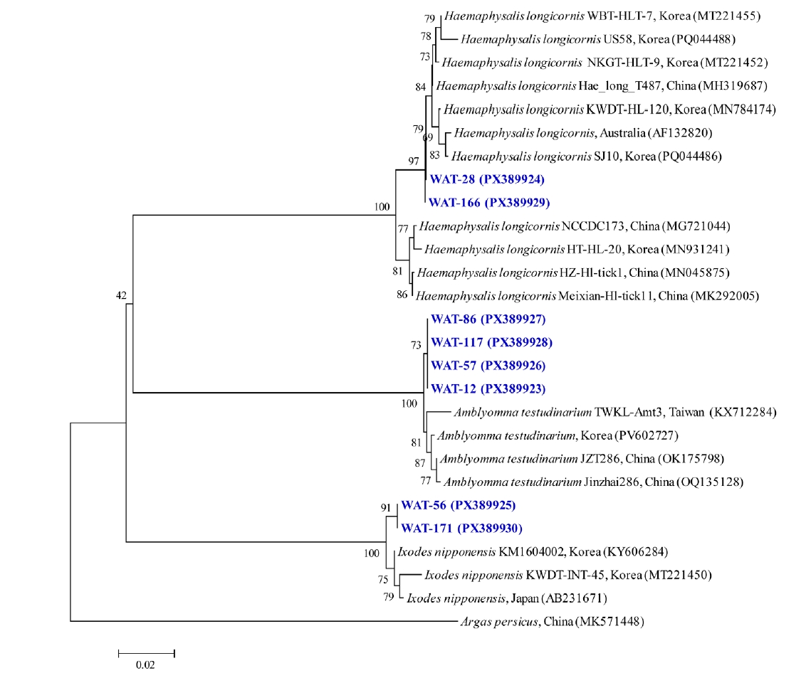
Two
cox1 sequences of
H. longicornis obtained in this study were identical (100% sequence identity) (
Fig. 1) and showed 98.8%–100% identity with reference sequences retrieved from the GenBank database. These reference sequences corresponded to the same gene and species and were selected based on high similarity identified through BLAST analysis. Similarly, 2
cox1 sequences of
I. nipponensis were identical to each other and shared 99.2%–99.7% identity with corresponding GenBank sequences. Four
cox1 sequences of
A. testudinarium also showed complete identity (100%) among themselves and exhibited 99.7%–100% identity when compared with reference sequences in GenBank. All
cox1 sequences generated in this study were deposited in GenBank under accession numbers PX389923–PX389930.
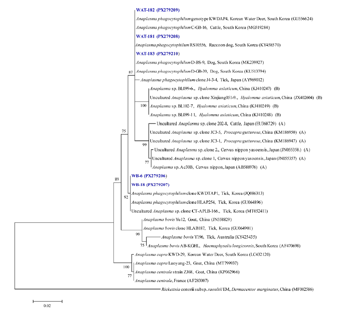
Phylogenetic analysis of 5 samples based on the 16S rRNA gene confirmed their classification as
A. phagocytophilum in both wildlife hosts and ticks (
Fig. 2). These sequences shared 98.8%–100% identity among themselves and showed 98.8%–100% identity with previously reported
A. phagocytophilum isolates in GenBank. The sequences obtained in this study were deposited in GenBank under accession numbers PX279206–PX279210.
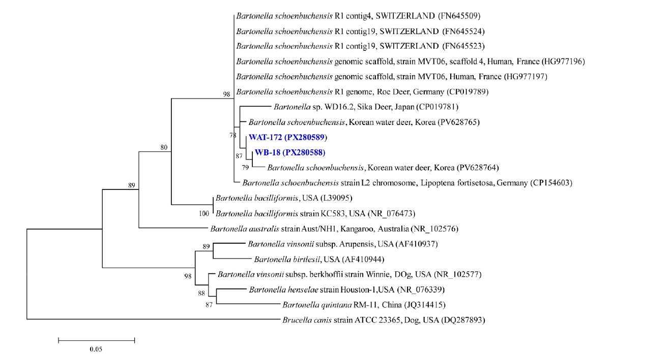
Phylogenetic analysis of the 23S rRNA gene sequences identified 2 samples as
B. schoenbuchensis (
Fig. 3). These sequences showed 99.6%–100% identity to each other and 98.1%–100% identity with previously reported
B. schoenbuchensis isolates in GenBank. The sequences were deposited under accession numbers PX280588–PX280589.
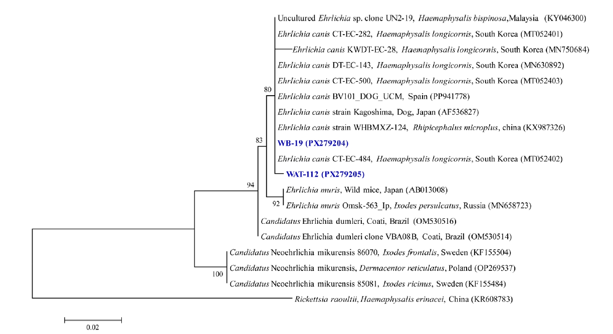
Similarly, phylogenetic analysis of the 16S rRNA gene sequences classified 2 samples as
E. canis (
Fig. 4). These sequences shared 99.7%–100% identity with each other and with previously reported
E. canis isolates in GenBank. The sequences obtained in this study were deposited under accession numbers PX279204–PX279205.
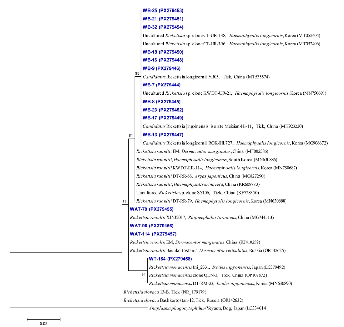
Phylogenetic analysis of the 16S rRNA gene sequences of Rickettsia revealed 3 sequences belonging to
R. raoultii, eleven sequences belonging to
Ca. Rickettsia longicornii, and one sequence belonging to
R. monacensis (
Fig. 5). The
R. raoultii sequences shared 100% identity with each other and showed 99.6%–100% identity with reference sequences in GenBank. The
Ca. R. longicornii sequences shared 99.6%–100% identity among themselves and with previously reported isolates. The
R. monacensis sequence showed 100% identity with previously reported sequences.
These sequences were deposited in GenBank under accession numbers PX279455–PX279457 (R. raoultii), PX279444–PX279454 (Ca. R. longicornii), and PX279458 (R. monacensis).
Comparison of pathogen detection between host blood and attached ticks in paired samples
Paired samples consisting of host blood and corresponding attached ticks were analyzed to evaluate host-tick associations. A total of 8 host individuals had paired samples (8 blood samples and 105 tick pools), allowing direct comparison between host blood and parasitizing ticks. In contrast, 26 of 34 host samples and 81 of 186 tick pools were not paired, limiting individual-level comparisons. Among the paired samples, simultaneous detection of identical pathogens in both host blood and attached ticks was observed in 2 Korean water deer individuals (Korean water deer #1 and #2) (
Table 3). In these cases,
A. phagocytophilum and
E. canis were detected in both host blood and corresponding tick pools. In contrast, in the remaining 6 host individuals, although several pathogens were detected either in tick pools or in host blood, no matched detection was observed. Notably,
R. raoultii showed high positivity in tick pools across most hosts, whereas it was not detected in the corresponding host blood samples. Conversely,
Ca. Rickettsia longicornii was consistently detected in host blood samples but was not identified in the attached ticks.
Sequence comparison revealed high nucleotide identity between host- and tick-derived sequences, ranging from 98.8% to 100% for A. phagocytophilum and 99.7% for E. canis. In addition, phylogenetic analysis showed that sequences obtained from host blood and ticks clustered closely together, indicating high genetic similarity.
Discussion
In this study, ticks and blood samples collected from wildlife in Korea were analyzed to investigate the distribution of major tick-borne bacterial pathogens, including Anaplasma, Ehrlichia, Bartonella, and Rickettsia species. The wildlife examined included Korean water deer, raccoon dogs, and wild boars. A total of 246 ticks were collected, comprising 129 H. longicornis, 100 A. testudinarium, and 17 I. nipponensis.
Among the 3 tick species identified,
A. testudinarium is primarily distributed in the southern regions of Korea, whereas reports from other regions remain relatively limited [
17]. Because all samples in this study were collected in Jeonnam Province, the regional distribution of this tick species may have contributed to its detection. In addition,
A. testudinarium was found exclusively in wild boars in this study, which is consistent with previous reports indicating that this species commonly infests large wild mammals such as wild boars [
18]. Wild boars frequently move between forested areas and agricultural lands, increasing opportunities for contact with diverse tick species. Such ecological characteristics likely influence the host association patterns of
A. testudinarium.
I. nipponensis showed the highest prevalence of
R. monacensis, which is consistent with previous studies identifying this species as a primary vector of
R. monacensis [
9]. Notably, all
R. monacensis–positive specimens in this study were detected exclusively in ticks collected from raccoon dogs, representing the first report of this pathogen associated with raccoon dogs in Korea, to our knowledge. This finding emphasizes the importance of region-specific surveillance considering host ecology and tick–pathogen interactions.
H. longicornis is the most commonly encountered tick species in Korea and is widely recognized as an important vector of multiple pathogens affecting both humans and animals [
18,
19]. This species is known to transmit a wide range of pathogens, such as
Rickettsia,
Anaplasma,
Borrelia,
Babesia,
Bartonella,
Coxiella, and severe fever with thrombocytopenia syndrome virus [
20]. Consistent with previous studies,
H. longicornis was the predominant tick species identified in this study [
21]. Therefore,
H. longicornis may function as a key vector linking diverse hosts and pathogens, potentially increasing the risk of pathogen transmission among humans, livestock, and wildlife. These findings highlight the importance of continuous surveillance of tick-borne pathogens from a One Health perspective.
In Korea,
A. phagocytophilum—the etiological agent of human granulocytic anaplasmosis—has been detected across diverse tick species [
3]. In the present study,
A. phagocytophilum were detected only in
H. longicornis and
I. nipponensis ticks, with
I. nipponensis showing the highest prevalence. These findings are consistent with previous studies conducted in Korea [
22]. The overall MIR of
A. phagocytophilum in ticks in this study was 6.1%. Similarly, a previous study in Korea reported a positivity rate of 24.5% (89/363) based on the MIR in ticks collected from Korean water deer carcasses [
7]. The detection of
A. phagocytophilum in ticks associated with wildlife suggests that these hosts may play an important role in maintaining the natural enzootic cycle of tick-borne pathogens. In addition, the prevalence of
A. phagocytophilum in blood samples from Korean water deer reached 75.0% in the present study. Previous investigations have also reported
A. phagocytophilum in Korean water deer [
10,
23,
24]. Taken together, these findings suggest that Korean water deer may act as important reservoir hosts of
A. phagocytophilum in the Republic of Korea, providing valuable evidence for assessing the risk of tick-borne zoonoses and for developing effective control strategies.
Bacteria of the genus
Bartonella are Gram-negative organisms that parasitize erythrocytes and endothelial cells, among which
B. schoenbuchensis has been identified as a pathogen with zoonotic potential [
25]. This bacterium has been detected in various wildlife hosts, including European roe deer (
Capreolus capreolus) in Germany [
26], and in larvae of the ectoparasitic deer ked
Lipoptena cervi collected from red deer (
Cervus elaphus) [
27]. In the present study, the MIR of
B. schoenbuchensis was 1.0% in
A. testudinarium and 52.9% in
I. nipponensis, with an overall MIR of 5.4%. In addition,
B. schoenbuchensis was detected in 6.3% of blood samples from Korean water deer. To date, no studies have reported the detection of this pathogen in ticks in Korea. However, a previous study reported a detection rate of 12.9% in Korean water deer collected between 2008 and 2009 [
28], and another study identified
B. schoenbuchensis with a prevalence of 6.8% (13/192) in Korean water deer [
16]. This study therefore provides the first evidence of
B. schoenbuchensis detection in ticks in Korea, highlighting the potential role of ticks as vectors of this pathogen. These findings highlight the need for comprehensive molecular epidemiological studies and long-term surveillance incorporating broader geographic coverage, diverse host species, and seasonal factors to clarify the transmission ecology and assess the public health risks associated with
B. schoenbuchensis.
E. canis is the causative agent of canine monocytic ehrlichiosis [
29]. In the present study,
E. canis was detected in ticks collected from wild animals. Based on MIR values, the positivity rates were 2.4% in Korean water deer, 1.6% in raccoon dogs, and 22.4% in wild boars, with an overall MIR of 26.4%. In animal blood samples, the prevalence was 12.5% in Korean water deer, 28.6% in raccoon dogs, and 9.1% in wild boars, with an overall prevalence of 14.7%. A previous study conducted in Korea reported
E. canis positivity in 20% (2/10) of Korean water deer [
23], whereas another investigation reported a prevalence of 0.5% (1/192) in Korean water deer [
16]. However, previous studies conducted in Korea did not detect
E. canis in blood samples from raccoon dogs [
30] or wild boars [
13]. In the present study,
E. canis was detected in blood samples from raccoon dogs and wild boars for the first time in Korea. Furthermore, the infections detected in Korean water deer indicate that the pathogen may circulate not only through tick-mediated transmission but also within wildlife host populations. Therefore, further studies are required to identify the specific tick species involved in the transmission of
E. canis. Broader epidemiological investigations involving wildlife hosts and their associated ticks will be essential to clarify the ecological dynamics of this zoonotic pathogen and to assess its potential public health significance.
Rickettsia species, particularly those belonging to the spotted fever group, have become an important public health concern as tick-borne rickettsioses continue to occur and re-emerge worldwide [
5]. In Korea,
R. raoultii was identified in 40.9% of ticks collected from dogs between 2010 and 2015 [
8]. In addition, a study conducted in Korea in 2021 reported the detection of
R. raoultii in 20% of ticks collected from Korean water deer [
6]. Similarly, in the present study, this pathogen was detected in all 3 tick species. These findings suggest that
R. raoultii may circulate among wildlife hosts and ticks in Korea. Because
R. raoultii is a zoonotic pathogen capable of causing human disease, continuous molecular epidemiological surveillance is required to assess the potential risk of human infection and to establish appropriate public health response strategies.
R. monacensis is widely distributed across Europe, Asia, and Africa and is mainly transmitted by
Ixodes spp. [
31]. In Korea, a study conducted between 2005 and 2006 reported the detection of
R. monacensis in
I. nipponensis collected from small mammals [
9,
32]. More recently, a domestic study conducted in 2024 also reported a high detection rate of
R. monacensis in
I. nipponensis [
33]. Similarly, in the present study,
I. nipponensis was the only tick species in which the pathogen was detected, with a MIR of 94.1%. Notably,
R. monacensis has also been isolated directly from human patient blood samples in Korea, highlighting its importance as a zoonotic pathogen [
34]. The high detection rate of
R. monacensis in
I. nipponensis observed in this study suggests that this pathogen is maintained within local tick populations. These findings highlight the need for continuous surveillance to assess the potential risk of infection in both humans and animals.
Candidatus Rickettsia longicornii has been identified in
H. longicornis in East Asia and is increasingly recognized as an emerging tick-borne pathogen [
6]. Most previous studies in Korea have focused on the detection of
Ca. R. longicornii in ticks. For example, this pathogen was detected at prevalences of 21.5% in ticks in 2021 [
35], and 31% in ticks collected from Korean water deer between 2013 and 2017 [
6]. In contrast, information on the occurrence of
Ca. R. longicornii in animal blood samples remains limited. A recent study reported the detection of this pathogen in only 0.2% (2/906) of dog blood samples in Korea [
11]. In the present study, however,
Ca. R. longicornii was detected in 94.1% of wildlife blood samples, including Korean water deer, raccoon dogs, and wild boars, representing an unexpectedly high prevalence. Interestingly,
Ca. R. longicornii was not detected in tick samples in the present study. This discrepancy between animal blood and tick samples may be related to several ecological factors, including transient bacteremia in wildlife hosts, host-associated circulation of the pathogen, or limitations in tick sample size. It is also possible that certain wildlife species may act as reservoir hosts, maintaining the pathogen within local ecosystems even when detection in ticks is relatively low. Taken together, these findings suggest that
Ca. R. longicornii may circulate widely among wildlife hosts in Korea and may represent an underrecognized component of the local tick–wildlife transmission cycle. Future studies should expand molecular epidemiological surveillance using multilocus genetic markers such as
gltA,
ompA/
ompB, and
sca4 to better clarify the transmission dynamics and potential zoonotic risk of this pathogen from a One Health perspective.
To investigate the association between pathogens in wildlife hosts and their parasitizing ticks, paired samples were analyzed at the individual level. Among 8 host individuals with paired samples, simultaneous detection of identical pathogens in both host blood and attached ticks was observed in only 2 Korean water deer individuals. In these cases, A. phagocytophilum and E. canis were detected in both host blood and tick pools, suggesting potential host–tick sharing of these pathogens. However, for the majority of hosts, no matched detection was observed. Notably, R. raoultii was widely detected in tick pools but was not identified in the corresponding host blood samples, whereas Ca. Rickettsia longicornii was consistently detected in host blood but not in attached ticks. These contrasting patterns may reflect differences in pathogen transmission dynamics, host specificity, or temporal variation in infection status. Overall, these findings suggest that direct concordance between host infection and tick infection is not consistently observed at the individual level. Although the matched cases provide limited molecular evidence supporting potential host–tick pathogen sharing, the low number of such cases and the use of pooled tick samples warrant cautious interpretation. Further studies based on larger sample sizes and individual-level analyses of both ticks and hosts are required to better elucidate transmission dynamics within wildlife–tick systems.
This study provides a molecular epidemiological overview of multiple tick-borne bacterial pathogens in wildlife and their associated ticks in Korea. H. longicornis was identified as the predominant tick species, and several pathogens—including Anaplasma, Ehrlichia, Bartonella, and Rickettsia—were detected in Korean water deer, raccoon dogs, and wild boars. Notably, some pathogens were identified for the first time in wildlife or ticks in Korea, highlighting previously unrecognized transmission dynamics within the wildlife-tick interface. These findings suggest that wildlife may serve as important reservoirs of tick-borne pathogens and may contribute to the maintenance of zoonotic cycles in natural ecosystems. Continued molecular surveillance and broader epidemiological studies are therefore required to better understand the ecology and potential public health implications of tick-borne pathogens in Korea.
Notes
-
Author contributions
Conceptualization: Lee YJ, Kim KT, Seo MG. Data curation: Kim B, Woo HM, Choi JW. Formal analysis: Kim B, Woo HM, Choi JW. Investigation: Lee YJ, Choi I. Methodology: Choi I. Project administration: Kang YM, Rhee MH, Kwak D. Resources: Hyun YS, Heo JW. Software: Choi I. Supervision: Kang YM, Rhee MH, Kwak D. Validation: Hyun YS, Heo JW. Visualization: Choi I, Hyun YS, Heo JW. Writing – original draft: Lee YJ, Kim KT, Seo MG. Writing – review & editing: Kim KT, Seo MG.
-
Conflict of interest
Dongmi Kwak serves as an editor of Parasites, Hosts and Diseases but had no involvement in the decision to publish this article. No other potential conflicts of interest relevant to this study were reported.
Fig. 1.Phylogenetic tree of tick species based on mitochondrial cytochrome c oxidase subunit I sequences constructed using the maximum likelihood method. Sequences obtained in this study are highlighted in blue. GenBank accession numbers and collection countries are shown for reference sequences. Argas persicus was used as the outgroup. Bootstrap values based on 1,000 replicates are indicated at the nodes, and the scale bar represents genetic distance.
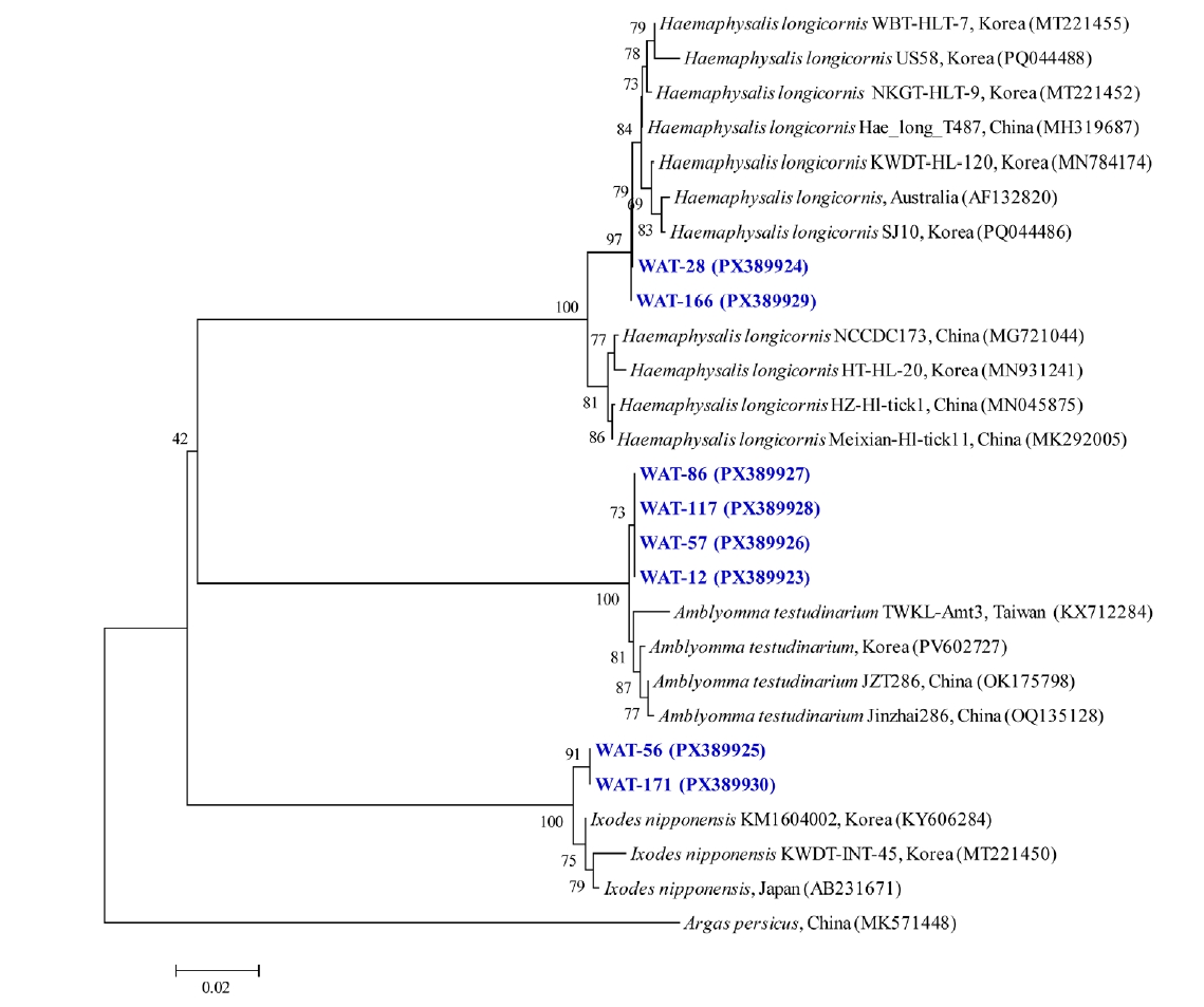
Fig. 2.Phylogenetic tree of Anaplasma species based on 16S rRNA gene sequences constructed using the maximum likelihood method. Sequences obtained in this study are labeled as “WB” (wildlife blood) and “WAT” (wildlife-attached ticks) to indicate their origin and are highlighted in blue. GenBank accession numbers are provided for reference sequences. Rickettsia raoultii was used as the outgroup. Bootstrap values (1,000 replicates) are shown at the nodes, and the scale bar represents genetic distance.
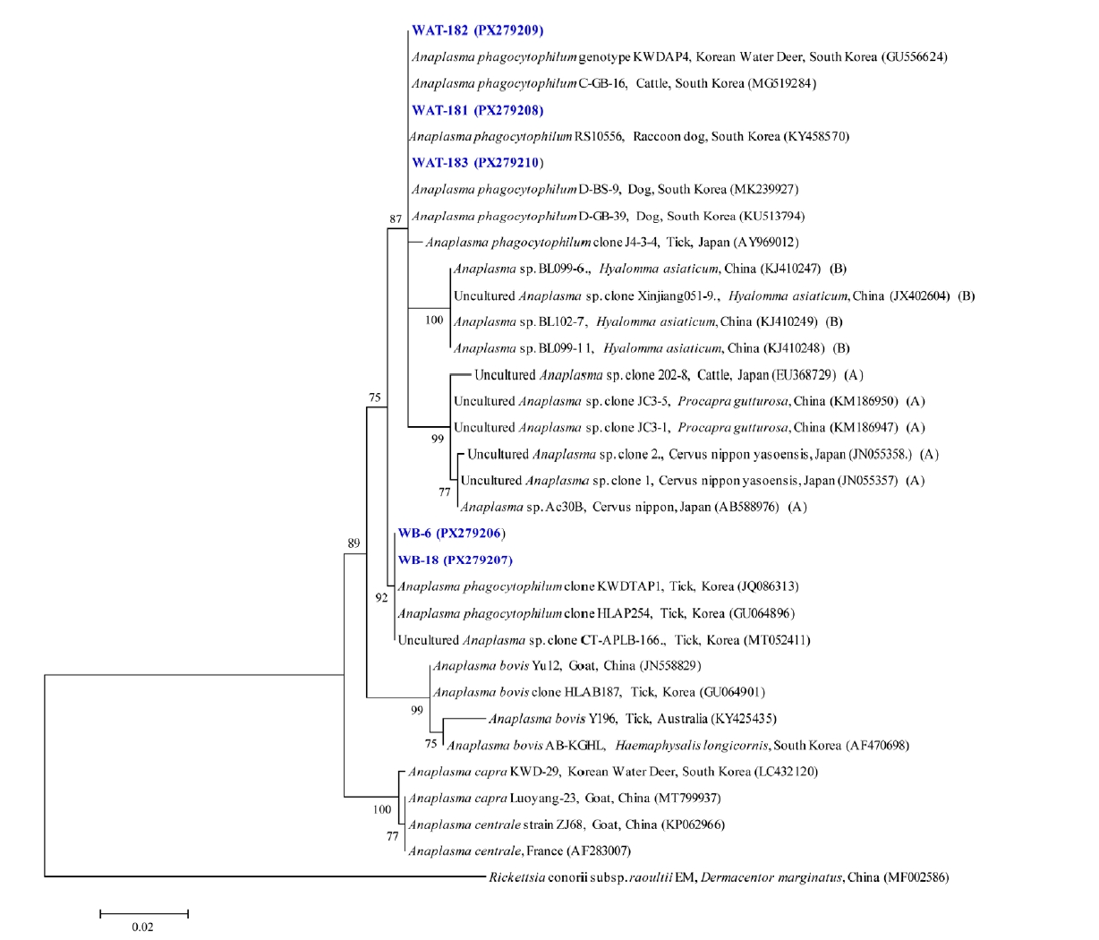
Fig. 3.Phylogenetic tree of Bartonella species based on 23S rRNA gene sequences constructed using the maximum likelihood method. Sequences obtained in this study are labeled as “WB” (wildlife blood) and “WAT” (wildlife-attached ticks) to indicate their origin and are highlighted in blue. GenBank accession numbers are indicated for reference sequences. Brucella canis was used as the outgroup. Bootstrap values (1,000 replicates) are shown at the nodes, and the scale bar represents genetic distance.
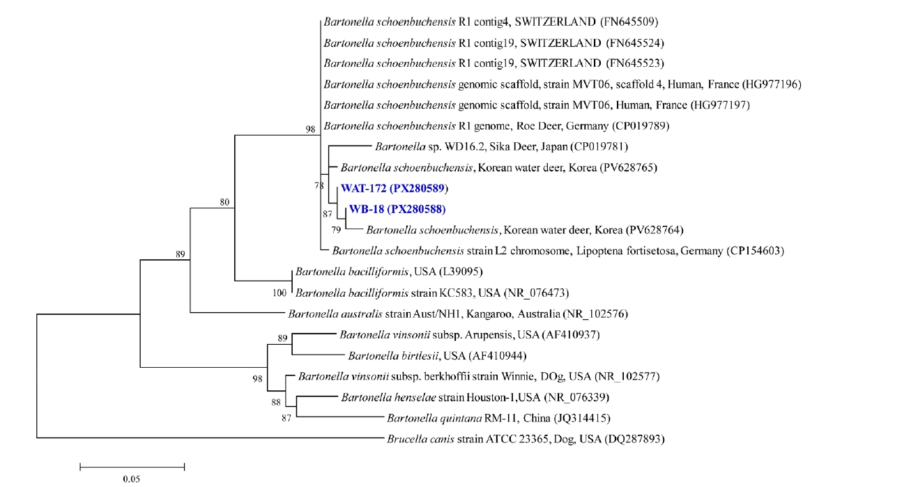
Fig. 4.Phylogenetic tree of Ehrlichia species based on 16S rRNA gene sequences constructed using the maximum likelihood method. Sequences obtained in this study are labeled as “WB” (wildlife blood) and “WAT” (wildlife-attached ticks) to indicate their origin and are highlighted in blue. GenBank accession numbers are indicated for reference sequences. Rickettsia raoultii was used as the outgroup. Bootstrap values (1,000 replicates) are shown at the nodes, and the scale bar represents genetic distance.
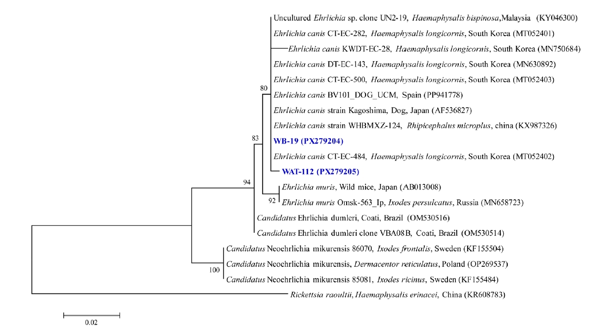
Fig. 5.Phylogenetic tree of Rickettsia species based on 16S rRNA gene sequences constructed using the maximum likelihood method. Sequences obtained in this study are labeled as “WB” (wildlife blood) and “WAT” (wildlife-attached ticks) to indicate their origin and are highlighted in blue. GenBank accession numbers are indicated for reference sequences. Anaplasma phagocytophilum was used as the outgroup. Bootstrap values (1,000 replicates) are shown at the nodes, and the scale bar represents genetic distance.
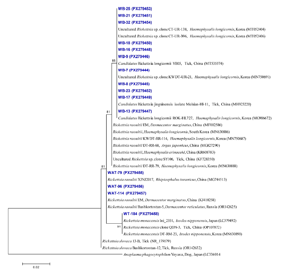
Table 1.Molecular detection of tick-borne pathogens in ticks collected from wild animals in Korea
Table 1.
|
Species |
Stage |
Tested ticks (pool) |
No. positive tick pool/tick pool tested (minimum infection ratea) |
|
Korean water deer |
Raccoon dog |
Wild boar |
Total |
|
A.p |
B.s |
E.c |
R.r |
R.m |
A.p |
B.s |
E.c |
R.r |
R.m |
A.p |
B.s |
E.c |
R.r |
R.m |
A.p |
B.s |
E.c |
R.r |
R.m |
|
Amblyomma testudinarium
|
Adult male |
61 (61) |
- |
- |
- |
- |
- |
- |
- |
- |
- |
- |
0/61 |
1/61 |
37/61 |
61/61 |
0/61 |
0/61 |
1/61 (1.6) |
37/61 (60.7) |
61/61 (100) |
0/61 |
|
Adult female |
39 (39) |
- |
- |
- |
- |
- |
- |
- |
- |
- |
- |
0/39 |
0/39 |
18/39 |
39/39 |
0/39 |
0/39 |
0/39 |
18/39 (46.2) |
39/39 (100) |
0/39 |
|
Subtotal |
100 (100) |
- |
- |
- |
- |
- |
- |
- |
- |
- |
- |
0/100 |
1/100 |
55/100 |
100/100 |
0/100 |
0/100 |
1/100 (1.0) |
55/100 (55.0) |
100/100 (100) |
0/100 |
|
Haemaphysalis longicornis
|
Adult male |
22 (10) |
0/5 |
0/5 |
3/5 |
5/5 |
0/5 |
0/5 |
0/5 |
0/5 |
5/5 |
0/5 |
- |
- |
- |
- |
- |
0/10 |
0/10 |
3/10 (13.6) |
10/10 (45.5) |
0/10 |
|
Adult female |
62 (42) |
2/12 |
0/12 |
1/12 |
12/12 |
0/12 |
0/30 |
0/30 |
0/30 |
30/30 |
0/30 |
- |
- |
- |
- |
- |
2/42 (3.2) |
0/42 |
1/42 (1.6) |
42/42 (67.7) |
0/42 |
|
Nymph |
24 (9) |
0/3 |
0/3 |
2/3 |
3/3 |
0/3 |
0/6 |
0/6 |
0/6 |
6/6 |
0/6 |
- |
- |
- |
- |
- |
0/9 |
0/9 |
2/9 (8.3) |
9/9 (37.5) |
0/9 |
|
Larva |
21 (8) |
0/1 |
0/1 |
0/1 |
1/1 |
0/1 |
0/7 |
0/7 |
0/7 |
7/7 |
0/7 |
- |
- |
- |
- |
- |
0/8 |
0/8 |
0/8 |
8/8 (38.1) |
0/8 |
|
Subtotal |
129 (69) |
2/21 |
0/21 |
6/21 |
21/21 |
0/21 |
0/48 |
0/48 |
0/48 |
48/48 |
0/48 |
- |
- |
- |
- |
- |
2/69 (1.6) |
0/69 |
6/69 (4.7) |
69/69 (53.5) |
0/69 |
|
Ixodes nipponensis
|
Adult male |
2 (2) |
- |
- |
- |
- |
- |
1/2 |
0/2 |
0/2 |
0/2 |
2/2 |
- |
- |
- |
- |
- |
1/2 (50.0) |
0/2 |
0/2 |
0/2 |
2/2 (100) |
|
Adult female |
15 (15) |
- |
- |
- |
- |
- |
12/15 |
9/15 |
4/15 |
1/15 |
14/15 |
- |
- |
- |
- |
- |
12/15 (80.0) |
9/15 (60.0) |
4/15 (26.7) |
1/15 (6.7) |
14/15 (93.3) |
|
Subtotal |
17 (17) |
- |
- |
- |
- |
- |
13/17 |
9/17 |
4/17 |
1/17 |
16/17 |
- |
- |
- |
- |
- |
13/17 (76.5) |
9/17 (52.9) |
4/17 (23.5) |
1/17 (5.9) |
16/17 (94.1) |
|
Total |
246 (186) |
2/21 (0.8) |
0/21 |
6/21 (2.4) |
21/21 (8.5) |
0/21 |
13/61 (5.3) |
9/61 (3.7) |
4/61 (1.6) |
49/61 (19.9) |
16/61 (6.5) |
0/100 |
1/100 (0.4) |
55/100 (22.4) |
100/100 (40.7) |
0/100 |
15/186 (6.1) |
10/186 (4.1) |
65/186 (26.4) |
170/186 (69.1) |
16/186 (6.5) |
Table 2.Prevalence of tick-borne pathogens detected in blood samples of wild animals
Table 2.
|
Animal species |
No. tested |
Positive samples, No. (%) |
|
Anaplasma phagocytophilum
|
Bartonella schoenbuchensis
|
Ehrlichia canis
|
Candidatus Rickettsia longicornii |
Total |
|
Korean water deer |
16 |
12 (75.0)a
|
1 (6.3) |
2 (12.5) |
14 (87.5) |
16 (100) |
|
Raccoon dog |
7 |
0 |
0 |
2 (28.6) |
7 (100) |
7 (100) |
|
Wild boar |
11 |
0 |
0 |
1 (9.1) |
11 (100) |
11 (100) |
|
Total |
34 |
12 (35.3) |
1 (2.9) |
5 (14.7) |
32 (94.1) |
34 (100) |
Table 3.Detection of tick-borne pathogens in paired host blood and attached ticks from the same individuals
Table 3.
|
Host ID |
No. positive tick pools/total tick pools |
No. positive host blood |
Matched detectiona
|
|
A.p |
B.s |
E.c |
R.r |
R.m |
A.p |
B.s |
E.c |
Ca.R.l |
|
Wild boar #1 |
0/27 |
1/27 |
0/27 |
27/27 |
0/27 |
0 |
0 |
0 |
1 |
No |
|
Raccoon dog #1 |
0/21 |
0/21 |
0/21 |
21/21 |
0/21 |
0 |
0 |
0 |
1 |
No |
|
Raccoon dog #2 |
0/8 |
0/8 |
0/8 |
8/8 |
0/8 |
0 |
0 |
0 |
1 |
No |
|
Wild boar #2 |
0/20 |
0/20 |
17/20 |
20/20 |
0/20 |
0 |
0 |
0 |
1 |
No |
|
Korean water deer #1 |
0/5 |
0/5 |
2/5 |
5/5 |
0/5 |
1 |
0 |
1 |
1 |
Yes |
|
Raccoon dog #3 |
0/5 |
0/5 |
0/5 |
5/5 |
0/5 |
0 |
0 |
1 |
1 |
No |
|
Raccoon dog #4 |
0/7 |
0/7 |
0/7 |
7/7 |
0/7 |
0 |
0 |
0 |
1 |
No |
|
Korean water deer #2 |
2/12 |
0/12 |
1/12 |
12/12 |
2/12 |
1 |
0 |
1 |
1 |
Yes |
References
- 1. de la Fuente J, Estrada-Pena A, Venzal JM, Kocan KM, Sonenshine DE. Overview: ticks as vectors of pathogens that cause disease in humans and animals. Front Biosci 2008;13:6938-46. https://doi.org/10.2741/3200
- 2. Low VL, Tan TK, Khoo JJ, Lim FS, AbuBakar S. An overview of rickettsiae in Southeast Asia: vector-animal-human interface. Acta Trop 2020;202:105282. https://doi.org/10.1016/j.actatropica.2019.105282
- 3. Seo MG, Lee H, Alkathiri B, et al. Tick populations and molecular analysis of Anaplasma species in ticks from the Republic of Korea. Microorganisms 2023;11:820. https://doi.org/10.3390/microorganisms11040820
- 4. Demma LJ, Holman RC, McQuiston JH, Krebs JW, Swerdlow DL. Epidemiology of human ehrlichiosis and anaplasmosis in the United States, 2001-2002. Am J Trop Med Hyg 2005;73:400-9.
- 5. Parola P, Paddock CD, Raoult D. Tick-borne rickettsioses around the world: emerging diseases challenging old concepts. Clin Microbiol Rev 2005;18:719-56. https://doi.org/10.1128/cmr.18.4.719-756.2005
- 6. Seo MG, Kwon OD, Kwak D. Molecular detection of Rickettsia raoultii, Rickettsia tamurae, and associated pathogens from ticks parasitizing water deer (Hydropotes inermis argyropus) in South Korea. Ticks Tick Borne Dis 2021;12:101712. https://doi.org/10.1016/j.ttbdis.2021.101712
- 7. Kang JG, Ko S, Kim HC, et al. Prevalence of Anaplasma and Bartonella spp. in ticks collected from Korean water deer (Hydropotes inermis argyropus). Korean J Parasitol 2016;54:87-91. https://doi.org/10.3347/kjp.2016.54.1.87
- 8. Seo MG, Kwon OD, Kwak D. High prevalence of Rickettsia raoultii and associated pathogens in canine ticks, South Korea. Emerg Infect Dis 2020;26:2530-2. https://doi.org/10.3201/eid2610.191649
- 9. Shin SH, Seo HJ, Choi YJ, et al. Detection of Rickettsia monacensis from Ixodes nipponensis collected from rodents in Gyeonggi and Gangwon Provinces, Republic of Korea. Exp Appl Acarol 2013;61:337-47. https://doi.org/10.1007/s10493-013-9699-1
- 10. Han YJ, Park J, Lee YS, et al. Molecular identification of selected tick-borne pathogens in wild deer and raccoon dogs from the Republic of Korea. Vet Parasitol Reg Stud Reports 2017;7:25-31. https://doi.org/10.1016/j.vprsr.2016.12.001
- 11. Kim KT, Seo MG. Molecular analysis of Rickettsia spp. and related tick-borne pathogens detected in dogs in Korea. Acta Trop 2023;243:106934. https://doi.org/10.1016/j.actatropica.2023.106934
- 12. Miranda EA, Han SW, Rim JM, et al. Serological evidence of Anaplasma spp., Borrelia burgdorferi and Ehrlichia canis in dogs from the Republic of Korea by rapid diagnostic test kits. J Vet Sci 2022;23:e20. https://doi.org/10.4142/jvs.21215
- 13. Byun HR, Ji SR, Kang JG, et al. Circulation of tick-borne pathogens in wildlife of the Republic of Korea. One Health 2024;19:100913. https://doi.org/10.1016/j.onehlt.2024.100913
- 14. Truong AT, Yun BR, Lim J, et al. Real-time PCR biochip for on-site detection of Coxiella burnetii in ticks. Parasit Vectors 2021;14:239. https://doi.org/10.1186/s13071-021-04744-z
- 15. Folmer O, Black M, Hoeh W, Lutz R, Vrijenhoek R. DNA primers for amplification of mitochondrial cytochrome c oxidase subunit I from diverse metazoan invertebrates. Mol Mar Biol Biotechnol 1994;3:294-9.
- 16. Kim B, Chae SJ, Lee YJ, et al. Expanding the wildlife reservoir landscape: a high-throughput molecular surveillance study of zoonotic pathogens in Korean water deer. One Health 2025;21:101187. https://doi.org/10.1016/j.onehlt.2025.101187
- 17. Seo JW, Han SY, Sung SH, et al. Survey on tick distribution and tick-borne pathogens in Daejeon and adjacent areas in South Korea. Ticks Tick Borne Dis 2021;12:101711. https://doi.org/10.1016/j.ttbdis.2021.101711
- 18. Kim CM, Yi YH, Yu DH, et al. Tick-borne rickettsial pathogens in ticks and small mammals in Korea. Appl Environ Microbiol 2006;72:5766-76. https://doi.org/10.1128/aem.00431-06
- 19. Lee MJ, Chae JS. Molecular detection of Ehrlichia chaffeensis and Anaplasma bovis in the salivary glands from Haemaphysalis longicornis ticks. Vector Borne Zoonotic Dis 2010;10:411-3. https://doi.org/10.1089/vbz.2008.0215
- 20. Rosenberg R, Lindsey NP, Fischer M, et al. Vital signs: trends in reported vectorborne disease cases: United States and Territories, 2004-2016. MMWR Morb Mortal Wkly Rep 2018;67:496-501. https://doi.org/10.15585/mmwr.mm6717e1
- 21. Chae JB, Cho YS, Cho YK, et al. Epidemiological investigation of tick species from near domestic animal farms and cattle, goat, and wild boar in Korea. Korean J Parasitol 2019;57:319-24. https://doi.org/10.3347/kjp.2019.57.3.319
- 22. Truong AT, Noh J, Park Y, et al. Molecular detection and phylogeny of tick-borne pathogens in ticks collected from dogs in the Republic of Korea. Pathogens 2021;10:613. https://doi.org/10.3390/pathogens10050613
- 23. Seong G, Han YJ, Oh SS, et al. Detection of tick-borne pathogens in the Korean water deer (Hydropotes inermis argyropus) from Jeonbuk Province, Korea. Korean J Parasitol 2015;53:653-9. https://doi.org/10.3347/kjp.2015.53.5.653
- 24. Kim B, Chae SJ, Lee YJ, et al. Nationwide geographical and temporal distribution of tick-borne diseases in Korean water deer (Hydropotes inermis argyropus). Animals (Basel) 2025;15:1499. https://doi.org/10.3390/ani15101499
- 25. Vayssier-Taussat M, Moutailler S, Féménia F, et al. Identification of novel zoonotic activity of Bartonella spp., France. Emerg Infect Dis 2016;22:457-62. https://doi.org/10.3201/eid2203.150269
- 26. Wijburg SR, Fonville M, de Bruin A, et al. Prevalence and predictors of vector-borne pathogens in Dutch roe deer. Parasit Vectors 2022;15:76. https://doi.org/10.1186/s13071-022-05195-w
- 27. de Bruin A, van Leeuwen AD, Jahfari S, et al. Vertical transmission of Bartonella schoenbuchensis in Lipoptena cervi. Parasit Vectors 2015;8:176. https://doi.org/10.1186/s13071-015-0764-y
- 28. Ko S, Kim SJ, Kang JG, et al. Molecular detection of Bartonella grahamii and B. schoenbuchensis-related species in Korean water deer (Hydropotes inermis argyropus). Vector Borne Zoonotic Dis 2013;13:415-8. https://doi.org/10.1089/vbz.2012.1105
- 29. Perez M, Rikihisa Y, Wen B. Ehrlichia canis-like agent isolated from a man in Venezuela: antigenic and genetic characterization. J Clin Microbiol 1996;34:2133-9. https://doi.org/10.1128/jcm.34.9.2133-2139.1996
- 30. Taili I, Kim S, Jeong DH, Na KJ. Prevalence of Rickettsiales in raccoon dogs (Nyctereutes procyonoides koreensis) in South Korea: a three-year survey. J Vet Sci 2025;26:e57. https://doi.org/10.4142/jvs.25046
- 31. Parola P, Paddock CD, Socolovschi C, et al. Update on tick-borne rickettsioses around the world: a geographic approach. Clin Microbiol Rev 2013;26:657-702. https://doi.org/10.1128/cmr.00032-13
- 32. Lee KM, Choi YJ, Shin SH, et al. Spotted fever group rickettsia closely related to Rickettsia monacensis isolated from ticks in South Jeolla province, Korea. Microbiol Immunol 2013;57:487-95. https://doi.org/10.1111/1348-0421.12062
- 33. Kim KG, Hwang DJ, Park JW, et al. Distribution and pathogen prevalence of field-collected ticks from south-western Korea: a study from 2019 to 2022. Sci Rep 2024;14:12336. https://doi.org/10.1038/s41598-024-61126-y
- 34. Kim YS, Choi YJ, Lee KM, et al. First isolation of Rickettsia monacensis from a patient in South Korea. Microbiol Immunol 2017;61:258-63. https://doi.org/10.1111/1348-0421.12496
- 35. Park HJ, Kim J, Choi YJ, et al. Tick-borne rickettsiae in Midwestern region of Republic of Korea. Acta Trop 2021;215:105794. https://doi.org/10.1016/j.actatropica.2020.105794


 , Beoul Kim1