Abstract
Enteric protozoan infections—cryptosporidiosis, giardiasis, microsporidiosis, and coccidiosis—constitute a significant global burden on human and animal health. Yet comprehensive parasitological surveys of wildlife in the eastern Tibetan Plateau, where anthropogenic, livestock, and wildlife activities extensively intersect, remain deficient. We collected 359 fecal specimens from wild carnivores across 98 random quadrats and employed molecular techniques for host identification and protozoan screening. Seven carnivore species were identified, with Tibetan fox (Vulpes ferrilata) predominating. Intestinal protozoa were exclusively recovered from Vulpes species, yielding a prevalence of 19.37% (68/351; 95% CI, 15.24%–23.51%). Eimeria falciformis (7.41%, 26/351) and Eimeria falciformis (7.12%, 25/351) were the most frequently detected, followed by Enterocytozoon bieneusi (3.70%, 13/351). Cryptosporidium was confirmed in one V. ferrilata, and Giardia intestinalis in 2 samples (V. ferrilata and Vulpes vulpes). This investigation furnishes the first molecular evidence that wild carnivores, especially V. ferrilata, harbor zoonotic protozoa including G. intestinalis assemblage AII and E. bieneusi genotypes PigEBITS5 and a D-like variant. The concurrent detection of Cryptosporidium yak genotype and E. bieneusi subgroup 1i in foxes, pikas, yaks, and sheep implies potential interspecies transmission at the wildlife–livestock interface. These findings fill knowledge gaps in regional parasite diversity and emphasize the need for enhanced surveillance of wildlife-borne protozoa under a One Health framework, thereby providing scientific evidence for zoonotic risk assessment and public health planning.
-
Key words: Protozoa, Tibetan Plateau, Carnivores, Cryptosporidium, Giardia intestinalis, Enterocytozoon bieneusi
Introduction
Protozoa comprise a highly diverse group of single-celled eukaryotic microorganisms, many of which are pathogenic and exhibit a global distribution. Diseases caused by these protozoa are especially prevalent in tropical and subtropical regions due to conducive environmental conditions and socioeconomic factors [
1]. Zoonotic protozoan infections pose serious threats to human health worldwide. Notable examples include cryptosporidiosis, giardiasis, and microsporidiosis, which can lead to chronic diarrhea, severe emaciation, and elevated mortality among immunocompromised individuals. In children, these infections are associated with growth retardation and impaired cognitive development [
2]. In China, intestinal protozoan diseases have emerged as a significant public health challenge, predominantly caused by such pathogens as
Cryptosporidium spp. and
Giardia intestinalis [
3]. With the expansion of animal husbandry and increased human encroachment into wildlife habitats, the risk of pathogens carried by wildlife contaminating water sources or the food chain and transmitting to humans and livestock has risen significantly [
4]. Studies conducted in the Americas and Europe have confirmed that wild canids such as coyotes (
Canis latrans) and red foxes (
Vulpes vulpes) serve as reservoirs for various zoonotic protozoa, including
Cryptosporidium spp. and
G. intestinalis. Some detected strains show high genetic homology with those found in human clinical cases, suggesting possible cross-species transmission and shared infection sources [
5]. Additionally, other protozoan species such as
Enterocytozoon bieneusi and
Eimeria spp. are highly prevalent in wildlife populations. These pathogens can be widely disseminated into the environment through fecal excretion, thereby posing continued risks to human and animal health [
6]. These findings collectively indicate that wildlife-origin protozoa may increasingly spill over into human populations through ecological interfaces, thereby creating multifaceted and evolving public health concerns. Understanding the transmission dynamics and host range of these protozoans is critical for assessing emerging zoonotic risks and developing effective control strategies.
The eastern Tibetan Plateau, a global biodiversity hotspot, sustains a unique ecosystem with extensive overlap between wildlife habitats and human activities. Dominated by pastoral economies, this region exposes herders and livestock (e.g., yaks and sheep) to wildlife-inhabited areas, amplifying risks of cross-species pathogen transmission. Previous investigations revealed that the area’s complex topography and climatic diversity support rich carnivore communities, including canids like the Tibetan fox (
Vulpes ferrilata), red fox, and wolf (
Canis lupus), alongside rare species such as the Tibetan brown bear (
Ursus arctos pruinosus) and snow leopard (
Panthera uncia). These animals harbor diverse parasites, including helminths transmissible to humans or livestock [
7]. Since wildlife and domestic animals in this region share grazing routes and water sources, a “bridge” for bidirectional pathogen transmission may form [
8]. However, current parasite surveillance systems primarily focus on humans and livestock, resulting in fragmented data on wildlife parasites, which hinders the assessment of their role as hosts and their zoonotic potential.
This study utilizes molecular methods to systematically characterize the species composition of wild carnivores in the eastern Tibetan Plateau, along with the genetic characteristics, prevalence, and public health risks of high-priority zoonotic protozoa (Cryptosporidium spp. and G. intestinalis) and highly prevalent protozoa with cross-species transmission potential (Eimeria spp. and E. bieneusi). The research aims to fill the gap in baseline parasitological data for this region, provide a scientific basis for assessing zoonotic disease risks, and offer theoretical support for formulating One Health-oriented prevention and control strategies.
Methods
Ethics statement
Ethical approval was not applicable because this study involved only the collection of fecal samples from free-ranging wild carnivores, with no direct contact with or disturbance to the animals.
Sample collection and processing
This study was conducted in November 2021 and November 2023 in the border region of Sichuan, Xizang, and Qinghai provinces on the eastern Tibetan Plateau, with Shiqu County, Sichuan Province serving as the primary sampling area (
Fig. 1). Using a randomized sampling design, 98 environmental quadrats measuring 50 m × 200 m were established. Within each quadrat, all visible fecal samples of wild carnivores were systematically collected using disposable sampling forceps, transferred to 50 ml centrifuge tubes, and preserved with 95% ethanol. Samples were transported to the laboratory and stored at -80°C in an ultra-low temperature freezer for at least 1 week to inactivate potential pathogens. After initial morphological screening, weathered or fragmented samples were excluded, resulting in 436 intact samples retained for subsequent analysis.
Genomic DNA was extracted from fecal samples using the QIAamp Fast DNA Stool Mini Kit (Qiagen). Host species were identified by amplifying the vertebrate-specific 16S rRNA gene fragment using universal primers (V-16S). Primer sequences are listed in
Table 1 [
9-
13]. The PCR mixture (25 μl) consisted of 1 μl each of forward and reverse primers, 12 μl of Ex Taq PCR Premix (RR902A, Takara), 0.5 μl of bovine serum albumin (Takara), 2 μl of template DNA, and RNase-free water (Takara) to adjust the final volume. Amplification conditions were as follows: initial denaturation at 94°C for 5 min; 35 cycles of denaturation at 94°C for 30 sec, annealing at 55°C for 30 sec, and extension at 72°C for 30 sec; followed by a final extension at 72°C for 10 min. PCR products were stored at 4°C. PCR products were visualized by 1% agarose gel electrophoresis (120 V, 30 min), and positive products were sent to Sangon Biotech for molecular cloning (10 clones per sample) and sequencing. Host and prey species were identified by comparing the sequencing results with the NCBI database (
https://blast.ncbi.nlm.nih.gov/Blast.cgi). The percent coverage and percent identity of the BLAST hits were above 99%.
Protozoa were detected using specific primers targeting the small subunit rRNA (
SSU rRNA) gene for
Cryptosporidium spp. and
Eimeria spp., the triosephosphate isomerase (
TPI) gene for
G. intestinalis, and the internal transcribed spacer 1 (
ITS1) region for
E. bieneusi (
Table 1) [
9-
13]. The reaction systems were consistent with those used for the host identification. The amplification conditions of each protozoan are listed in
Table 1 [
9-
13], for example, for the detection of
Cryptosporidium spp., a nested PCR approach was employed: the first round used primers CRY-F1/R1 (amplicon size: ~1,325 bp), and the second round used CRY-F2/R2 (about 830 bp), both with an annealing temperature of 55°C. PCR products were purified and subjected to molecular cloning (5 clones per sample) when overlapping peaks were observed in sequencing chromatograms. Otherwise, products were directly sequenced by Sangon Biotech. Species or genotypes were determined by comparing obtained sequences with reference sequences in the NCBI database.
Sequences were assembled and edited using DNAStar 7.0 (DNASTAR). Multiple sequence alignment and editing were performed using the ClustalW algorithm alongside reference sequences retrieved from GenBank. Haplotypes were identified using DnaSP version 6.12. The optimal nucleotide substitution model was selected with jModelTest 2.1.10 under the Akaike Information Criterion. The
TVM+
I+
G,
TIM3+
I+
G,
TrN+
I+
G, and
TPM1uf+
G models were applied for phylogenetic analyses of
Cryptosporidium sp.,
G. intestinalis,
Eimeria spp., and
E. bieneusi, respectively. Bayesian inference trees were constructed using MrBayes 3.2.4, running Markov Chain Monte Carlo simulations for 1,000,000 generations with sampling every 1,000 generations. The first 25% of samples were discarded as burn-in before summarizing the tree topology. Final trees were visualized and annotated using iTOL v7.2.1 (
https://itol.embl.de/itol.cgi).
Data on host identification and protozoan detection were compiled and analyzed using R version 4.3.2 (
https://www.r-project.org). Differences in detection rates among groups were assessed using chi-square tests. Data were visualized using the Hiplot Pro platform (Shanghai Tengyun Biotechnology,
https://hiplot.com.cn/).
Results
Host identification
A total of 436 wild carnivore fecal samples were collected from the eastern Tibetan Plateau. Host species were successfully identified for 359 samples (82.34%) via 16S rRNA gene sequencing, representing 7 carnivore species across 4 families: Canidae (354/359, 98.61%), Mustelidae (3/359, 0.84%), Ursidae (1/359, 0.28%), and Felidae (1/359, 0.28%). The Tibetan fox (V. ferrilata) was the predominant species (307/359, 85.52%), followed by the red fox (V. vulpes; 44/359, 12.26%) and the wolf (C. lupus; 3/359, 0.84%). Mustelid samples included 2 Asian badgers (Meles leucurus) and one mountain weasel (Mustela altaica). Single samples were identified for the Tibetan brown bear (U. arctos pruinosus) and leopard cat (Prionailurus bengalensis).
Protozoan haplotype identification
Eight distinct protozoan haplotypes were identified from 359 samples.
Cryptosporidium was confirmed in
V. ferrilata feces (haplotype CHSQ.C.36.7, 100% identical to sequence KF971356 from GenBank).
Giardia intestinalis (haplotype CHSQ.G.13.9, 99.09% similarity to MN844148) was detected in both
V. ferrilata and
V. vulpes.
Eimeria banffensis (CHSQ.E.25.5, 100% match to PP594267) occurred in both
Vulpes species, while
Eimeria falciformis (CHSQ.E.23.8, 100% match to MH752019) and a novel
Eimeria sp. (CHSQ.E.25.4, 99.25% similarity to OP352330) were only detected in
V. ferrilata. Three
E. bieneusi haplotypes (CHSQ.E.4.8, CHSQ.E.8.3, and CHSQ.E.4.2) were identified solely in
V. ferrilata (
Table 2). All sequences were deposited in GenBank under accession numbers PX249759, PX262650, PX249765–PX249767, and PX262653–PX262655.
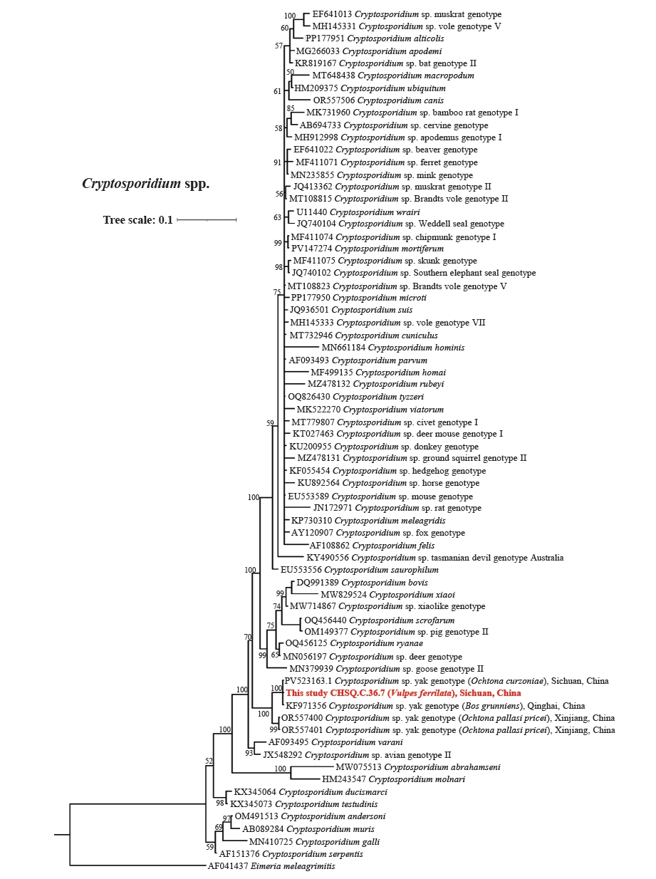
Phylogenetic analysis revealed that the only
Cryptosporidium sp. strain detected in
V. ferrilata samples (CHSQ.C.36.7), together with isolates from yak (
Bos grunniens) in Qinghai (GenBank accession No. KF971356) and plateau pika (
Ochotona curzoniae) in Sichuan (PV523163), formed a distinct clade (100% bootstrap support). This clade corresponds to the previously described
Cryptosporidium sp. yak genotype (
Fig. 2). The phylogenetic tree of
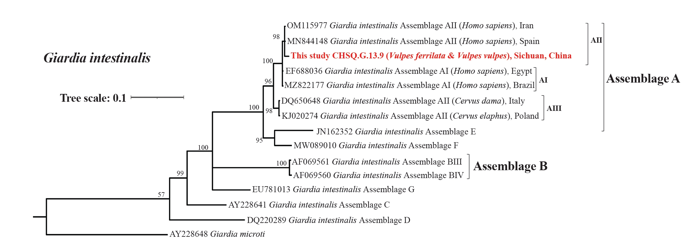
G. intestinalis displayed 7 well-supported clades corresponding to Assemblages A–G. The 2
G. intestinalis strains identified in this study (both from
V. ferrilata and
V. vulpes samples and belonging to a single haplotype CHSQ.G.13.9), were placed within the subclade AII of Assemblage A (98% support) (
Fig. 3). A phylogenetic tree constructed using
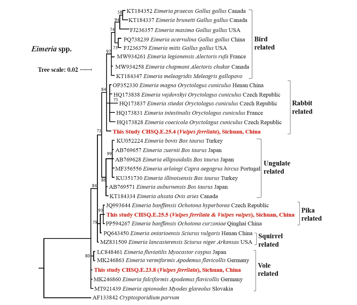
Eimeria sp. sequences from NCBI and haplotype sequences from this study resolved into 6 major clades, each associated with a specific group of host animals (e.g., birds, ungulates, and rodents). Among these,
E. banffensis from this study (CHSQ.E.25.5) clustered with 2 other
E. banffensis strains (both from pika hosts) with 93% support. Another strain,
E. falciformis (CHSQ.E.23.8), clustered within a clade comprising other
Eimeria species (such as
E. fluviatilis,
E. vermiformis,
E. falciformis, and
E. apionodes), all of which are known to infect voles (e.g.,
Myocastor coypus,
Apodemus flavicollis, and
Myodes glareolus). Additionally, CHSQ.E.25.4 clustered (84% support) with
E. magna,
E. vejdovskyi,
E. stiedai,
E. intestinalis, and
E. coecicola, species primarily reported in European rabbits (
Oryctolagus cuniculus) (
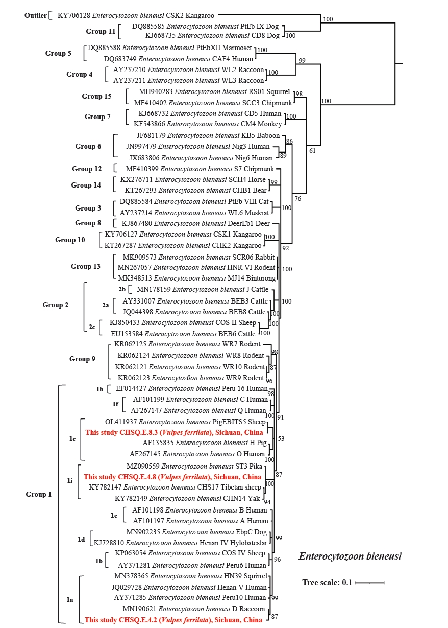
Fig. 4). The phylogenetic tree of
E. bieneusi was divided into 15 genetic groups (Groups 1–15), with Group 1 further subdivided into 9 subgroups (a–i). All
E. bieneusi haplotypes in this study belonged to Group 1: CHSQ.E.4.8 (99.59% similarity to ST3, MZ090559) formed subgroup 1i with genotypes from yak and sheep on the Tibetan Plateau; CHSQ.E.8.3 (100% identity to PigEBITS5, OL411937) clustered in subgroup 1e (hosts: pigs, sheep, humans); CHSQ.E.4.2 (99.19% similarity to genotype D, MN190621) fell within subgroup 1a, which includes known human-pathogenic genotypes, such as Henan V and Peru10 (
Table 2;
Fig. 5).
No protozoa were detected in
C. lupus,
M. leucurus,
M. altaica,
U. arctos pruinosus or
P. bengalensis. Six protozoan species were identified exclusively in canids (
V. ferrilata and
V. vulpes), and the detection rate in fecal samples was 19.37% (68/351; 95% CI, 15.24%–23.51%) in foxes. Detection rates varied significantly among protozoan species (
χ²=64.31,
P<0.01).
E. falciformis (7.41%, 26/351; 95% CI, 4.67%–10.15%) and
E. banffensis (7.12%, 25/351; 95% CI, 4.43%–9.81%) exhibited the highest rates, followed by
E. bieneusi (3.70%, 13/351; 95% CI, 1.73%–5.68%). A single
Cryptosporidium sp.-positive and one
G. intestinalis-positive sample occurred in
V. ferrilata, while one additional
G. intestinalis was detected in
V. vulpes.
V. ferrilata was the only host for all identified protozoa, showing a significantly higher detection rate (21.17%, 65/307; 95% CI, 16.60%–25.74%) than
V. vulpes (3/44;
χ²=5.08,
P=0.02) (
Table 3).
Discussion
The eastern Tibetan Plateau, situated at the convergence of Sichuan, Qinghai, and Xizang, features complex terrain shaped by dual monsoon systems from the Indian and Pacific Oceans. This creates a humid climate (400–800 mm annual precipitation) supporting the plateau’s most complete vertical vegetation spectrum, which offers diverse microhabitats for wildlife [
14]. Canids dominated the carnivore community (98.61%), with
V. ferrilata representing 85.52% of identified samples. Compared to large carnivores (e.g., snow leopards, brown bears), small carnivores like foxes and weasels exhibit lower individual spatial requirements but higher population densities. While ecologically vital for ecosystem stability, they may act as reservoirs for zoonotic pathogens, amplifying disease circulation across wildlife-domestic-human interfaces and elevating regional public health risks [
15].
Cryptosporidium, a major diarrheal pathogen second only to rotavirus, infects diverse vertebrates and persists in environmental matrices. Over 49 species and 120 genotypes exist, with 23 genotypes confirmed as human-infective, including
C. hominis,
C. parvum,
C. meleagridis,
C. canis,
C. felis,
C. ubiquitum,
C. cuniculus,
C. viatorum,
C. muris,
C. andersoni,
C. erinacei,
C. tyzzeri,
C. bovis,
C. suis,
C. scrofarum,
C. occultus,
C. xiaoi,
C. fayeri,
C. ditrichi,
C. equi (horse genotype),
C. wrairi,
C. mortiferum (chipmunk genotype I),
C. baileyi, mink genotype, and skunk genotype [
16]. To the best of our knowledge, there have been no reports of
Cryptosporidium sp. yak genotype infecting the human populations within the study area. Although the plateau’s harsh climate was historically considered unfavorable for
Cryptosporidium spp. [
17], we detected it in
V. ferrilata, with the strain showing 100% identity to the
Cryptosporidium sp. yak genotype previously reported in
B. grunniens from Qinghai Province [
18]. Identical genotypes were also found in
O. curzoniae (PV523163) in this region [
19]. Phylogenetic analysis (
Fig. 2) revealed this genotype forms a distinct clade closely related to strains from Pallas's pika (
Ochotona pallasi pricei, OR557400 and OR557401) in Xinjiang, suggesting it may represent a novel plateau-adapted genotype circulating among sympatric foxes, pikas, and livestock via shared pastures and water. Its zoonotic potential warrants further investigation.
This study detected
G. intestinalis in the feces of both
V. ferrilata and
V. vulpes. This flagellate is an intestinal parasitic protozoan with complex host specificity, featuring a life cycle that includes infectious cysts and pathogenic trophozoites [
20]. In China,
G. intestinalis and
Cryptosporidium spp. are the only 2 parasites prioritized as microbial indicators explicitly listed in the Standards for Drinking Water Quality (GB 5749–2022), underscoring their public health significance [
21]. Genetic analyses divide
G. intestinalis into host-adapted assemblages: A and B infect humans and diverse mammals, while C/D (canids), E (livestock), F (felids), and G (rodents) show stricter host specificity [
22]. The assemblage AII identified here (
Fig. 3) matches dominant human-infective strains in China [
23]. Environmental contamination by fox-derived cysts poses health risks, particularly for pastoral communities reliant on natural water sources.
For
E. bieneusi, over 500 genotypes (including 52 zoonotic types) are recognized [
24]. ITS-based phylogeny classifies genotypes into Groups 1–15, with Groups 1 and 2 demonstrating broad host ranges and zoonotic potential, while Groups 3–15 are typically host-specific (e.g., Group 3 in wild rodents, Group 4 in carnivores/rodents) [
25]. In this study, CHSQ.E.4.2 (99.19% similarity to genotype D, MN190621) belongs to subgroup 1a, a major human-infective group; CHSQ.E.8.3 (identical to PigEBITS5, OL411937) clusters in subgroup 1e, confirmed to infect humans. In particular, CHSQ.E.4.8 (predominant genotype, 11/13) aligns with genotypes of ST3, CHS17, and CHN14 in subgroup 1i from pika, sheep, and yak, respectively, in Qinghai [
26,
27]. Therefore, it is suggested that subgroup 1i is a dominant genotype group circulating among wild and domestic animals in the eastern Tibetan Plateau, and further research is required to verify its human-infective capacity (
Fig. 5).
The intestinal parasites common to these animals also pose a potential persistent threat to human health.
Cryptosporidium infection is one of the most common causes of diarrhea in humans worldwide. As of 2021, incomplete statistics indicate that at least 29 provinces in China have reported human
Cryptosporidium infections, involving 107 cities and a total of 4,975 clearly confirmed cases [
28]. Although this disease is not a statutory infectious disease in China, it is easy ignored. With regard to
G. intestinalis, an estimated 280 million people worldwide suffer from clinically diagnosable giardiasis. High infection rates of giardiasis have been documented in developing countries. In China, approximately 28.5 million human cases of giardiasis are estimated to occur annually. According to the limited genotyping studies available, both assemblage A (subtypes AI and AII) and assemblage B of
G. intestinalis have been identified in China, with assemblage AII and assemblage B being the predominant genotypes [
23]. It is worth noting that
E. bieneusi, by far the most frequently reported microsporidian species causing human disease, is responsible for intestinal illness in both non- and immunocompromised patients. A recent meta-analysis estimated the overall prevalence of
E. bieneusi infection in humans at 7.9% (95% CI, 6.9%–8.8%). Furthermore, in some low-income countries, nearly 10% of children presenting with diarrhea test positive for
E. bieneusi [
29].
On the other hand, it should be noted that the most frequently detected parasite in canid fecal samples was
Eimeria spp. However, existing data indicate that the parasitism of Eimeriidae in canids is a complex and controversial issue. Although oocysts of species such as
E. canis,
E. aurei, and
E. lomarii have been found in canid feces or intestines, their endogenous developmental stages (e.g., schizonts, gametocytes) have not been confirmed within canid hosts. Thus,
Eimeria are suspected to be spurious parasites in canids, with their presence likely due to the consumption of infected true hosts by canids [
30]. Nonetheless, although these oocysts are likely not replicating in foxes, they remain intact and sporulation-competent, posing a potential transmission risk. In this study,
E. banffensis (pika-associated) and
E. falciformis (vole-associated) dominated in fox feces, aligning with dietary analyses showing pikas and voles as primary prey [
31]. The rare detection of a rabbit-associated
Eimeria sp. (CHSQ.E.25.4) (
Table 3), which likely originated from the native plateau hare (
Lepus oiostolus), and is distinctly separated from those derived from
O. cuniculus in the phylogenetic tree, further supports prey-derived transmission. The findings indicate the presence of these
Eimeria species in the environment or food chain of the eastern Tibetan Plateau, indirectly reflecting the complex flow of protozoan parasites within the local ecosystem.
Although this study detected multiple intestinal protozoans in carnivore fecal samples from the eastern Tibetan Plateau using molecular methods, the inherent limitations of fecal material make it difficult to confirm whether these parasites truly colonized the wildlife hosts or induced pathological effects. Detection of this pathogen in carnivore feces may suggest a potential food chain transmission risk, as exemplified by
Mastophorus muris. This parasite utilizes insects (e.g., beetles, locusts, grasshoppers, cockroaches) as intermediate hosts and rodents as definitive hosts [
7]. Although
M. muris does not require
V. ferrilata in its life cycle, ingestion of infected rodents by
V. ferrilata results in excretion of parasite eggs in the carnivore's feces, thereby posing a potential transmission risk. These findings heighten concerns regarding carnivores as potential vectors of zoonotic protozoans. Given the relatively scarce baseline research and insufficient host investigations in the Tibetan Plateau, this study underscores significant public health concerns.
As an ecological hotspot on the Qinghai-Tibet Plateau, Shiqu County harbors abundant wildlife resources. Animal husbandry serves as the primary source of income for local residents, with nomadism representing a traditional way of life. The considerable overlap between grazing areas and wildlife habitats, coupled with the sharing of water sources, poses a potential risk for the transmission of zoonotic enteric protozoan parasites from wildlife to humans [
7]. To improve the living conditions of pastoralists, the Chinese government-initiated settlement construction projects in 2001. However, smaller and more remote settlements often lack basic public facilities, leading to difficulties in accessing clean water [
32]. The combination of inadequate clean water access and local herders’ customs—such as not washing hands before meals and lying directly on the grass—in an area where water sources are shared with wildlife, may facilitate the spread of intestinal protozoa. Notably, the overlapping activity areas create opportunities for indirect contact between humans and wildlife, specifically through livestock. Previous studies have indicated that with the progression of human society, the boundaries between humans and wildlife are gradually blurring, and the living space of wildlife is increasingly compressed. Consequently, wildlife is forced to share resources and habitats with humans to a certain extent. Although wild animals tend to avoid direct human disturbance, they may occasionally come into contact with livestock for predation [
33]. During such interactions, pathogen spillover can occur among humans, livestock, and wildlife.
Zoonotic parasites pose a serious challenge to global public health due to their cross-species transmission potential, complex life cycles, and diverse host range, which complicate control efforts [
34]. These pathogens form intricate transmission networks through multi-host interactions within food webs, with their virulence and spread influenced by host ecology, behavior, and environmental factors [
35]. As apex predators, carnivores accumulate a diversity of parasites through bioaccumulation, posing a considerable health risk to herders who frequently interact with natural environments. Of particular concern are traditional local practices (such as consuming untreated water, raw or undercooked meat), which may serve as key risk factors for acquiring these zoonotic infections.
This study not only provides the first regional assessment of potentially transmissible protozoans carried by wildlife in the eastern Tibetan Plateau, but also highlights how grazing activities may foster transmission chains among humans, livestock, and wildlife. It thus calls for sustained attention to the cross-species mechanisms and ecological risks of zoonotic protozoan diseases.
Canids, particularly V. ferrilata, dominated the wild carnivore assemblage in the eastern Tibetan Plateau and showed a notable detection rate of protozoan infections (all protozoan species detected in this study were identified in V. ferrilata samples). This research represents the first documentation of intestinal protozoa detected in wildlife in this region, revealing the detection of 2 confirmed zoonotic pathogens: G. intestinalis assemblage AII (CHSQ.G.13.9) and E. bieneusi genotype CHSQ.E.8.3 (identical to PigEBITS5). Additionally, a potentially zoonotic genotype of E. bieneusi (CHSQ.E.4.2) was detected, underscoring the existence of wildlife-origin protozoan pathogens and their latent threat to human health. The discovery of the Cryptosporidium sp. yak genotype and E. bieneusi (CHSQ.E.4.8) in yaks, foxes, and rodents suggests that these genotypes are dominant locally and reveals potential pathogen transmission and spillover risks between wildlife and livestock. It is important to emphasize that protozoan infections in wildlife remain understudied across the Tibetan Plateau. Further investigation is needed to elucidate the transmission patterns of poorly characterized native protozoa (such as the Eimeria sp. reported here), and to clarify their life cycles and potential public health implications.
Notes
-
Data availability
All data generated or analyzed during this study are included in this published article.
-
Author contributions
Conceptualization: Deng X, Wang X. Data curation: Deng X. Formal analysis: Deng X. Funding acquisition: Wang X. Investigation: Deng X, Xue C, Chen Q, Zuo Q, Li M, Peng X, Wang X. Methodology: Deng X, Wang X. Project administration: Wang X. Resources: Wang X. Software: Deng X, Wang X. Supervision: Deng X, Wang X. Validation: Deng X, Wang X. Visualization: Deng X. Writing – original draft: Deng X. Writing – review & editing: Li P, Wang X.
-
Conflict of interest
The authors have no conflicts of interest to declare.
-
Funding
This work was supported by the Prevention and Control of Emerging and Major Infectious Diseases-National Science and Technology Major Project (grant No. 2025ZD01900100, 2025ZD01900112) and the National Natural Science Foundation of China (grant No. 82404325).
-
Acknowledgments
We gratefully acknowledge the Shiqu County Center of Disease Control for its support during the field study.
During the writing of this manuscript, we employed AI-based language tools (DeepSeek) solely for the purposes of language refinement, grammar correction, and stylistic improvement. These tools were not used to generate any scientific content, data, or conclusions. All AI-suggested changes were critically reviewed and either accepted or modified by the authors to ensure that the final manuscript accurately reflects our own findings and interpretations. The authors assume full responsibility for the originality, accuracy, and integrity of the content presented.
Fig. 1.Study area. The red points represent the coordinate points of the sample quadrats and the yellow lines represent the provincial boundaries.
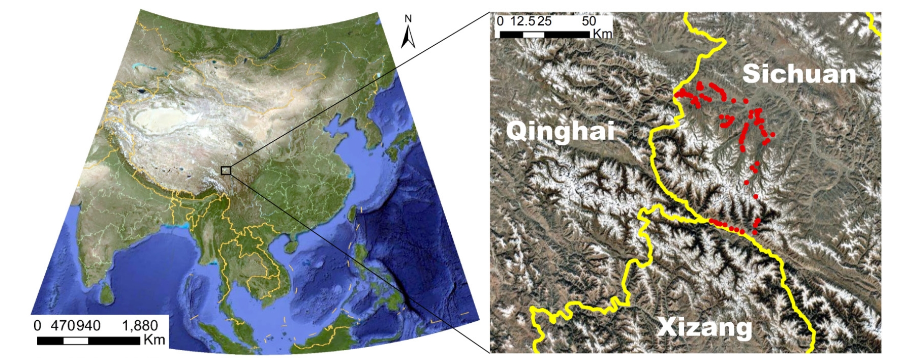
Fig. 2.Phylogenetic tree based on the Cryptosporidium spp. based on partial SSU rRNA gene sequences with the nucleotide substitution model of TVM+I+G.
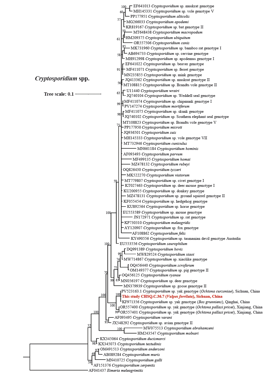
Fig. 3.Phylogenetic tree based on the Giardia intestinalis, based on partial TPI gene sequences with the nucleotide substitution model of TIM3+I+G.
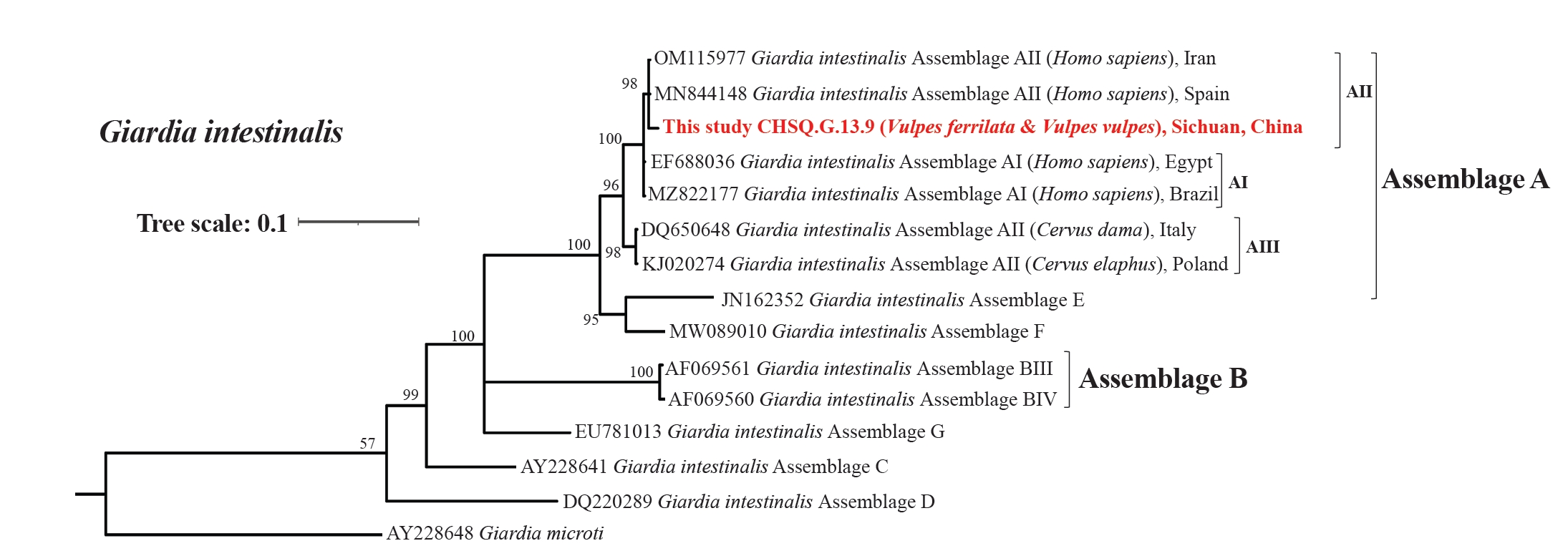
Fig. 4.Phylogenetic tree based on the Eimeria spp. based on partial SSU rRNA gene sequences with the nucleotide substitution model of TrN+I+G.
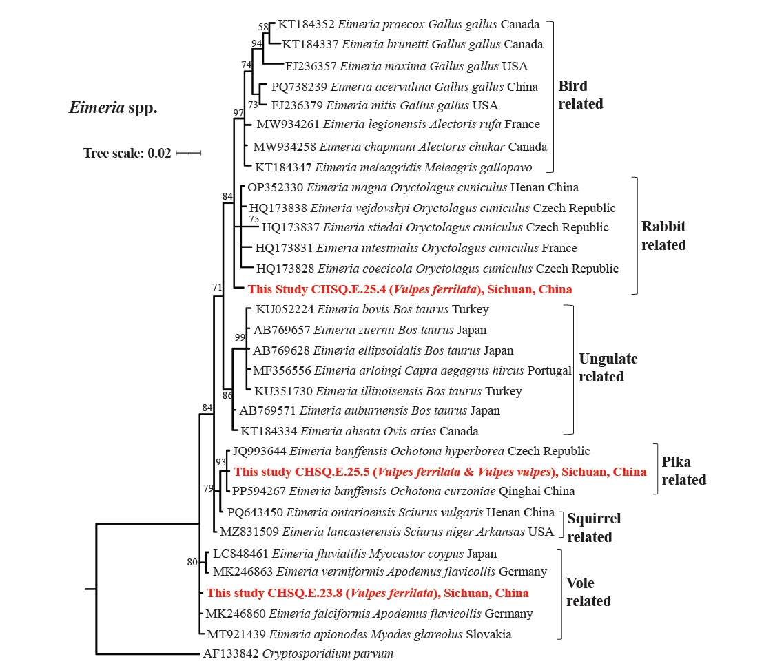
Fig. 5.Phylogenetic tree based on the Enterocytozoon bieneusi based on partial ITS1 gene sequences with the nucleotide substitution model of TPM1uf+G.
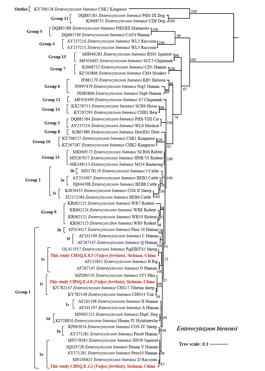
Table 1.Sequences of the host and protozoan primers and annealing temperature of PCR in this study
Table 1.
|
Primers |
Classification |
Primer names |
Target genes |
Primer sequences (from 5′→3′) |
Product length (bp) |
Annealing temperature (℃) |
References |
|
Universal primers for host recognition |
Vertebrate |
V-16S |
16S ribosomal RNA (16S rRNA) |
F: GAGAAGACCCTATGGAGC |
380 |
55 |
[9] |
|
R: ATAGAAACCGACCTGGAT |
|
Specific primers for parasite detection |
Cryptosporidium
|
CRY |
Small subunit ribosomal RNA (SSU rRNA) |
F1: TTCTAGAGCTAATACATGCG |
~1,325 |
55 |
[10] |
|
R1: CCCATTTCCTTCGAAACAGGA |
|
F2: GGAAGGGTTGTATTTATTAGATAAAG |
830 |
55 |
|
|
R2: AAGGAGTAAGGAACAACCTCCA |
|
Giardia intestinalis
|
GITS |
Triosephosphate isomerase (TPI) |
F1: AAATIATGCCTGCTCGTCG |
605 |
50 |
[11] |
|
R1: CAAACCTTITCCGCAAACC |
|
F2: CCCTTCATCGGIGGTAACTT |
530 |
50 |
|
|
R2: GTGGCCACCACICCCGTGCC |
|
Enterocytozoon bieneusi
|
EB |
Internal transcribed spacer 1 (ITS1) |
F1: GGTCATAGGGATGAAGAG |
410 |
57 |
[12] |
|
R1: TTCGAGTTCTTTCGCGCTC |
|
F2: GCTCTGAATATCTATGGCT |
392 |
55 |
|
|
R2: ATCGCCGACGGATCCAAGTG |
|
Eimeria
|
ESSU |
SSU rRNA
|
F1: TACCCAATGAAAACAGTTT |
636 |
53.6 |
[13] |
|
R1: CAGGAGAAGCCAAGGTAGG |
|
F2: CCTTCCGCGCTTCGCTGCGT |
294 |
66.5 |
|
|
R2: CGTCTTCAAACCCCCTACTG |
Table 2.Eight protozoan haplotypes from wild carnivores in the eastern Tibetan Plateau matched with NCBI GenBank
Table 2.
|
Haplotype names |
No. of samples |
Host (no. of samples) |
NCBI accession No. |
Coverage and similarity of the most similar sequences in GenBank (%) |
Species with the closest sequence in GenBank |
Genotype of the closest sequence in GenBank |
|
CHSQ.C.36.7 |
1 |
Vulpes ferrilata (1) |
PX249759 |
KF971356 (100%, 100%) |
Cryptosporidium
|
Yak genotype |
|
CHSQ.G.13.9 |
2 |
V. ferrilata (1) |
PX262650 |
MN844148 (100%, 99.09%) |
Giardia intestinalis
|
Assemblage AII |
|
Vulpes vulpes (1) |
|
CHSQ.E.25.5 |
25 |
V. ferrilata (23) |
PX249765 |
PP594267 (100%, 100%) |
Eimeria banffensis
|
- |
|
V. vulpes (2) |
|
CHSQ.E.23.8 |
26 |
V. ferrilata (26) |
PX249766 |
MH752019 (100%, 100%) |
Eimeria falciformis
|
- |
|
CHSQ.E.25.4 |
1 |
V. ferrilata (1) |
PX249767 |
OP352330 (100%, 99.25%) |
Eimeria magna
|
- |
|
CHSQ.E.4.2 |
11 |
V. ferrilata (11) |
PX262653 |
MN190621 (100%, 99.19%) |
Enterocytozoon bieneusi
|
D genotype |
|
CHSQ.E.4.8 |
1 |
V. ferrilata (1) |
PX262654 |
MZ090559 (100%, 99.59%) |
E. bieneusi
|
ST3 |
|
CHSQ.E.8.3 |
1 |
V. ferrilata (1) |
PX262655 |
OL411937 (100%, 100%) |
E. bieneusi
|
PigEBITS5 |
Table 3.Detection rate of protozoa parasites in main wild carnivores from Tibetan Plateau
Table 3.
|
Protozoan |
Detection rate (%) in carnivores, with 95% CI |
|
Vulpes ferrilata
|
Vulpes vulpes
|
Total |
|
Cryptosporidium sp. yak genotype |
0.33 (1/307, 0–0.96) |
0 (0/44) |
0.28a (1/351, 0–0.84) |
|
Giardia intestinalis
|
0.33 (1/307, 0–0.96) |
2 (1/44, 0–7) |
0.57a,b (2/351, 0–1.36) |
|
Eimeria banffensis
|
7.49 (23/307, 4.55–10.44) |
5 (2/44, 0–11) |
7.12c (25/351, 4.43–9.81) |
|
Eimeria falciformis
|
8.47 (26/307, 5.35–11.58) |
0 (0/44) |
7.41c (26/351, 4.67–10.15) |
|
Eimeria sp. |
0.33 (1/307, 0.00–0.96) |
0 (0/44) |
0.28a (1/351, 0–0.84) |
|
Enterocytozoon bieneusi
|
4.23 (13/307, 1.98–6.49) |
0 (0/44) |
3.70b,c (13/351, 1.73–5.68) |
|
Total |
21.17 (65/307, 16.60–25.74) |
7 (3/44, 0–14) |
19.37 (68/351, 15.24–23.51) |
References
- 1. Lv S, Tian LG, Liu Q, et al. Water-related parasitic diseases in China. Int J Environ Res Public Health 2013;10:1977-2016. https://doi.org/10.3390/ijerph10051977
- 2. Thompson RCA, Smith A. Zoonotic enteric protozoa. Vet Parasitol 2011;182:70-8. https://doi.org/10.1016/j.vetpar.2011.07.016
- 3. Li SA, Lin YL, Bai YP, et al. Prevalence and molecular characterization of Cryptosporidium spp. and Giardia duodenalis in humans and dogs from Fujian Province, Southeast China. Food Waterborne Parasitol 2025;40:e00278. https://doi.org/10.1016/j.fawpar.2025.e00278
- 4. Robertson LJ, Clark CG, Debenham JJ, et al. Are molecular tools clarifying or confusing our understanding of the public health threat from zoonotic enteric protozoa in wildlife? Int J Parasitol Parasites Wildl 2019;9:323-41. https://doi.org/10.1016/j.ijppaw.2019.01.010
- 5. Trout JM, Santín M, Fayer R. Giardia and Cryptosporidium species and genotypes in coyotes (Canis latrans). J Zoo Wildl Med 2006;37:141-4. https://doi.org/10.1638/05-06TYM-123005.1
- 6. Leśniańska K, Perec-Matysiak A. Wildlife as an environmental reservoir of Enterocytozoon bieneusi (Microsporidia): analyses of data based on molecular methods. Ann Parasitol 2017;63:265-81. https://doi.org/10.17420/ap6304.113
- 7. Chen Q, Wang X, Li C, et al. Investigation of parasitic nematodes detected in the feces of wild carnivores in the eastern Qinghai-Tibet Plateau, China. Pathogens 2022;11:1520. https://doi.org/10.3390/pathogens11121520
- 8. Jenkins EJ, Simon A, Bachand N, Stephen C. Wildlife parasites in a One Health world. Trends Parasitol 2015;31:174-80. https://doi.org/10.1016/j.pt.2015.01.002
- 9. Xiong M, Shao X, Long Y, et al. Molecular analysis of vertebrates and plants in scats of leopard cats (Prionailurus bengalensis) in southwest China. J Mammal 2016;97:1054-64. https://doi.org/10.1093/jmammal/gyw061
- 10. Khan A, Shaik JS, Grigg ME. Genomics and molecular epidemiology of Cryptosporidium species. Acta Trop 2018;184:1-14. https://doi.org/10.1016/j.actatropica.2017.10.023
- 11. Wang Y, Cao J, Chang Y, et al. Prevalence and molecular characterization of Cryptosporidium spp. and Giardia duodenalis in dairy cattle in Gansu, northwest China. Parasite 2020;27:62. https://doi.org/10.1051/parasite/2020058
- 12. Buckholt MA, Lee JH, Tzipori S. Prevalence of Enterocytozoon bieneusi in swine: an 18-month survey at a slaughterhouse in Massachusetts. Appl Environ Microbiol 2002;68:2595-9. https://doi.org/10.1128/AEM.68.5.2595-2599.2002
- 13. Orlandi PA, Carter L, Brinker AM, et al. Targeting single-nucleotide polymorphisms in the 18S rRNA gene to differentiate Cyclospora species from Eimeria species by multiplex PCR. Appl Environ Microbiol 2003;69:4806-13. https://doi.org/10.1128/AEM.69.8.4806-4813.2003
- 14. Zhuo G, Chen S, Zhou B. Spatio-temporal variation of vegetation coverage over the Tibetan Plateau and its responses to climatic factors. Acta Ecol Sin 2018;38:3208-18. https://doi.org/10.5846/stxb201705270985
- 15. Marneweck C, Butler AR, Gigliotti LC, et al. Shining the spotlight on small mammalian carnivores: global status and threats. Biol Conserv 2021;255:109005. https://doi.org/10.1016/j.biocon.2021.109005
- 16. Peng X, Wang X, Jian J, et al. Investigation of Cryptosporidium spp. and Enterocytozoon bieneusi in free-ranged livestock on the southeastern Qinghai-Xizang Plateau, China. BMC Infect Dis 2025;25:356. https://doi.org/10.1186/s12879-025-10737-5
- 17. Wang X, Jiang Y, Wu W, et al. Cryptosporidiosis threat under climate change in China: prediction and validation of habitat suitability and outbreak risk for human-derived Cryptosporidium based on ecological niche models. Infect Dis Poverty 2023;12:35. https://doi.org/10.1186/s40249-023-01085-0
- 18. Ma J, Cai J, Ma J, Feng Y, Xiao L. Occurrence and molecular characterization of Cryptosporidium spp. in yaks (Bos grunniens) in China. Vet Parasitol 2014;202:113-8. https://doi.org/10.1016/j.vetpar.2014.03.030
- 19. Zuo Q, Bianba Z, Xue C, et al. Infections and genetic diversity of Cryptosporidium spp. and Giardia spp. in small wild mammals on the Eastern Tibetan plateau: public health implications. BMC Microbiol 2025;25:616. https://doi.org/10.1186/s12866-025-04363-z
- 20. Einarsson E, Troell K, Hoeppner MP, et al. Coordinated changes in gene expression throughout encystation of Giardia intestinalis. PLoS Negl Trop Dis 2016;10:e0004571. https://doi.org/10.1371/journal.pntd.0004571
- 21. National Public Service Platform for Standards Information. GB 5749-2022 Standards for drinking water quality [Internet]. 2023 [cited 2026 Apr 10]. Available from: https://std.samr.gov.cn/gb/search/gbDetailed?id=DAB6B92C0764FC96E05397BE0A0A5F84
- 22. Sprong H, Cacciò SM, van der Giessen JW; ZOOPNET network and partners. Identification of zoonotic genotypes of Giardia duodenalis. PLoS Negl Trop Dis 2009;3:e558. https://doi.org/10.1371/journal.pntd.0000558
- 23. Li J, Wang H, Wang R, Zhang L. Giardia duodenalis infections in humans and other animals in China. Front Microbiol 2017;8:2004. https://doi.org/10.3389/fmicb.2017.02004
- 24. Zhang Y, Koehler AV, Wang T, Gasser RB. Enterocytozoon bieneusi of animals: with an 'Australian twist'. Adv Parasitol 2021;111:1-73. https://doi.org/10.1016/bs.apar.2020.10.001
- 25. Li W, Feng Y, Santin M. Host specificity of Enterocytozoon bieneusi and public health implications. Trends Parasitol 2019;35:436-51. https://doi.org/10.1016/j.pt.2019.04.004
- 26. Zhang Q, Cai J, Li P, et al. Enterocytozoon bieneusi genotypes in Tibetan sheep and yaks. Parasitol Res 2018;117:721-7. https://doi.org/10.1007/s00436-017-5742-1
- 27. Liu X, Du S, Yang X, et al. First genotyping of Enterocytozoon bieneusi in plateau pikas (Ochotona curzoniae) from the Qinghai Plateau, Northwest China. Vet Res Commun 2021;45:453-7. https://doi.org/10.1007/s11259-021-09824-0
- 28. Wang X, Shen Y, Cao J. Epidemic status and prevention and control progress of cryptosporidiosis in China. J Trop Dis Parasitol 2022;20:136-48. https://doi.org/10.3969/j.issn.1672-2302.2022.03.004
- 29. Nourrisson C, Lavergne RA, Moniot M, Morio F, Poirier P. Enterocytozoon bieneusi, a human pathogen. Emerg Microbes Infect 2024;13:2406276. https://doi.org/10.1080/22221751.2024.2406276
- 30. Gherman CM, Mihalca AD. A synoptic overview of golden jackal parasites reveals high diversity of species. Parasit Vectors 2017;10:419. https://doi.org/10.1186/s13071-017-2329-8
- 31. Wang X, Xue C, Deng X, et al. Seasonal population dynamics and dietary switching of Vulpes spp. amplify Echinococcus spp. transmission in the Eastern Tibetan plateau: implications for wildlife-mediated zoonotic risks. BMC Vet Res 2025;21:475. https://doi.org/10.1186/s12917-025-04907-5
- 32. Wang Q, Wang Y, Luo Z, et al. Settlement characteristics and transmission of echinococcosis: a cross-sectional study in nomadic communities on the Qinghai-Tibet Plateau, China. Infect Dis Poverty 2025;14:47. https://doi.org/10.1186/s40249-025-01316-6
- 33. Mohan M, Sathyakumar S, Krishnamurthy R. Predator in proximity: how does a large carnivore respond to anthropogenic pressures at fine-scales? Implications for interface area management. PeerJ 2024;12:e17693. https://doi.org/10.7717/peerj.17693
- 34. Thompson RC. Parasite zoonoses and wildlife: One Health, spillover and human activity. Int J Parasitol 2013;43:1079-88. https://doi.org/10.1016/j.ijpara.2013.06.007
- 35. Han BA, Schmidt JP, Bowden SE, Drake JM. Rodent reservoirs of future zoonotic diseases. Proc Natl Acad Sci U S A 2015;112:7039-44. https://doi.org/10.1073/pnas.1501598112


 , Chuizhao Xue2,3,4,5