Abstract
The present study was performed to estimate the seroprevalence of larval Anisakis simplex infection among the residents health-examined in 3 hospitals in southern parts of Korea. A total of 498 serum samples (1 serum per person) were collected in 3 hospitals in Busan Metropolitan city, Masan city, and Geoje city in Gyeongsangnam-do (Province) and were examined by IgE-ELISA and IgE-western blotting with larval A. simplex crude extract and excretory-secretory products (ESP). The prevalence of antibody positivity was 5.0% and 6.6% with ELISA against crude extracts and ESP, respectively. It was also revealed that infection occurred throughout all age groups and higher in females than in males. A specific protein band of 130 kDa was detected from 10 patients with western blot analysis against crude extract and ESP among those who showed positive results by ELISA. Our study showed for the first time the seroprevalence of anisakiasis in Korea. The allergen of 130 kDa can be a candidate for serologic diagnosis of anisakiasis.
-
Key words: Anisakis simplex, seroepidemiology, excretory-secretory product, crude extract, ELISA, western blotting
INTRODUCTION
The nematode
Anisakis simplex is a representative parasite for marine mammals. Almost all marine fish and cephalopods can become infected with the third stage larva (L3) of
A. simplex. Human infections occur upon the ingestion of marine fish or cephalopods. The ingested
A. simplex larvae by humans penetrate the stomach wall causing acute abdominal pain, nausea, and vomiting within a few hours. When the larvae invade the gastric or intestinal mucosa, inflammatory reaction often results in ulcer or eosinophilic granuloma. Recently, it has become clear that anisakiasis is often associated with strong allergic reactions ranging from urticaria to anaphylactic shock [
1-
4].
Chai et al. [
5] reported that the prevalence of anisakiasis has remarkably increased throughout the world in the last 30 years. One of the main reasons for this increase is attributed to the preference for raw and slightly cooked food. This trend will bring about the rise of infectious diseases caused by parasitic infections, like
A. simplex larvae, through marine fishery products. Although anisakiasis might occur in any country where the people eat raw or undercooked fish or squids, the majority of cases have been diagnosed in Korea, Japan, Spain, and some other countries because of their eating habits [
6-
9].
The prevalence of
A. simplex infection has been reported to be different depending on the countries and areas. Several studies have reported that more than 10% of gastrointestinal anisakiasis showed allergic symptoms related to fish consumption. The anisakid infection rate in Spain was known to be around 0.43% in Galicia area and 12.4% in the population of Madrid [
10,
11]. An epidemiological study in Japan has shown that anisakiasis was more frequent in costal populations and among 20- to 50-year-old males. In addition, these patients were reported to engage in fishing industry or inhabit coastal areas [
12]. The high prevalence of
A. simplex larval infection of fish and cephalopods has been reported in various fish species and squids [
13-
15]. Favored fishes of the Korean people, such as mackerels, cods, Alaska pollacks, scabbard fish, and squids were heavily infected with
A. simplex L3 [
16,
17]. These reports suggested possible association between fish consumption and allergic responses in Korean people. Nevertheless, seroepidemiologic surveys among Koreans have not been accomplished.
To obtain data of seroprevalence against A. simplex allergens among Koreans, we analyzed blood samples from residents in the southern parts of Korea by ELISA and Western blot analysis using crude extract and excretory-secretory products (ESP) from A. simplex L3.
MATERIALS AND METHODS
Subjects of investigation
We prepared 498 sera from blood samples collected in 3 hospitals, each located in Busan Metropolitan city, Masan city, and Geoje city. The study population were selected from the patients admitted for health examinations. They had no history of allergic symptoms, including allergic rhinitis, urticaria, atopic dermatitis, and asthma (total IgE <100 IU/ml). Serum samples were obtained by clotting and centrifugation of the blood at room temperature and were stored at -70℃. The collection of sera for use in these studies was approved by the Human Subjects Investigation Committee of Kosin University, Busan, Korea.
Preparation of Anisakis antigens
A. simplex L3 larvae were collected manually from the viscera, flesh, and body cavities of naturally infected mackerels (Scomber japonicus) and thoroughly washed with PBS. The crude extract of A. simplex L3 larvae was prepared from 300 larvae (about 1 g). Larvae were frozen in liquid nitrogen and smashed by mortar. To extract proper amounts of proteins, the protein extraction solution was added and stored on ice for 30 min according to the manufacturer's instructions (PRO_PREP™, iNtRON Biotechnology, Seoul, Korea). The supernatant was collected after centrifugation at maximum speed for 10 min at 4℃. The amount of protein was measured by the Bradford method.
ESP were prepared as described below. The collected larvae were incubated in PBS for 24 hr to remove salts and other foreign bodies. Larvae were incubated in DMEM (Dulbecco's Modified Eagle Medium) with gentamycin (150 mg/ml) and vancomycin (10 mg/ml) at 0.5 ml/larva, 37℃ for 48 hr. The media were collected and centrifuged at 2,500 rpm for 20 min and concentrated with Amicon stirred cells , cut off molecular weight less than 10,000 (Millipore Corp, Massachusetts, USA).
IgE ELISA
The crude extract and ESP of A. simplex L3 larvae were used as antigens. Both of them were diluted for the ELISA Starter Kit (Koma Biotech, Seoul, Korea) as 1 g/ml and 100 µl of aliquots were added each well of 96 well ELISA plate. Antigens were incubated overnight at 4℃ and washed with Tris-Buffered Saline with tween-20 (TBST) 3 times. Blocking solution was added each well and incubated for 1 hour at room temperature. Samples were incubated with 1:5 diluted patients' sera and 1:500 diluted peroxidase conjugated goat anti-human IgE (Sigma Aldrich, St. Louis, Missouri, USA) as the secondary antibody. For color reactions, 100 µl of TMB substrate solution was added and stopped the reactions when color reaction was enough to measure. Absorbance was measured at 450 nm by ELISA reader (Emax, Molecular Devices, Downingtown, Pennsylvania, USA). A mean OD value±3SD of the negative control sera was set as a cut-off value. Negative control sera were collected from 10 healthy persons aged 20-25 years who had no allergic history and the total IgE level was less than 100 IU/ml.
Immunoblot
Antigens from crude extracts and ESP were run through protein gel electrophoresis. Proteins were diluted with dissociation buffer (3:1) and boiled at 100℃ for 5 min. Boiled proteins were run through 12% SDS-polyacrylamide gel and transferred on a nitrocellulose membrane by Mini-Protean II (Bio-Rad, Richmond, California, USA) at 25 volt for 1.5 hr. Transferred proteins were checked by ponceau staining and blocked with PBS containing 5% skim milk at room temperature for 1 hr. After 3 times washing with PBST for 10 min, membranes were incubated with diluted ELISA positive patients' sera (1:10) at 4℃ overnight. Blotted membranes were washed 3 times with PBST for 10 min and incubated with horseradish peroxidase-labeled goat anti-human IgE (1:500) (Sigma) at room temperature for 1 hr. After 3 times washing with PBST for 10 min, color reaction was conducted by adding 4-chloro-1-naphtol (Sigma) or DAB substrate (Pierce, Rockford, Illinois, USA) on the blotted membrane.
RESULTS
The study population consisted of 269 females and 229 males. The ages of patients were from teens to 98 years old. Among them, 107 were in their twenties, and was the largest group. The numbers of thirties, forties, fifties, sixties, seventies, and eighties and older were 81, 71, 55, 55, 45, and 50, respectively (
Table 1). To analyze the pattern of antigens from crude extracts and ESP of
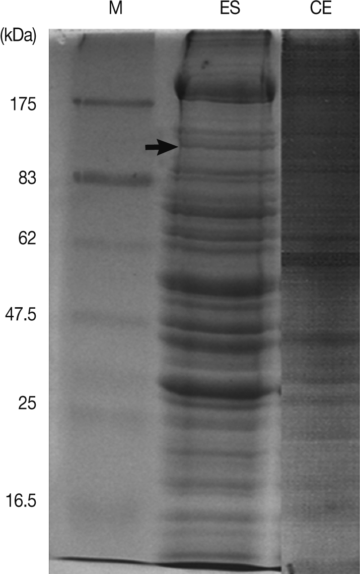
A. simplex L3, proteins were run through SDS-PAGE. Proteins were distributed from molecular weight of 200 kDa to 10 kDa (
Fig. 1).
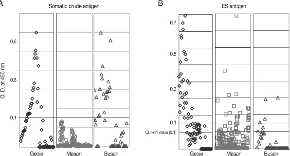
The optical density (OD) by ELISA of the 498 patients revealed 0.03±0.084 (mean±SD), and 0.01±0.108 for antigens from crude extract and ESP, respectively. The seropositive rate was 5.0% with crude extract antigen while that with ESP antigen was 6.6% (33 positives) by ELISA among the 498 patients (by cut-off OD value of 0.10). Twenty were females among 33 positive patients (
Table 1). The OD value of the positive group was 0.28±0.114 and 0.23±0.117, and that of negative group was 0.01±0.042 and 0.01±0.079 for antigens from crude extracts and ESP, respectively. The specific serum IgE level against
A. simplex crude extracts and ESP showed various distributions between the study populations. The serum samples from Geoje city showed more extensive distribution and higher values than those from the other 2 cities (
Fig. 2).
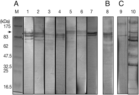
Western blotting carried out for ELISA positive sera revealed a specific band of about 130 kDa in 10 patients in both crude extract and ESP (
Fig. 3). The mean OD of ELISA for 10 patients who showed the specific band in western blot was 0.28±0.221. The regional distribution of positive serum samples by Western blotting showed that 8 positive patients were in Geoje city, and 2 patients were in Masan city. Eight were females among all 10 positive sera.
DISCUSSION
In our study, we analyzed the serum samples obtained from residents of Busan Metropolitan city, Masan city, and Geoje city by ELISA and western blotting for the seroepidemiologic study of
A. simplex L3 infection. The positive rates by ELISA showed 5.0% and 6.6% for antigens from crude extracts and ESP, respectively. Western blot analysis of ELISA positive sera revealed the specific band of about 130 kDa in 10 patients in both crude extract and ESP. This protein is considered similar to Ani s 7 (139 kDa) which was proposed by Rodriguez et al. [
18], which was reported to be the typical antigen for
A. simplex L3. Our work provided the first data on the seroprevalence of anti-
Anisakis IgE sensitization in Korea.
In Spain, the prevalence of anisakiasis varied depending on location, such as 12.4% in Madrid and 0.43% in Galicia [
10,
11]. Valinas et al. [
10] suggested that the low positive infection rate (0.43%) of anisakis allergy in Galicia, Spain, was due to the difference between live or dead larvae. They postulated that only live
A. simplex L3 can cause anisakis allergy. However, other reports set forth a counterargument that it can be different according to the habit of fish consumption, genetic background, and diagnostic methods [
19].
Many factors can affect the seroepidemiological prevalence. The types of antigens and diagnostic methods, however, are the most important factors influencing seroepidemiological investigations. Although the crude extracts and ESP are frequently used for serologic diagnosis, ESP were reported as the more potent and clinically important allergens for diagnosis [
20-
23]. Our results also showed that ESP was more potent allergen than the crude extract. However, cross reactions with other allergens, such as intestinal and blood-tissue nematodes, should be considered in regard to seropositive prevalence [
24,
25].
Many excretory-secretory and somatic
Anisakis-specific antigens have been reported, including Ani s 1 (21 kDa) [
26], Ani s 2 (100 kDa, paramyosin) [
27], Ani s 3 (41 kDa, tropomyosin) [
28-
29], Ani s 4 (9 kDa, cysteine protease inhibitor) [
30-
31], Ani s 5 (15 kDa protein homologous with the SXP/RAL-2 family proteins), and Ani s 6 (7 kDa, serine protease inhibitor) [
32]. Recent studies also reported functionally unknown antigens, including Ani s 8 (15 kDa), Ani s 9 (14 kDa), and Ani s 10 (21 kDa protein with unknown function; Allergen Nomenclature Sub-Committee;
http://www.allergen.org). We identified a 130 kDa protein with western blotting among ELISA positive patients. This protein showed a similar molecular weight with Ani s 7. Ani s 7 (139 kDa) was identified to have a 1096-amino acid fragment 7 (GenBank: EF158010) and 19 repeats of a novel CX17-25 CX 9-22 CX8 CX6 tandem repeat motif [
33]. Ani s 7 was reported probably the most important major excretory-secretory allergens since they were recognized by 100% of infected patients. In addition, Anti-Ani s 7 IgE antibodies were reported that they were induced by live
Anisakis larvae peaked at about day 30 post-infection and then decreased slowly over the course of 2 months [
33]. Our results also suggested that Ani s 7 can be a potent serodiagnostic antigen in Korean patients.
In conclusion, we recognized that the seropositive rate of anisakiasis was 5.0% and 6.6% with ELISA against crude extracts and ESP, respectively. The specific 130 kDa protein was confirmed by Western blot analysis among ELISA positive serum samples. This protein was similar with Ani s 7 in molecular weight and can be a candidate for diagnosis of anisakiasis among Koreans.
ACKNOWLEDGEMENTS
This work was supported by the Korea Research Foundation Grant funded by the Korean Government (MOEHRD, Basic Research Promotion Fund) (KRF-2006-E00037).
References
- 1. Kasuya S, Hamano H, Izumi S. Mackerel induced urticaria and Anisakis. Lancet 1990;335:665.
- 2. Daschner A, Pascual CY. Anisakis simplex: sensitization and clinical allergy. Curr Opin Allergy Clin Immunol 2005;5:281-285.
- 3. Moneo I, Caballero ML, Rodriguez-Perez R, Rodriguez-Mahillo AI, Gonzalez-MuÇoz M. Sensitization to the fish parasite Anisakis simplex: clinical and laboratory aspects. Parasitol Res 2007;101:1051-1055.
- 4. Audicana MT, Kennedy MW. Anisakis simplex: from obscure infectious worm to inducer of immune hypersensitivity. Clin Microbiol Rev 2008;21:360-379.
- 5. Chai JY, Murrell KD, Lymbery AJ. Fish-borne parasitic zoonoses; status and issues. Int J Parasitol 2005;35:1233-1254.
- 6. Im KI, Shin HJ, Kim BH, Moon SI. Gastric anisakiasis cases in Cheju-do, Korea. Korean J Parasitol 1995;33:179-186.
- 7. López-Serrano MC, Gomez AA, Daschner A, Moreno-Ancillo A, de Parga JM, Caballero MT, Barranco P, Cabañas R. Gastroallergic anisakiasis: findings in 22 patients. J Gastroenterol Hepatol 2000;15:503-506.
- 8. Nawa Y, Hatz C, Blum J. Sushi delights and parasites: the risk of fishborne and foodborne parasitic zoonoses in Asia. Clin Infect Dis 2005;41:1297-1303.
- 9. Choi SJ, Lee JC, Kim MJ, Hur GY, Shin SY, Park HS. The clinical characteristics of Anisakis allergy in Korea. Korean J Intern Med 2009;24:160-163.
- 10. Valinas B, Lorenzo S, Eiras A, Figueriras A, Sanmartin ML, Ubeira FM. Prevalence and risk factors for IgE sensitization to Anisakis simplex in a Spanish population. Allergy 2001;56:667-671.
- 11. Puente P, Anadon AM, Rodero M, Romaria F, Ubeira FM, Cuellar C. Anisakis simplex; the high prevalence in Madrid (Spain) and its relation with fish consumption. Exp Parasitol 2008;118:271-274.
- 12. Kimura S. Positive ratio of allergen specific IgE antibodies in serum, from a large scale study. Rinsho Byori 2001;49:376-380.
- 13. Chun SK, Chung BK, Ryu BS. Studies on Anisakis sp. (1) on the infection state of anisakiasis like larvae isolated from various marine fishes. Korean J Fish Aquat Sci 1968;1:99-105. (in Korean).
- 14. Abollo E, Gestal C, Pascual S. Anisakis infestation in marine fish and cephalopods from Galician waters: an updated perspective. Parasitol Res 2001;87:492-499.
- 15. Shih HH, Ku CC, Wang CS. Anisakis simplex (Nematoda: Anisakidae) third-stage larval infections of marine cage cultured cobia, Rachycentron canadum L., in Taiwan. Vet Parasitol 2010;171:277-285.
- 16. Ma HW, Jiang TJ, Quan FS, Chen XG, Wang HD, Zhang YS, Cui MS, Zhi WY, Jiang DC. The infection status of anisakid larvae in marine fish and cephalopods from the Bohai Sea, China and their taxonomical consideration. Korean J Parasitol 1997;35:19-24.
- 17. Choi SH, Kim J, Jo JO, Cho MK, Yu HS, Cha HJ, Ock MS. Anisakis simplex larvae: infection status in marine fish and cephalopods purchased from the Cooperative Fish Market in Busan, Korea. Korean J Parasitol 2011;49:39-44.
- 18. Rodríguez E, Anadón AM, García-Bodas E, Romarís F, Iglesias R, Gárate T, Ubeira FM. Novel sequences and epitopes of diagnostic value derived from the Anisakis simplex Ani s 7 major allergen. Allergy 2008;63:219-225.
- 19. Falcão H, Lunet N, Neves E, Barros H. Do only live larvae cause Anisakis simplex sensitization? Allergy 2002;57:44.
- 20. Arlian LG, Morgan MS, Quirce S, Marañón F, Fernández-Caldas E. Characterization of allergens of Anisakis simplex. Allergy 2003;58:1299-1303.
- 21. Baeza ML, Rodríguez A, Matheu V, Rubio M, Tornero P, de Barrio M, Herrero T, Santaolalla M, Zubeldia JM. Characterization of allergens secreted by Anisakis simplex parasite: clinical relevance in comparison with somatic allergens. Clin Exp Allergy 2004;34:296-302.
- 22. Kim JS, Kim KH, Cho S, Park HY, Cho SW, Kim YT, Joo KH, Lee JS. Immunochemical and biological analysis of allergenicity with excretory-secretory products of Anisakis simplex third stage larva. Int Arch Allergy Immunol 2005;136:320-328.
- 23. Moneo I, Caballero ML, Rodriguez-Perez R, Rodriguez-Mahillo AI, Gonzalez-Muñoz M. Sensitization to the fish parasite Anisakis simplex; clinical and laboratory aspects. Parasitol Res 2007;101:1051-1055.
- 24. Sakanari JA, Mckerrow JH. Anisakiasis. Clin Microbiol Rev 1989;2:278-284.
- 25. Cho JK, Cho SW. Shared epitope for monoclonal IR162 between Anisakis simplex larvae and Clonorchis sinensis and cross-reactivity between antigens. J Parasitol 2000;86:1145-1149.
- 26. Moneo I, Caballero ML, Gómez F, Ortega E, Alonso MJ. Isolation and characterization of a major allergen from the fish parasite Anisakis simplex. J Allergy Clin Immunol 2000;106:177-182.
- 27. Shimakura K, Miura H, Ikeda K, Ishizaki S, Nagashima Y, Shirai T, Kasuya S, Shiomi K. Purification and molecular cloning of a major allergen from Anisakis simplex. Mol Biochem Parasitol 2004;135:69-75.
- 28. Asturias JA, Eraso E, Martínez A. Cloning and high level expression in Escherichia coli of an Anisakis simplex tropomyosin isoform. Mol Biochem Parasitol 2000;108:263-267.
- 29. Asturias JA, Eraso E, Moneo I, Martínez A. Is tropomyosin an allergen in Anisakis? Allergy 2000;55:898-899.
- 30. Moneo I, Caballero ML, González-Muñoz M, Rodríguez-Mahillo AI, Rodríguez-Perez R, Silva A. Isolation of a heat-resistant allergen from the fish parasite Anisakis simplex. Parasitol Res 2005;96:285-289.
- 31. Rodriguez-Mahillo AI, Gonzalez-Muñoz M, Gomez-Aguado F, Rodriguez-Perez R, Corcuera MT, Caballero ML, Moneo I. Cloning and characterization of the Anisakis simplex allergen Ani s 4 as a cysteine-protease inhibitor. Int J Parasitol 2007;37:907-917.
- 32. Kobayashi Y, Ishizaki S, Shimakura K, Nagashima Y, Shiomi K. Molecular cloning and expression of two new allergens from Anisakis simplex. Parasitol Res 2007;100:1233-1241.
- 33. Anadón AM, Romarís F, Escalante M, Rodríguez E, Gárate T, Cuéllar C, Ubeira FM. The Anisakis simplex Ani s 7 major allergen as an indicator of true Anisakis infections. Clin Exp Immunol 2009;156:471-478.
Fig. 1SDS-PAGE of excretory-secretory products (ES) from Anisakis simplex L3 larvae and somatic crude extract (CE). M: Molecular marker. Arrow: 130 kDa.

Fig. 2Distribution of specific serum IgE against Anisakis simplex somatic crude extracts (CE) and excretory-secretory products (ES). It was expressed as the OD value of ELISA in residents of Geoje city, Masan city, and Busan Metropolitan city. Each point represents a single serum. Cut-off OD value is 0.10.

Fig. 3Western blot analysis of Anisakis simplex L3 excretory-secretory products (A, C) and crude extracts (B) with sera of patients. A,B: sera from Geoje city, C: sera from Masan city. The 130 kDa protein (arrow) was detected in 10 sera among ELISA positive samples. M: Molecular marker.

Table 1.Age group distribution of subjects and results of ELISA and western blot with Anisakis simplex L3 larva crude extract antigen and excretory-secretory antigen
Table 1.
|
Age |
No. of subjects
|
ELISA positive
|
WBa positive
|
|
Total |
Female |
Male |
CEb
|
ESc
|
CE |
ES |
|
10-19 |
34 |
23 |
11 |
4 |
3 |
0 |
0 |
|
20-29 |
107 |
67 |
40 |
6 |
5 |
0 |
1 |
|
30-39 |
81 |
54 |
27 |
5 |
6 |
0 |
4 |
|
40-49 |
71 |
26 |
45 |
3 |
4 |
0 |
1 |
|
50-59 |
55 |
17 |
38 |
2 |
1 |
1 |
1 |
|
60-69 |
55 |
15 |
40 |
1 |
3 |
0 |
1 |
|
70-79 |
45 |
29 |
16 |
4 |
7 |
1 |
0 |
|
80- |
50 |
38 |
12 |
0 |
4 |
0 |
0 |
|
Total |
498 |
269 |
229 |
25 |
33 (20d) |
2e
|
8e
|


