Abstract
Toxoplasma gondii is a zoonotic parasite resulting in human infections and one of the infectious pathogens leading to uveitis and retinochoroiditis. The present study was performed to assess T. gondii infection in 20 ocular patients with chronic irregular recurrent uveitis (20 aqueous humor and 20 peripheral blood samples) using PCR. All samples were analyzed by nested PCR targeting a specific B1 gene of T. gondii. The PCR-positive rate was 25% (5/20), including 5% (1) in blood samples, 25% (5) in aqueous humor samples, and 5% (1) in both sample types. A molecular screening test for T. gondii infection in ocular patients with common clinical findings of an unclear retinal margin and an inflammatory membrane over the retina, as seen by fundus examination, may be helpful for early diagnosis and treatment.
-
Key words: Toxoplasma gondii, toxoplasmosis, uveitis, nested PCR, B1 gene
Toxoplasma gondii is an important cause of zoonotic infection worldwide and may lead to uveitis, pneumonia, pericarditis, and neurologic disorders in immunocompetent hosts [
1]. As ocular infection is one of the main manifestations of
T. gondii infection, ocular toxoplasmosis is one of the most common types of infectious uveitis and a leading cause of posterior uveitis, also called retinochoroiditis [
2-
4], which may occur either immediately or long after initial infection. Recently, the importance of
T. gondii infection in ocular patients has been recognized in view of etiologic and clinical findings. The global prevalence of ocular toxoplasmosis is variable, with 1.8% in Japan, 39.8% in Colombia, 22.2% in Thailand, and 8.4% in the U.S. [
2,
5-
7]. Cases of ocular toxoplasmosis have been reported intermittently since the first case was described in Korea [
8-
11]. In 2003,
T. gondii parasites were successfully isolated from a Korean ocular patient [
12]. Recently, clinical features of ocular toxoplasmosis in Korean patients were described by Park et al. [
13]. However, general information on ocular toxoplasmosis in Korea is insufficient because clinical reports of ocular toxoplasmosis are rare and no epidemiologic survey has been conducted.
This study was carried out to assess
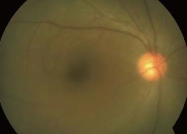
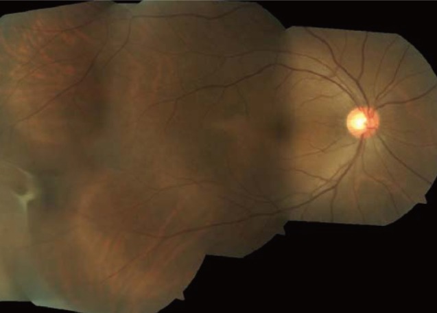
T. gondii infection in 40 clinical samples (20 peripheral blood samples and 20 aqueous humor samples) from ocular patients with irregular recurrent uveitis by PCR. Most of the included patients did not show typical ocular toxoplasmic retinochoroidal lesions, but complained of blurry vision and floaters; common clinical findings included an unclear retinal margin and an inflammatory membrane over the retina, as shown by fundus photography (
Figs. 1,
2). Samples were collected by an ophthalmologist from April to August 2009 and transferred from the Nune Eye Hospital to the Division of Malaria and Parasitic Disease at the Korea National Institute of Health for detection of
T. gondii. Genomic DNA was extracted from aqueous humor and peripheral blood with the commercially available DNeasy Mini column kit (Qiagen, Hilden, Germany) according to the manufacturer's instructions. DNA concentrations were measured using the Quant-iT™ dsDNA HS Assay Kit (Invitrogen, Carlsbad, California, USA) and read on a Qubit™ Fluorometer (Invitrogen). The average DNA concentration was 8.05 ng/µL (3.3-16.9 ng/µL). The DNA samples were stored at -22℃ until use. Two pairs of oligonucleotide primers, specific to the B1 gene of
T. gondii, were used to perform nested PCR using purified blood cell DNA as a template. The published B1 gene sequence was amplified using the Maxime PCR premix Kit (Intron, Seongnam, Korea) according to the method described by Hurtado et al. [
14]. The PCR program was modified as follows: 94℃ for 3 min, 15 cycles of 94℃ for 30 sec, 65℃ for 45 sec, 72℃ for 1 min, 35 cycles of 94℃ for 20 sec, 53℃ for 30 sec, 72℃ for 30 sec and a final extension of 72℃ for 5 min.
The overall positive rate for
T. gondii detection was 5% (1/20) of blood samples and 25% (5/20) of aqueous humor samples by PCR and 5% (1/20) in both blood and aqueous humor samples (
Table 1). This was a little lower than that of other countries [
15-
17]. It was reported that if aqueous humor samples are withdrawn too early after the onset of symptoms, false-negative by PCR results may be produced [
17] or may be attributable to late sampling [
18]. Therefore, PCR has previously been shown to be a useful tool for early diagnosis of
T. gondii infection in patients who test negative by ELISA [
19,
20] and PCR detection of parasites within aqueous humor samples formally confirms the diagnosis of either primary or reactivated ocular toxoplasmosis [
21].
T. gondii was found in the blood of acutely and chronically infected patients regardless of toxoplasmic retinochoroiditis [
20]. Although the examination with light microscopy in peripheral blood samples was not carried out, the specific B1 gene of
T. gondii in the blood was identified by nested PCR and confirmed by sequencing. The PCR-positive patients with chronic
T. gondii infection in the present study had an irregular recurrence of symptoms, as previously stated, and the infection may thus be regarded as atypical chronic ocular toxoplasmosis. Additionally, the supplementary diagnostic tests for other infections, including syphilis (VDRL), HIV (ELISA), tuberculosis (chest X-ray), and autoimmune diseases, such as Vogt-Koyanagi-Harada disease and Behcet disease, were negative in all patients.
The population with ocular toxoplasmosis vary by geographic location [
2,
5-
7,
22]. Moreover, an atypical presentation of ocular toxoplasmosis has been reported [
4]. Recently, there has been an increase in the reports of ocular toxoplasmosis, addressing the influence of patient age, frequency of inflammation, clinical features, diagnosis, and patterns of uveitis [
7]. Therefore, it is necessary to further study the prevalence of ocular toxoplasmosis in a larger population of ocular patients with typical and atypical uveitis or retinochoroiditis in Korea.
In conclusion, we suggest that PCR analysis for diagnosis of ocular toxoplasmosis may be needed for the earlier diagnosis and treatment of ocular patients presenting with blurry vision and floaters caused by an unclear retinal margin and an inflammatory membrane over the retina.
ACKNOWLEDGMENT
This work was supported by a grant (4847-302-210-13, 2009) from the Korea National Institute of Health, Republic of Korea.
References
- 1. Bossi P, Bricaire F. Severe acute disseminated toxoplasmosis. Lancet 2004;364:579.
- 2. Keino H, Nakashima C, Watanabe T, Taki W, Hayakawa R, Sugitani A, Okada AA. Frequency and clinical features of intraocular inflammation in Tokyo. Clin Experiment Ophthalmol 2009;37:595-601.
- 3. Rothova A, de Boer JH, Ten Dam-van Loon NH, Postma G, de Visser L, Zuurveen SJ, Schuller M, Weersink AJ, van Loon AM, de Groot-Mijnes JD. Usefulness of aqueous humor analysis for the diagnosis of posterior uveitis. Ophthalmology 2008;115:306-311.
- 4. Smith JR, Cunningham ET Jr. Atypical presentations of ocular toxoplasmosis. Curr Opin Ophthalmol 2002;13:387-392.
- 5. De-la-Torre A, Lopez-Castillo CA, Rueda JC, Mantilla RD, Gomez-Marin JE, Anaya JM. Clinical patterns of uveitis in two ophthalmology centres in Bogota, Colombia. Clin Experiment Ophthalmol 2009;37:458-466.
- 6. Sirirungsi W, Pathanapitoon K, Kongyai N, Weersink A, de Gerro-Mijnes JD, Leechanachai P, Ausayakhun S, Rothova A. Infectious uveitis in Thailand: Serologic analyses and clinical features. Ocul Immunol Inflamm 2009;17:17-22.
- 7. London NJ, Hovakimyan A, Cubillan LD, Siverio CD Jr, Cunningham ET Jr. Prevalence, clinical characteristics, and causes of vision loss in patients with ocular toxoplasmosis. Eur J Ophthalmol 2011;21:811-819.
- 8. Kong SM, Lee TS, Kim CW. 3 Cases of ocular toxoplasmosis. J Korean Ophthalmol Soc 1975;16:141-145.
- 9. Yoo HY, Cho BC. 2 Cases of ocular toxplasmosis. J Korean Ophthalmol Soc 1979;20:231-237.
- 10. Shyn KH, Kim KY, Park SC. A clinical experience of ocular toxoplasmosis, treated with acetyl spiramycin. J Korean Ophthalmol Soc 1979;20:427-431.
- 11. Kim MH, Choi YK, Park YK, Nam HW. A toxoplasmic uveitis case of a 60-year-old male in Korea. Korean J Parasitol 2000;38:29-31.
- 12. Chai JY, Lin A, Shin EH, Oh MD, Han ET, Nam HW, Lee SH. Laboratory passage and characterization of an isolate of Toxoplasma gondii from an ocular patient in Korea. Korean J Parasitol 2003;41:147-154.
- 13. Park YH, Han JH, Nam HW. Clinical features of ocular toxoplasmosis in Korean patients. Korean J Parasitol 2011;49:167-171.
- 14. Hurtado A, Aduriz G, Moreno B, Barandika J, García-Pérez AL. Single tube nested PCR for detection of Toxoplasma gondii in fetal tissues from naturally aborted ewes. Vet Parasitol 2001;102:17-27.
- 15. Fardeau C, Romand S, Rao NA, Cassoux N, Bettembourg O, Thulliez P, Lehoang P. Diagnosis of toxoplasmic retinochoroiditis with atypical clinical features. Am J Ophthalmol 2002;134:196-203.
- 16. Figueroa MS, Bou G, Marti-Belda P, Lopez-Velez R, Guerrero A. Diagnostic value of polymerase chain reaction in blood and aqueous humor in immunocompetent patients with ocular toxoplasmosis. Retina 2000;20:614-619.
- 17. Villard O, Filisetti D, Roch-Deries F, Garweg J, Flament J, Candolfi E. Comparison of enzyme-linked immunosorbent assay, Immunoblotting, and PCR for diagnosis of Toxoplasmic chorioretinitis. J Clin Microbiol 2003;41:3537-3541.
- 18. Payeur G, Bijon JC, Pagano N, Kien T, Candolfi E, Penner MF. Diagnosis of ocular toxoplasmosis by the ELISA method applied to the determination of immunoglobulins of the aqueous humor. J Fr Ophtalmol 1988;11:75-79.
- 19. Bidgoli S, Koch P, Caspers L. Toxoplasmic chorioretinitis: positive PCR on vitreous with negative serology for Toxoplasma gondii. J Fr Ophtalmol 2011;34:384.e1-384.e5.
- 20. Silveira C, Vallochi AL, Rodrigues da Silva UR, Muccioli C, Holland GN, Nussenblatt RB, Belfort R, Rizzo LV. Toxoplasma gondii in the peripheral blood of patients with acute and chronic toxoplasmosis. Br J Ophthalmol 2011;95:396-400.
- 21. Bastien P. Molecular diagnosis of toxoplasmosis. Trans R Soc Trop Med Hyg 2002;96:S205-S215.
- 22. Accorinti M, Bruscolini A, Pirraglia MP, Liverani M, Caggiano C. Toxoplasmic retinochoroiditis in an Italian referral center. Eur J Ophthalmol 2009;19:824-830.
Fig. 1The margin of the retinal vessels and disc was unclear due to the vitreous opacity in the patient.

Fig. 2Inflammatory membrane over the retina as seen by fundus photography in the patient.

Table 1.PCR results for blood and aqueous humor samples from 20 patients with irregular recurrent uveitis
Table 1.
|
Nested PCR
|
|
Blood |
Aqueous humor |
Both |
|
No. of positive/Total |
1/20 |
5/20 |
1/20 |
|
% |
5 |
25 |
5 |
Citations
Citations to this article as recorded by

- Molecular diagnosis of toxoplasmosis in immunocompromised patients
Florence Robert-Gangneux, Sorya Belaz
Current Opinion in Infectious Diseases.2016; 29(4): 330. CrossRef - IgG Avidity Antibodies against Toxoplasma gondii in High Risk Females of Reproductive Age Group in India
Naushaba Siddiqui, Fatima Shujatullah, Haris M. Khan, Tamkin Rabbani, Parvez A. Khan
The Korean Journal of Parasitology.2014; 52(5): 487. CrossRef - Establecimiento de una reacción en cadena de la polimerasa para la detección de bacterias y hongos
Héctor Javier Pérez-Cano
Revista Mexicana de Oftalmología.2014; 88(2): 67. CrossRef - PCR-Based Detection of Toxoplasma gondii DNA in Blood and Ocular Samples for Diagnosis of Ocular Toxoplasmosis
C. Bourdin, A. Busse, E. Kouamou, F. Touafek, B. Bodaghi, P. Le Hoang, D. Mazier, L. Paris, A. Fekkar, M. J. Loeffelholz
Journal of Clinical Microbiology.2014; 52(11): 3987. CrossRef - Prevalence and Genetic Characterization of Toxoplasma gondii in House Sparrows (Passer domesticus) in Lanzhou, China
Wei Cong, Si-Yang Huang, Dong-Hui Zhou, Xiao-Xuan Zhang, Nian-Zhang Zhang, Quan Zhao, Xing-Quan Zhu
The Korean Journal of Parasitology.2013; 51(3): 363. CrossRef - Serologic Survey of Toxoplasmosis in Seoul and Jeju-do, and a Brief Review of Its Seroprevalence in Korea
Hyemi Lim, Sang-Eun Lee, Bong-Kwang Jung, Min-Ki Kim, Mi Youn Lee, Ho-Woo Nam, Jong-Gyun Shin, Cheong-Ha Yun, Han-Ik Cho, Eun-Hee Shin, Jong-Yil Chai
The Korean Journal of Parasitology.2012; 50(4): 287. CrossRef


