Abstract
Acanthotrema felis is an intestinal trematode of cats originally reported from the Republic of Korea. Only 1 human case infected with a single adult worm has been previously recorded. In the present study, we report 4 human cases infected with a total of 10 worms recovered after anthelmintic treatment and purging. All 4 patients reside in coastal areas of Jeollanam-do, Korea, and have consumed brackish water fish including the gobies, Acanthogobius flavimanus. The worms averaged 0.47 mm in length and 0.27 mm in width, and had 3 sclerites on the ventrogenital sac; 1 was short and thumb-like, another was long and blunt-ended, and the 3rd was long and broad-tipped. They were identified as A. felis Sohn, Han, & Chai, 2003. Surveys on coastal areas to detect further human cases infected with A. felis are required.
-
Key words: Acanthotrema felis, human infection, intestinal fluke, heterophyid fluke
INTRODUCTION
The genus
Acanthotrema (Digenea: Heterophyidae) are intestinal parasites of fish-eating birds and mammals [
1].
Acanthotrema acanthotrema, the type species, was originally described from a bird (
Sterna maxima) in 1928 in Brazil [
2], and other species were subsequently documented. Currently, 8 species are included in this genus;
A. acanthotrema,
A. armata,
A. cursitans,
A. felis,
A. hancocki,
A. martini,
A. tanayensis, and
A. tridactyla [
1]. The newest species is
A. felis, which was described in 2003 from the small intestine of stray cats in the Republic of Korea [
1]. Various brackish water fish have been reported to be the second intermediate host for
Acanthotrema species including the mud sucker
Gillichthys mirabilis and the Southern California killifish for
A. hancocki [
3] and the goby
Acanthogobius flavimanus for
A. felis [
4].
The geographical locality of
Acanthotrema spp. is wide. For example,
A. acanthotrema has been reported from Brazil [
2],
A. hancocki,
A. martini, and
A. cursitans from the United States [
3,
5,
6],
A. tridactyla from Egypt [
7] and Kuwait [
8],
A. armata from Spain [
2],
A. tanayensis from the Philippines [
9], and
A. felis from Korea [
1]. The definitive hosts for
A. acanthotrema,
A. armata,
A. hancocki, and
A. martini are birds, including sea gulls, and those of
A. felis,
A. cursitans, and
A. tanayensis are mammals, particularly cats [
1].
A. tridactyla can experimentally infect kittens [
8] and chicks [
7].
Human infections have never been reported in
Acanthotrema spp., with the exception of
A. felis [
10]. A 70-year-old man residing in Muan-gun, Korea, expelled a single adult worm of
A. felis after praziquantel treatment, together with 148
Heterophyes nocens, 65
Gymnophalloides seoi, 30
Stictodora fuscata, 1
Stictodora lari, and 1
Stellantchasmus falcatus [
10]. We describe here 4 additional human cases naturally infected with
A. felis in southern coastal areas of Korea.
CASE RECORD
During surveys of human intestinal flukes in Shinan-gun (1990), Muan-gun (1995), and Yeongam-gun (2003), Jeollanam-do, Korea, small trematode egg (STE) positive cases in Kato-Katz fecal smears were subjected to adult fluke collection after anthelmintic treatment and purging. Briefly, praziquantel at a dose of 10-15 mg/kg was orally administered and an hour later the patient was given 30 g of magnesium sulfate. All the diarrheic stools were collected from each case for 4-5 hr and examined under a stereomicroscope. The fluke specimens, if positive, were morphologically examined using a light microscope. Informed consent was obtained from each patient regarding the adult worm recovery.
Ten specimens of
A. felis were recovered from 4 patients (1-7 per person, average 2.5 worms) (
Table 1). All 4 patients were commonly infected with other heterophyid flukes including
H. nocens,
Pygidiopsis summa,
S. fuscata, and
Heterophyopsis continua. The number of specimens of the aforementioned fluke species was much higher than that of
A. felis (
Table 1). The patients were suffering from vague abdominal discomfort and indigestion, sometimes accompanied by frequent stool, lethargy, and anorexia. However, it was considered that these symptoms were not necessarily due to
A. felis infection. The patients had habitually eaten raw flesh of brackish water fish, such as the mullet and goby.
The morphological characteristics and measurements (µm) of the recovered
A. felis adults are as follows (
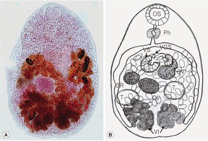
Table 2). The worms were pear-shaped and were covered with fine tegumental spines, except near the posterior extremity (
Fig. 1A, B). Their average body size was 472 in length (range, 405-510) and 269 (230-300) in width. These dimensions were slightly smaller than previously described [
1]. The ventral sucker was embedded in the ventrogenital sac which had a distinct membranous covering. The ventrogenital sac was located in the median line, usually at the anterior end of the middle third of the body. Inside the ventrogenital sac, 3 sclerites were observed; 1 was short and thumb-like, 1 was long and blunt-ended, and 1 was long and broadly tipped (
Fig. 1). In some specimens, minute spines were seen at the base of the sclerite complex. The gonotyl was U-shaped, unspined, located right to the sclerite complex, and was included in the ventrogenital sac. The ovary was spherical, submedian, and posterolateral to the ventrogenital sac. The seminal receptacle, anterolateral to the ovary, was barely observed. Two testes were ovoid, posterolateral to the ovary, and a little obliquely side-by-side, and were located at the posterior half of the body. The bipartite seminal vesicle was connected to the genital sinus and then to the genital opening in the ventrogenital sac. Vitellaria distributed from the post-testicular level to the posterior extremity, forming 2 large groups of follicles. The uterus formed a transverse loop immediately in front of the ovary, and nearly filled up the posterior half of the body. Eggs elongate oval and light brown. Specimens have been deposited in the Seoul National University Parasite Collection (SNUPC nos. 14011-14020).
DISCUSSION
Since the first discovery of
A. felis in Korea, its presence has been documented several times in the country. Adult flukes were reported from feral cats caught near Pusan [
1] and Shinan-gun [
11] and from a human in Muan-gun [
10]. The present study adds Shinan-gun and Yeongam-gun as new localities for human infections. Metacercariae of
A. felis were discovered in 7 (35.0%) of 20
A. flavimanus in Haenam-gun [
4], and in 31 (91.2%) of 34
A. flavimanus in Muan-gun [
12], with the total number of metacercariae being 116 (17 per fish) and 7,292 (235 per fish), respectively.
Among the 8 species of
Acanthotrema, only
A. felis is so far known to distribute in Korea [
1], and is the only species infecting humans around the world, based on a previous report [
10] and the present study. However, humans seem to be a less susceptible host than birds or mammals, and the pathogenicity of Acanthotrema worms to humans is unknown. In the 5 known human cases, the number of worms recovered per individual was usually 1 with the present exception of 1 patient who expelled 7 worms. This strongly suggests that the human cases may be accidental infections. This low number of infected worms probably would not cause any harmful effect to humans. Humans may have innate resistance against this fluke, although this issue needs further clarification.
A. felis is morphologically close to other heterophyids such as
Stictodora spp. However,
Acanthotrema has fewer than 12 spines or sclerotizations, and
Stictodora has more than 12 spines on the ventral sucker or ventrogenital sac [
13]. It was not difficult to assign our specimens to
Acanthotrema because they typically had 3 sclerotizations in the ventrogenital sac [
1]. Other morphological characters included a submedian ovary, a bipartite seminal vesicle, a short prepharynx, absence of spines on the gonotyl, and the presence of 3 sclerites on the ventrogenital sac.
A. tanayensis has 4 sclerites which are unarmed [
9] and the sclerites of
A. acanthotrema consist of 2 armed pieces and a third unarmed piece [
2].
A. tridactyla has 3 sclerites having minute spines on the tip [
7].
A. armata has 2 armed sclerites [
2], and
A. cursitans has 2 unarmed, branched sclerites [
6].
A. hancocki and
A. martini have 1 U-shaped sclerite [
3,
5]. The present specimens displayed 3 sclerites with spines on the tip, differing from
A. acanthotrema and
A. tridactyla, which have 2-3 clerites with minute spines on the tip. The shape of the sclerites in our specimens was slightly different from that in the original report of this species [
1]. The sclerites in the original report consisted of 1 short, thumb-like piece and 2 long and pointed ones [
1]. However, in our specimens, the sclerites consisted of 1 short and thumb-like piece, 1 long and broadly tipped piece, and 1 long and blunt-ended piece. We regard this as an intraspecific variation and identify our specimens as
A. felis.
The present study is the second report on human
A. felis infection following the report of Cho et al. [
10]. This fluke should be included among the list of human-infecting foodborne intestinal flukes in Korea [
14] and Asia [
15]. Further studies are required to clarify the geographical distribution and host-parasite relationships of
A. felis infection.
Notes
-
We have no conflict of interest related to this study.
References
- 1. Sohn WM, Han ET, Chai JY. Acanthotrema felis n. sp. (Digenea: Heterophyidae) from the small intestine of stray cats in the Republic of Korea. J Parasitol 2003;89:154-158.
- 2. Lafuente M, Roca V, Carbonell E. Description of Acanthotrema armata n. sp. (Trematoda: Heterophyidae) from Larus audouinii (Aves: Laridae), with an amended diagnosis of the genus Acanthotrema Travassos, 1928. Syst Parasitol 2000;45:131-134.
- 3. Martin WE. Parasitictodora hancocki n. gen., n. sp. (Trematoda: Heterophyidae), with observation on its life cycle. J Parasitol 1950;36:360-370.
- 4. Sohn WM, Han ET, Seo M, Chai JY. Identification of Acanthotrema felis (Digenea: Heterophyidae) metacercariae encysted in the brackish water fish Acanthogobius flavimanus. Korean J Parasitol 2003;41:101-105.
- 5. Sogandares-Bernal F. Four trematodes from the Black Skimmer, Rynchops nigra Linn. (Aves: Rynchopidae), in Gasparilla Sound, Florida, including the description of a new genus and two new species. Quart J Florida Acad Sci 1959;22:125-132.
- 6. Kinsella JM, Heard RW III. Morphology and life cycle of Stictodora cursitans n. comb. (Trematoda: Heterophyidae) from mammals in Florida salt marshes. Trans Am Microsc Soc 1974;93:408-412.
- 7. Martin WE, Kuntz RE. Some Egyptian heterophyid trematodes. J Parasitol 1955;41:374-382.
- 8. Abdul-Salam J, Sreelatha BS, Ashkanani H. Surface ultrastructure of Stictodora tridactyla (Trematoda: Heterophyidae) from Kuwait Bay. Parasitol Int 2000;49:1-7.
- 9. Velasquez CC. Observations on some Heterophyidae (Trematoda: Digenea) encysted in Philippine fishes. J Parasitol 1973;59:77-84.
- 10. Cho SH, Cho PY, Lee DM, Kim TS, Kim IS, Hwang EJ, Na BK, Sohn WM. Epidemiological survey on the infection of intestinal flukes in residents of Muan-gun, Jeollanam-do, the Republic of Korea. Korean J Parasitol 2010;48:133-138.
- 11. Shin EH, Park JH, Guk SM, Kim JL, Chai JY. Intestinal helminth infections in feral cats and a raccoon dog on Aphae Island, Shinan-gun, with a special note on Gymnophalloides seoi infection in cats. Korean J Parasitol 2009;47:189-191.
- 12. Cho SH, Kim IS, Hwang EJ, Kim TS, Na BK, Sohn WM. Infection status of estuarine fish and oysters with intestinal fluke metacercariae in Muan-gun, Jeollanam-do, Korea. Korean J Parasitol 2012;50:215-220.
- 13. Pearson JC. A revision of the subfamily Haplorchinae Looss, 1899 (Trematoda: Heterophyidae). II. Genus Galactosomum. Philos Trans R Soc Lond B Biol Sci 1973;266:341-447.
- 14. Chai JY, Lee SH. Food-borne intestinal trematode infections in the Republic of Korea. Parasitol Int 2002;51:129-154.
- 15. Chai JY, Shin EH, Lee SH, Rim JH. Foodborne intestinal flukes in Southeast Asia. Korean J Parasitol 2009;47(suppl):S69-S102.
Fig. 1(A) An adult fluke of Acanthotrema felis recovered from a patient. Acetocarmine stained. Bar=45 µm. (B) Line drawing of the worm in Fig. 1A showing its internal organs. The same magnification as Fig. 1A. OS, oral sucker; Ph, pharynx; VS, ventral sucker; VGS, ventrogenital sac; SV, seminal vesicle; SR, seminal receptacle; Ov, ovary; T, testis; Vt, vitellaria.

Table 1.
Acanthotrema felis and other trematode specimens recovered from the patients
Table 1.
|
Patient code |
Age/sex |
Residence |
Date |
No. of A. felis specimens recovered |
Other flukes recovered (no. of specimens) |
|
A |
36/M |
Shinan-gun |
Feb. 1990 |
7 |
H. nocens (86), P. summa (33), S. fuscata (5), H. continua (1) |
|
B |
56/M |
Shinan-gun |
Feb. 1990 |
1 |
H. nocens (155), P. summa (16), S. fuscata (14) |
|
C |
50/M |
Muan-gun |
Jan. 1995 |
1 |
H. nocens (81), P. summa (70), S. fuscata (34) |
|
D |
54/M |
Yeongam-gun |
Nov. 2003 |
1 |
H. nocens (146), S. fuscata (9) |
Table 2.Measurements (μm) of Acanthotrema felis specimens compared to those of the original description
Table 2.
|
Organ |
The present specimens |
Sohn et al. (2003) |
|
Body length × width |
472 × 269 |
495 × 263 |
|
Oral sucker |
58 × 59 |
49 × 58 |
|
Pharynx |
41 × 37 |
43 × 39 |
|
Ventral sucker |
50 × 41 |
90 × 58 |
|
Testis (right) |
88 × 43 |
68 × 50 |
|
Testis (left) |
83 × 50 |
67 × 48 |
|
Sclerite 1 |
27 |
|
|
Sclerite 2 |
38 |
|
|
Sclerite 3 |
37 |
|
|
Egg |
26 × 16 |
27 × 15 |

