Abstract
Trichinellosis transmission to humans via the consumption of reptile meat is rare worldwide. In Korea, however, 2 such outbreaks, possibly via consumption of soft-shelled turtle meat, have occurred in 2 successive years. In 17 August 2014, 6 patients were admitted to Wonju Severance Christian Hospital complaining of myalgia, fever, and headache. Eosinophilia was the indication of the initial laboratory results, and they were eventually diagnosed as trichinellosis by ELISA. All of the patients worked at the same company and had eaten raw soft-shelled turtle meat at a company dinner 10 days prior to their admission. They were treated with albendazole for 2 weeks, upon which all of their symptoms disappeared. This is the 8th report on human trichinellosis in Korea, and the second implicating raw soft-shelled turtle meat.
-
Key words: Trichinella, trichinellosis, soft-shelled turtle, ELISA, Korea
INTRODUCTION
Although pork remains the predominant source of trichinellosis throughout the world, other meats occasionally have been implicated as well [
1]. In recent years, the
Trichinella spp. host range has been widening, and new species and genotypes have emerged [
1]. A wider host spectrum is especially evident in non-encapsulated species, such as
Trichinella pseudospiralis, infecting birds [
2]. Furthermore, it has been shown that
Trichinella papuae and
Trichinella zimbabwensis can infect reptiles, the species living in equatorial regions [
2]. It has also been established that the life cycle of
Trichinella spiralis can be reproduced in reptiles when maintained at higher temperatures [
3].
Human trichinellosis transmission via consumption of reptile meat was first reported in Thailand in 2008, where the sources of infection were turtle and brown lizard meat [
4]. The second outbreak occurred in 2008 in Taiwan, where
T. papuae was the likely causative agent [
5]. Prior to the onset of trichinellosis symptoms, all 8 patients had eaten raw soft-shelled turtles (
Pelodiscus sinensis), which were tentatively identified as the source of infection [
5]. Korea has proved to be an endemic area of trichinellosis, with 7 outbreaks having occurred. Almost all of them resulted from consumption of raw wild boar meat, whereas the infection source of the most recent outbreak was suspected to be raw soft-shelled turtle meat [
6]. Herein we report an additional outbreak of human trichinellosis, in which case consumption of raw soft-shelled turtle meat was the probable route of infection.
CASE RECORD
A 42-year-old woman residing in Wonju, Gangwon-do was admitted to the Department of Internal Medicine, Wonju Severance Christian Hospital on 17 August with 5 of her colleagues, all complaining of myalgia, headache, and fever. Prior to the onset of their symptoms (7 days before their admission), they had been healthy. Along with the aforementioned symptoms, some of them also complained of periorbital edema, diarrhea, skin rash, and pruritus. Eosinophilia was observed in all of their blood (1,225-10,760/μl), and biochemical examinations showed that creatinine phosphokinase (CPK) and lactic dehydrogenase (LDH) were elevated in some of the patients (
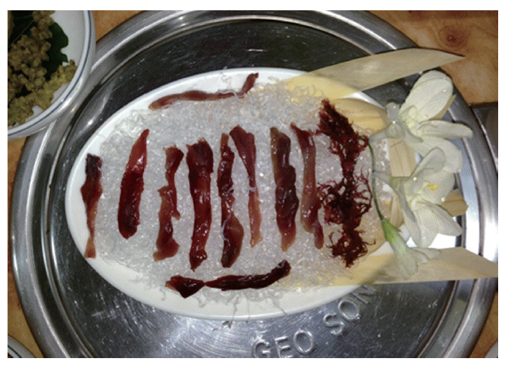
Table 1). They worked at the same company, and had all consumed raw soft-shelled turtle meat at a company dinner on 1 August (
Fig. 1). On 6 September, at the Department of Parasitology and Tropical Medicine, Seoul National University College of Medicine, ELISA was performed under the suspicion of trichinellosis. For use in the formulation of a crude antigen,
T. spiralis larvae were recovered from laboratory-maintained mice. The sera collected from the 5th Korean outbreak were used as a positive control, and the other ELISA procedures were the same as those in a previous
Clonorchis sinensis ELISA [
7]. Three of the present serum samples showed positivity against
T. spiralis larval antigen (
Table 2). An additional ELISA was performed on 24 September, which confirmed the positivity of the remaining 3 sera. Since all the 6 patients had eaten the turtle meat at the same time and they had no experience of wild boar meat in raw, the turtle meat was strongly suspected as the source of infection. They were treated with albendazole (800 mg/day) for 14 days, and their symptoms were gradually resolved. At the follow-up visit 3 months later, all of them were healthy without any clinical signs of trichinellosis. This is the 8th report on human trichinellosis in Korea and the second one implicating raw soft-shelled turtle meat.
DISCUSSION
The results of the present study strongly suggest that trichinellosis had been spread in Korea by consumption of soft-shelled turtle meat. This fact has added weight given that soft-shelled turtle meat has been used medicinally for many decades in Korea [
8] and that some Koreans still believe that soft-shelled turtle meat helps hemopoiesis. Thus, possible mechanisms by which soft-shelled turtles harbor
Trichinella sp. larvae should be explored.
Among various species of soft-shelled turtle, only Trionyx sinensis has been distributed in Korean waters [
9]. Korean soft-shelled turtles usually eat aquatic animals such as fish, crabs, and frogs. Many of them, as were those in the present case, are raised in aquatic farms [
8]. In order to trace the route of infection, contact with the farm owner was attempted, though the effort proved unsuccessful due to the sudden closure of the farm. Ruling out infection by formulated feeds, we speculated that it might have been transmitted through the consumption of other animals, or less likely, by cannibalism. Cannibalism among farmed crocodiles, for example, has been implicated as playing a central role in transmission of
T. papuae infection [
10]. In any case, it is known that without proper management of domestic animals,
Trichinella infection can be transmitted from the sylvatic to the domestic environment [
11]. In Korea, a systematic survey on the relation between
Trichinella infection and soft-shelled turtles is urgently needed.
Indeed, despite the 8 trichinellosis outbreaks (including the present one) that have occurred in Korea, efforts to arrange for and complete epidemiological surveys of relevant wild life are still lacking. A serological surveillance was performed on pig breeding farms, showing them to be trichinellosis free, though the animals were investigated under controlled housing conditions [
12]. A survey of 521 wild boars (
Sus scrofa) revealed 1.7% positivity for
T. spiralis larvae, though this investigation also had a limitation, specifically in having proceeded only by serology [
12]. Yet another research lacuna is the absence of any comprehensive survey of the number of
Trichinella species distributed in Korea. Thus far, the presence of
T. spiralis only has been confirmed, by a PCR-RFLP analysis performed on 2 Korean trichinellosis patients [
13]. The presence of other species in Korea is possible, especially in light of the existence of
T. nativa in China,
T. britovi and
T. nativa in Europe, and
T. nativa and
T. murrelli in North America [
14-
17]. For the purpose of molecular identification, recovery of larvae is necessary. However, unfortunately, muscle biopsy could not be performed in all cases. Hence, in order to broaden the known
Trichinella fauna in Korea, further investigation of wild life is required.
For the diagnosis of trichinellosis, ELISA is currently the most frequently used tool [
18]. However, ELISA can deliver false-negative results during the early stage of infection. With low doses of
T. spiralis larvae, IgG antibodies appeared at day 40 post-infection (PI) in inbred mice [
19]. In the previous outbreak of trichinellosis, the ELISA positivity usually took at least at day 34 PI, but negativity still occurred even at day 42 PI [
20]. In the present cases, however, half of the patients showed positivity to
Trichinella antigen even at day 21 PI. Considering that the specific IgG responses to
T. spiralis infection are dose-dependent [
21], the infection dose in this outbreak may have been heavier than those of the previous ones. In experimentally infected goats, the optical densities (OD) remained elevated until week 10 PI [
22], and it was continually increased until day 80 PI in another experiment using mice [
19]. In this respect, it seems strange that the OD decreased from 0.559 to 0.192 in 1 patient, while ODs showed much increment in other patients. Additionally, although ELISA is the most commonly used method for the detection of
Trichinella infection, it might result in less specificity due to false positive reactions [
23]. In the present study, ELISA was performed using the crude antigen of
Trichinella larvae. Of course, a recent study revealed that the crude antigen demonstrated a good specificity of 91.8% and had the potential for detection of swine trichinellosis [
24]. However, the specificity increased to 97.9% in ELISA by using excretory-secretory (ES) antigen, and even increased to 100% in western blot (WB) analysis [
25]. Hence, ELISA by ES antigen or WB analysis should be considered for a definitive diagnosis of trichinellosis, in case of muscle biopsy being unavailable.
In the spread of trichinellosis, globalization might play a role.
T. papuae, which has been proved, along with
T. zimbabwensis, to infect mammals and reptiles, was suspected as the causative species in the 2008 Taiwan outbreak among people who had eaten raw soft-shelled turtle meat [
5]. If established as the source of infection in the case,
T. papuae known to be distributed in Thailand and Malaysia [
26,
27] could have been spread to Taiwan synanthropically in rats traveling with humans [
28]. In the case of encapsulated species, larvae can survive in decaying muscles over long periods of time: 3 months for
T. nativa and
T. britovi in rat carcasses, for example [
29]. In frozen carnivore muscle,
T. nativa larvae can survive, in fact, for years [
30]. The non-encapsulated species,
T. papuae and
T. pseudospiralis, meanwhile, can retain their infectivity up to 9 days at 20-24˚C, and up to 40 days at 5˚C [
29]. Hence, in addition to widely disseminating information on the trichinellosis risk incurred in the consumption of raw soft-shelled turtle meat, significant investment in veterinary public health services should be made to prevent foodborne trichinellosis in Korea.
Notes
-
We have no conflict of interest related to this work.
Fig. 1.The raw soft-shelled turtle meat eaten by the present patients.

Table 1.Laboratory findings of patients in the present case
Table 1.
|
No. |
Sex |
Age |
WBC |
EOS (%) |
CK |
LDH |
AST |
ALT |
|
1 |
M |
40 |
23,190 |
46.4 |
7,332 |
874 |
101 |
115 |
|
2 |
M |
28 |
9,010 |
13.6 |
456 |
292 |
21 |
18 |
|
3 |
M |
31 |
10,320 |
20 |
261 |
454 |
32 |
45 |
|
4 |
M |
32 |
16,110 |
28.5 |
164 |
349 |
21 |
28 |
|
5 |
M |
43 |
13,350 |
20.5 |
488 |
363 |
25 |
20 |
|
6 |
F |
42 |
9,810 |
25.6 |
77 |
277 |
36 |
35 |
Table 2.ELISA results in sera of 6 patients
a
Table 2.
|
No. |
ELISA lgG (3 week)b
|
ELISA lgG (5 week)c
|
|
1 |
0.458 (positive) |
0.678 (positive) |
|
2 |
0.177 (negative) |
0.442 (positive) |
|
3 |
0.053 (negative) |
0.553 (positive) |
|
4 |
0.382 (positive) |
0.565 (positive) |
|
5 |
0.559 (positive) |
0.192 (negative) |
|
6 |
0.254 (borderline) |
0.733 (positive) |
References
- 1. Pozio E. The broad spectrum of Trichnella hosts: from cold- to warm-blooded animals. Vet Parasitol 2005;132:3-11.
- 2. Pozio E, Marucci G, Casulli A, Sacchi L, Mukaratirwa S, Foggin CM, La Rosa G. Trichinella papuae and Trichinella zimbabwensis induce infection in experimentally infected varans, caimans, pythons and turtles. Parasitology 2004;128:333-342.
- 3. Asatrian A, Movsessian S, Gevorkian A. Experimental infection of some reptiles with T. spiralis and T. pseudospiralis. Abstract Book of the 10th International Conference on Trichinellosis, 20-24 August, 2000. Fontainebleau, France. 2000. 154.
- 4. Khamboonruang C. The present status of trichinellosis in Thailand. Southeast Asian J Trop Med Public Health 1991;22:312-315.
- 5. Lo YC, Hung CC, Lai CS, Wu Z, Nagano I, Maeda T, Takahashi Y, Chiu CH, Jiang DD. Human trichinosis after consumption of soft-shelled turtles, Taiwan. Emerg Infect Dis 2009;15:2056-2058.
- 6. Lee SR, Yoo SH, Kim HS, Lee SH, Seo M. Trichinosis caused by ingestion of raw soft-shelled turtle meat in Korea. Korean J Parasitol 2013;51:219-221.
- 7. Choi MH, Park IC, Li S, Hong ST. Excretory-secretory antigen is better than crude antigen for the serodiagnosis of clonorchiasis by ELISA. Korean J Parasitol 2003;41:35-39.
- 8. Oh YN. tudy on artificial incubation of fresh-water soft-shelled turtle, Trionyx sinensis Strauch, of Korea. Ph.D. thesis. Cheonbuk National University; 1993. 166.
- 9. Kim KS, Rho YG, Kim YC, Lee BI, Shon JK. Seedling production of the soft-shelled turtle, Trionyx sinensis. Bulletin of National Fisheries Research and Development Institute 1999;56:119-132.
- 10. Owen IL, Awui C, Langelet E, Soctine W, Reid S. The probable role of cannibalism in spreading Trichinella papuae infection in a crocodile farm in Papua New Guinea. Vet Parasitol 2014;203:335-338.
- 11. Pozio E. Searching for Trichinella: not all pigs are created equal. Trends Parasitol 2014;30:4-11.
- 12. Wee SH, Lee CG, Joo HD, Kang YB. Enzyme-linked immunosorbent assay for detection of Trichinella spiralis antibodies and the surveillance of selected pig breeding farms in the Republic of Korea. Korean J Parasitol 2001;39:261-264.
- 13. Sohn WM, Huh S, Chung DI, Pozio E. Molecular identification of Korean Trichinella isolates. Korean J Parasitol 2003;41:125-127.
- 14. Li F, Cui J, Wang ZQ, Jiang P. Sensitivity and optimization of artificial digestion in the inspection of meat for Trichinella spiralis. Foodborne Pathog Dis 2010;7:879-885.
- 15. Pozio E, Serrano FJ, La Rosa G, Reina D, Perez-Martin E, Navarrete I. Evidence of potential gene flow in Trichinella spiralis and in Trichinella britovi in nature. J Parasitol 1997;83:163-166.
- 16. Oivanen L, Kapel CM, Pozio E, La Rosa G, Mikkonen T, Sukura A. Associations between Trichinella species and host species in Finland. J Parasitol 2002;88:84-88.
- 17. Liciardi M, Marucci G, Addis G, Ludovisi A, Gomez Morales MA, Deiana B, Cabaj W, Pozio E. Trichinella britovi and Trichinella spiralis mixed infection in a horse from Poland. Vet Parasitol 2009;161:345-348.
- 18. Dupouy-Camet J, Kociecka W, Bruschi F, Bolas-Fernandez F, Pozio E. Opinion on the diagnosis and treatment of human trichinellosis. Expert Opin Pharmacother 2002;3:1117-1130.
- 19. Reiterová K, Antolová D, Hurníková Z. Humoral immune response of mice infected with low doses of Trichinella spiralis muscle larvae. Vet Parasitol 2009;159:232-235.
- 20. Rhee JY, Hong ST, Lee HJ, Seo M, Kim SB. The fifth outbreak of trichinosis in Korea. Korean J Parasitol 2011;49:405-408.
- 21. Bień J. The usefulness of ELISA test for early serological detection of Trichinella spp. infection in pigs. Wiad Parazytol 2007;53:149-151. (in Polish).
- 22. Korínková K, Pavlícková Z, Kovarcík K, Koudela B. Distribution of muscle larvae and antibody dynamics in goats experimentally infected with Trichinella spiralis. Parasitol Res 2006;99:643-647.
- 23. Gamble HR, Pozio E, Bruschi F, Nöckler K, Kapel CM, Gajadhar AA. International Commission on Trichinellosis: recommendations on the use of serological tests for the detection of Trichinella infection in animals and man. Parasite 2004;11:3-13.
- 24. Tattiyapong M, Chaisri U, Vongpakorn M, Anantaphruti MT, Dekumyoy P. Comparison of three antigen preparations to detect trichinellosis in live swine using IgG-ELISA. Southeast Asian J Trop Med Public Health 2011;42:1339-1350.
- 25. Nöckler K, Reckinger S, Broglia A, Mayer-Scholl A, Bahn P. Evaluation of a Western Blot and ELISA for the detection of anti-Trichinella-IgG in pig sera. Vet Parasitol 2009;163:341-347.
- 26. Khumjui C, Choomkasien P, Dekumyoy P, Kusolsuk T, Kongkaew W, Chalamaat M, Jones JL. Outbreak of trichinellosis caused by Trichinella papuae, Thailand, 2006. Emerg Infect Dis 2008;14:1913-1915.
- 27. Intapan PM, Chotmongkol V, Tantrawatpan C, Sanpool O, Morakote N, Maleewong W. Molecular identification of Trichinella papuae from a Thai patient with imported trichinellosis. Am J Trop Med Hyg 2011;84:994-997.
- 28. Gottstein B, Pozio E, Nöckler K. Epidemiology, diagnosis, treatment and control of trichinellosis. Clin Microbiol Rev 2009;22:127-145.
- 29. Pozio E, Zarlenga DS. New pieces of the Trichinella puzzle. Int J Parasitol 2013;43:983-997.
- 30. Dick TA, Pozio E. Trichinella spp. and trichinellosis. In Samuel WM, Pybus MJ, Kocan AA eds, Parasitic Diseases of Wild Mammals. 2nd ed. Ames, Iowa, USA. Iowa State University Press; 2001, pp 380-396.
