Abstract
A man with only yellowing of the skin and eye sclera was diagnosed with clonorchiasis, which rarely manifested jaundice as the initial symptom. However, because of a lack of evidence for a diagnostic gold standard, the time until definitive diagnosis was more than a week. The diagnostic process relied on inquiring about the patient’s history, including the place of residence, dietary habits, and symptoms, as well as on serological findings, an imaging examination, and pathological findings. MRCP and CT results showed mild dilatation of intrahepatic ducts and increased periductal echogenicity. The eggs were ultimately found in stool by water sedimentation method after the negative report through direct smear. DNA sequencing of PCR production of the eggs demonstrated 98-100% homology with ITS2 of Clonorchis sinensis. After anti-parasite medical treatment, the patient’s symptoms were gradually relieved. Throughout the diagnostic procedure, besides routine examinations, the sedimentation method or concentration method could be used as a sensitive way for both light and heavy C. sinensis infection in the definite diagnosis.
-
Key words: Clonorchis sinensis, painless jaundice, image, PCR, sedimentation method
INTRODUCTION
Clonorchiasis is a serious foodborne zoonotic disease in Southeast Asia, especially in China, Korea, and Vietnam. The clinical manifestations are intimately associated with the degree of infection and with complications. During infection,
Clonorchis sinensis parasitize the intrahepatic bile ducts in the liver, releasing eggs into the ducts and thickening the walls, which can cause jaundice [
1-
3]. The eggs, along with the bile were released into the intestine. Therefore, eggs can be found by stool examination, which is considered as the diagnostic standard. The main non-specific symptoms include fatigue, hepatomegaly, and certain abdominal symptoms, such as nausea, abdominal distention, vomiting, abdominal discomfort and dyspepsia. The patient may exhibit jaundice if the bile duct is obstructed, and complications include cholangitis, bile duct stones, suppurative cholangitis, and even cholangiocarcinoma [
1-
3]. In 2009, International Agency for Research on Cancer (IARC) classified
C. sinensis as a Group 1 carcinogen based on its involvement in the etiology of cholangiocarcinoma [
4]. Therefore, early diagnosis and timely treatment are very important. However, the disease is often neglected due to its non-specific and atypical symptoms. Once at the hospital, the patient may exhibit more obvious symptoms, although stool examination may occasionally cause a missed diagnosis because of low production of eggs. Thus, it is important to choose accurate methods of diagnosis for clonorchiasis. Several accessory methods are emphasized in addition to stool examinations, especially imaging examinations, including computed tomography (CT) and magnetic resonance (MR) imaging [
5,
6].
CASE RECORD
A 48-year-old man who lived in Songyuan city, Jilin province, China, presented to the hospital with yellowing of the skin and sclera of the eyes that had been present for 10 days. Physical examinations detected painless jaundice of the skin and sclera and light tenderness at the site of right upper quadrant pain. The patient was alert, without fever or acute illness. There was no history of alcohol consumption or smoking or of liver diseases in the past. However, the patient had a clear dietary history of eating raw fish.
The laboratory data showed white blood cells 11.61×10
9/L (WBCs, normal=3.5-9.5×10
9/L), with an eosinophil count of 5.78×10
9/L (normal=0.02-0.52×10
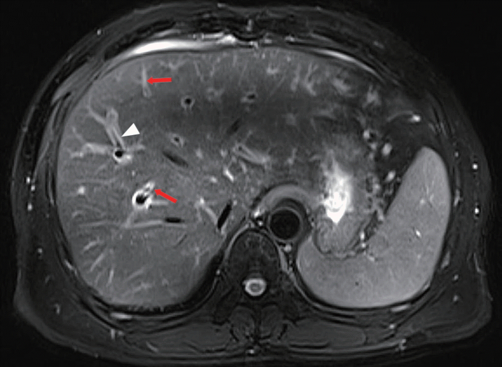
9/L) and an eosinophil percentage of 50% (normal=0.4-8%). AST 67 U/L (normal=15-40 U/L), ALT 211.9 U/L (normal=9-50 U/L), γ-GT 677 U/L (normal=10-60 U/L), total bilirubin 229.7 μmol/L (normal=6.8-30 μmol/L), and direct bilirubin 143.1 μmol/L (normal=0.0-8.6 μmol/L) were also detected. Viral marker assessment revealed HBs Ag (-), anti-HBs Ab (+), anti-HAV IgM (-), and anti-HCV Ab (-), and serum tumor markers were normal. Stool examinations for eggs by direct smear were negative 3 times. MR cholangiopancreatography (MRCP) showed cholecystitis, mild dilatation of the intrahepatic duct, and increased periductal echogenicity along the diffuse dilated bile ducts (
Fig. 1), but no space-occupying lesions. A CT scan revealed the same changes as on the MRCP.
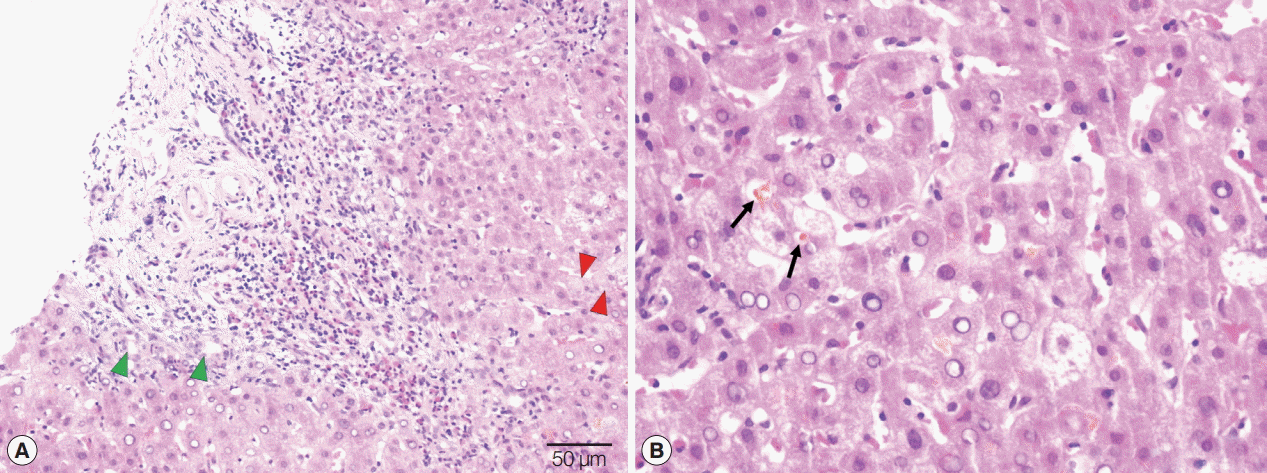
As there were no findings in stool examinations, other possible diagnoses were explored while the patient was treated with liver-protective treatment. Based on the results above, viral hepatitis could be excluded, but other kinds of hepatitis (such as autoimmune hepatitis, etc.) which could also cause jaundice were still suspicious. Therefore, to identify the etiology of jaundice, the patient agreed the liver biopsy under the guidance of ultrasound. Eosinophil infiltration, obvious proliferation in the bile ducts, hydropic degeneration of liver cells, and bile plug in bile capillaries were observed in pathological results, suggesting chronic hepatitis G3S1 (
Fig. 2).
Combining the history, symptoms, serological findings, pathological results, and imaging results, infection of
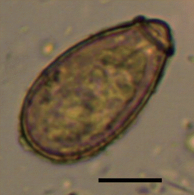
C. sinensis was considered as the most likely reason for the jaundice. Therefore, another method was carried out to test the feces. Finally, eggs were found in the stool by the water sedimentation method and then collected, but a low count (
Fig. 3). At last, the eggs were definitively identified as the eggs of
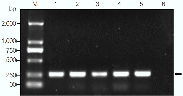
C. sinensis by PCR using primers from our laboratory (
Fig. 4). After sequencing, the DNA had 98-100% homology with internal transcribed spacer 2 (ITS2) of
C. sinensis China Shenyang isolates (GenBank no. AF217099) and North Korean isolates (GenBank no. AF217094). The patient was treated with praziquantel 1,200 mg 3 times per day (total of 3,600 mg), and the jaundice faded gradually. Fecal examination was negative after 1 month, and the liver function indices were nearly normal.
DISCUSSION
Clonorchiasis is a serious zoonosis, but it is often neglected because of its atypical manifestations. Although people have realized the danger of this disease, the trend of infection is not decreasing. One report on a national survey showed that China accounted for 80% of the 15.3 million cases of
C. sinensis infection worldwide [
7], and another national investigation showed that the prevalence of
C. sinensis infection increased by 75% between 1992 and 2004 [
8]. Therefore, clonorchiasis is still a large health threat for the Chinese people. The river basin of the Songhua River is a heavy endemic area of clonorchiasis in northeast China [
9], and Songyuan city, where the patient lives, is just one of the endemic areas along the Songhua River.
Regarded as an important risk factor, the habit of eating raw fish is a key clue leading to suspicion of clonorchiasis, including our report. The patient had eaten raw fish several times in the past, which could cause the infection. Furthermore, eosinophilia might be an allergic reaction in clonorchiasis and other human parasitic infections [
10]. The level of eosinophils in our report was 10 times higher than the reference range, and eosinophil infiltration was also observed in the biopsy. All of the results revealed a strong inflammatory reaction to a parasitic infection. However, jaundice as the initial symptom of clonorchiasis occurs occasionally, and it increases the difficulties of diagnosis. Once it occurs, hepatitis may always be firstly considered, and then relevant checks will be done to verify, which prolongs the diagnostic time. Thus, the role of the patient’s history and blood examinations in the diagnosis of endemic parasitic infection should be emphasized more [
11]. Along with the clues described previously, these findings supported a parasitic infection, and clonorchiasis should be considered first.
In addition, non-invasive radiologic examinations, such as MRCP and CT, make remarkable contributions to diagnosis of clonorchiasis. Dilation, increased periductal echogenicity, and stricture of the intrahepatic bile ducts can be attributed to changes due to
C. sinensis infection [
5,
12], but non-specific. Some of imaging findings in the present report were in line with reported presentations, without other causes of obstructive jaundice, such as stones or space-occupying lesions. The only possible explanation of jaundice was that the infiltration by inflammatory cells and the bile duct wall thickening might have caused the bile duct stricture, leading to painless jaundice. In this case, the stool examination by direct smear and images did not reveal the obvious diagnostic indications, while the patient’s jaundice was not relieved after liver-protective treatment. Based on these reasons, liver biopsy was performed to speed up diagnosis and avoid the missed diagnosis of serious diseases. However, waiting for the results of the other methods of stool examination may be more appropriate.
Finding eggs in the stool has been considered as the most significant diagnostic standard for definite diagnosis. However, the direct smear method led to a certain degree of missed diagnosis. The accuracy of the results depended not only on different degrees of infection, different amounts, and parts of the stool samples, but also on the skills and experiences of the operators. Fortunately, eggs were detected by the water sedimentation method when the results were negative after repeated direct smears in the suspected case of clonorchiasis. This method is a significant way to assist diagnosis, especially for mild infections, although it takes more time. In fact, the Kato-Katz (KK) method is a more ideal diagnostic test for clonorchiasis, owing to simple procedure, quick operation, high reliability, and low cost [
13]. Besides, formalin-ether (FE) concentration technique is also another sensitive way for light infections. According to the merits of the KK method, it should be the first choice in clinical work, and the FE concentration and water sedimentation techniques are recommended for mild infections. Additionally, PCR assays as molecular diagnostic methods for clonorchiasis have been studied extensively in recent years. Due to the similar morphologies of eggs of the Opisthorchiidae trematodes, this technology should be widely performed to assist the definite diagnosis. More sensitive and convenient molecular technologies are currently being explored.
In conclusion, because of a lack of typical symptoms, people with C. sinensis infection for many years are often not aware of the situation. Therefore, any clue or finding should be taken as reminders for the further clinical work, such as a detailed history, certain non-specific symptoms (such as jaundice), abnormal increase of eosinophils amount (an important symptom for parasitic infections), and characteristic imaging findings. The water sedimentation method or FE concentration method offers a supplementary way that could reduce the rate of missed diagnosis when eggs are not found under a microscope through KK method or direct smear, especially if clonorchiasis is highly suspected.
Notes
-
The authors declare that they have no competing interests.
Fig. 1.MRCP revealed the similar changes as on the CT scan. Mild dilatation of the intrahepatic bile duct (white arrowhead) and increased periductal echogenicity (red arrows).

Fig. 2.Pathologic findings of the liver biopsy. (A) A mass of eosinophil infiltration, small bile ducts hyperplasia in portal area (green arrowhead), and hydropic degeneration of liver cells (red arrowhead). Scale bar=50 μm. (B) Bile plug in bile capillaries (arrow). Scale bar=20 μm.

Fig. 3.An egg found in the patient's stool by a sedimentation method. Scale bar=10 μm.

Fig. 4.PCR products on Clonorchis sinensis eggs. M, DNA marker; lanes 1-5, PCR amplicons; lane 6, negative control. The arrow on the right side represents the amplicon of Clonorchis sinensis eggs.

References
- 1. Choi BI, Han JK, Hong ST, Lee KH. Clonorchiasis and cholangiocarcinoma: etiologic relationship and imaging diagnosis. Clin Microbiol Rev 2004;17:540-552.
- 2. Lim JH. Liver flukes: the malady neglected. Korean J Radiol 2011;12:269-279.
- 3. Sripa B, Kaewkes S, Sithithaworn P, Mairiang E, Laha T, Smout M, Pairojkul C, Bhudhisawasdi V, Tesana S, Thinkamrop B, Bethony JM, Loukas A, Brindley PJ. Liver fluke induces cholangiocarcinoma. PLoS Med 2007;4:e201.
- 4. Bouvard V, Baan R, Straif K, Grosse Y, Secretan B, El Ghissassi F, Benbrahim-Tallaa L, Guha N, Freeman C, Galichet L, Cogliano V; Group WHOIAfRoCMW. A review of human carcinogens-Part B: biological agents. Lancet Oncol 2009;10:321-322.
- 5. Jeong YY, Kang HK, Kim JW, Yoon W, Chung TW, Ko SW. MR imaging findings of clonorchiasis. Korean J Radiol 2004;5:25-30.
- 6. Choi D, Hong ST. Imaging diagnosis of clonorchiasis. Korean J Parasitol 2007;45:77-85.
- 7. Yang GJ, Liu L, Zhu HR, Griffiths SM, Tanner M, Bergquist R, Utzinger J, Zhou XN. China's sustained drive to eliminate neglected tropical diseases. Lancet Infect Dis 2014;14:881-892.
- 8. Li T, He S, Zhao H, Zhao G, Zhu XQ. Major trends in human parasitic diseases in China. Trends Parasitol 2010;26:264-270.
- 9. Choi MH, Park SK, Li Z, Ji Z, Yu G, Feng Z, Xu L, Cho SY, Rim HJ, Lee SH, Hong ST. Effect of control strategies on prevalence, incidence and re-infection of clonorchiasis in endemic areas of China. PLoS Negl Trop Dis 2010;4:e601.
- 10. Lai CH, Chin C, Chung HC, Liu H, Hwang JC, Lin HH. Clonorchiasis-associated perforated eosinophilic cholecystitis. Am J Trop Med Hyg 2007;76:396-398.
- 11. Klemencic S, Phelan M, Patrick R, Vahdat N. Oriental cholangiohepatitis (clonorchiasis infestation) caused by Clonorchis sinensis. J Emerg Med 2012;43:e107-9.
- 12. Kim BG, Kang DH, Choi CW, Kim HW, Lee JH, Kim SH, Yeo HJ, Lee SY. A case of clonorchiasis with focal intrahepatic duct dilatation mimicking an intrahepatic cholangiocarcinoma. Clin Endosc 2011;44:55-58.
- 13. Hong ST, Choi MH, Kim CH, Chung BS, Ji Z. The Kato-Katz method is reliable for diagnosis of Clonorchis sinensis infection. Diagn Microbiol Infect Dis 2003;47:345-347.


