Abstract
Thelazia callipaeda (Spirurida, Thelaziidae), a parasitic eye worm causing thelaziasis has been reported in humans and dogs in Korea. However, its occurrence in other potential reservoir hosts, including wild animals, remains unclear. In the present study, we described the 2 cases of thelaziasis from both of feral and captive wild animal in Korea. In August and November 2017, 2 cases of the parasitic infections were found in the third eyelid of rescued leopard cat Prionailurus bengalensis and reared raccoon dog Nyctereutes procyonoides at the Chungnam Wild Animal Rescue Center. A total of 20 and 24 worms were detected from the left and right eyes of leopard cat, respectively. In the left eye of the raccoon dog, 5 worms were recognized. Male worms were 969–11,860 μm long (10,600 μm on average) and 300–320 μm width (315 μm on average). Female worms were 13,430–15,330 (14,480) μm long and 320–370 (344) μm wide in size. They commonly had a characteristic scalariform buccal cavity and short esophagus. The vulva openings were located at the anterior of esophago-intestinal junction in females. The thelaziasis is reported in 2 species of wildlife, P. bengalensis and N. procyonoides, for the first time in Korea.
-
Key words: Prionailurus bengalensis, Nyctereutes procyonoides, Thelazia callipaeda, zoonoses
Introduction
The genus
Thelazia (Spirurida, Thelaziidae) is group of nematodes that infect eye of various kinds of mammals and birds [
1,
2]. The adult worm is parasitized at the conjunctival sac and its adjacent regions of host, and induce several symptoms such as conjunctivitis, epiphora, purulent ocular discharge, keratitis and corneal ulcers so called thelaziasis or thelaziosis [
2]. One of the species,
Thelazia callipaeda Railliet and Henry, 1910 is comparatively well-known parasite among the group. Likely with its common name, “the oriental eyeworm”, it is distributed wide ranges of East Asian countries with some European countries which are area that the species was introduced and have been inducing the thelaziasis [
3].
In addition to dog (
Canis familiaris), raccoon dog (
Nyctereutes procyonoides) and American red fox (
Vulpes fulva), several kinds of mammals such as cat (
Felis catus), European rabbit (
Oryctolagus cuniculus), rodents, monkey, and human are known to act as a definitive host of
T. callipaeda [
2]. Current studies also added wolf (
C. lupus), red fox (
Vulpes vulpes), European wildcat (
F. silvestris), beech marten (
Martes foina), sable (
M. zibellina), European badger (
Meles meles), and European hare (
Lepus europaeus) to the list of definitive hosts from European countries [
4–
7]. In the most recent papers, additional wildlife, i.e., raccoon (
Procyon lotor), Japanese raccoon dog (
N. viverrinus), Japanese red fox (
V. vulpes japonica), masked palm civet (
Paguma larvata), Japanese badger (
M. anakuma), and Japanese black bear (
Ursus thibetanus japonicus) were newly added to the host list [
8,
9]. Although the understanding of sylvatic life cycle is commonly recognized as one of the important factors for parasitic control, it is often neglected. In particular, information on the infection status and host range of
T. callipaeda remains scarce in Korea, despite the fact that the host diversity of the parasite has been comparatively well investigated in adjacent countries [
2,
8,
9].
The aim of the present study is to introduce 2 cases of thelaziasis which found in both of captive and free-living domestic wildlife and to identify the causative parasite through morphological analysis.
Case Record
Case I
A female, orphaned raccoon dog was found in Seosan-gun (county) (36°47′24.4″N, 126°27′33.9″E) and subsequently admitted to the Chungnam Wild Animal Rescue Center (CNWARC, 36°40′14.5″N, 126°51′43.3″E) in May 2011. The animal was evaluated to be unsuitable for release back into the wild due to significant human imprinting, which rendered it incapable of surviving independently in its natural habitat. As a result of the issue, the animal has since been cared for at the CNWARC located in Yesan-gun (36°40′14.1″N 126°51′41.6″E), where it fulfills a maternal role by nurturing orphaned raccoon dogs annually.
In August 2017, the left eye of the raccoon dog presented with symptoms including blood congestions, epiphora and ocular discharge. After removing foreign bodies on the eye using cotton swab and saline, revealing small moving objects within the conjunctival sac. Five worms were collected by using forceps, and the specimens were preserved in 70% ethanol for further analysis.
Following the removal of all parasites, an ophthalmic ointment (OcuFlox; Samil, Seoul, Korea) was prescribed for a duration of a week. Remarkable improvement in all clinical symptoms was observed after the application of the eye ointment. Furthermore, no worms were detected in the clinical follow-up conducted over ensuing month.
Case II
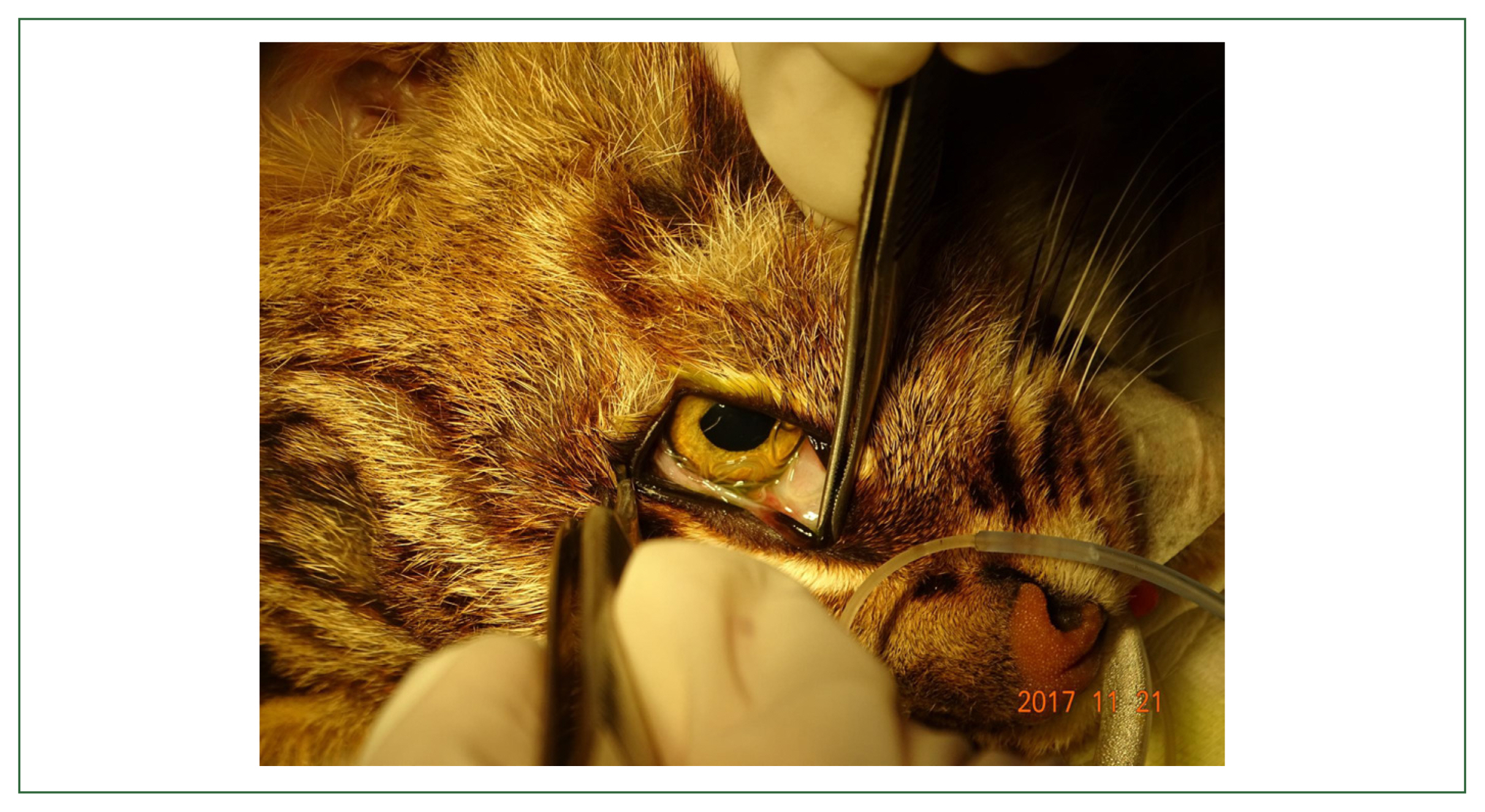
In November 2017, a leopard cat (Prionailurus bengalensis) exhibiting mobility problems was found in Asan-si (city) (36°48′29.3″N, 126°55′34.2″E). The animal was presumed to have been involved in a vehicle collision and was admitted to the CNWARC. It was observed that the leopard cat had significant difficulty moving its hind legs and was unable to stand, indicating potential neurological and/or musculoskeletal impairment. The animal’s demeanor was also notably subdued, lacking the typical alertness and responsiveness. The leopard cat was assessed to have a body condition score of 2 out of 9, indicating a concerning level of undernourishment accompanied by moderate to severe dehydration. For a thorough physical examination, the animal was anesthetizing using isoflurane (Ifran; Hana Pharm, Seoul, Korea). Subsequent radiographic examination was carried out for accurate diagnosis. Following anesthesia induction, meloxicam (Metacam; Boehringer Ingelheim, Mexico City, Mexico) was administered to provide analgesic action. Notably, the examination revealed the presence of hematuria, epiphora and ocular discharge. The radiographic findings clearly indicated a comminuted fracture on the pelvis.
After the removal of the ocular discharge and a detailed ophthalmic examination, thread-like, translucent worms were discovered beneath the third eyelids in both eyes of the leopard cat (
Fig. 1). Using forceps and a cotton swab, they were carefully collected and subsequently preserved in 70% ethanol for identification. A total of 20 and 24 specimens were retrieved from the left and right eyes, respectively. Given the grave prognosis related with the fractures, the decision was made to humanely euthanize the animal, and was carried out via the intravenous administration of 5 ml of T61 after anesthesia.
From the specimens collected, 5 males and 5 female nematodes obtained from the leopard cat, along with 2 males from the raccoon dog were selected for morphological identification. The specimens underwent several washes in 1× phosphate-buffered saline and were mounted with polyvinyl alcohol solution. The clearing process was followed by observation and measurement of the specimens using a light microscope (BX53; Olympus, Tokyo, Japan).
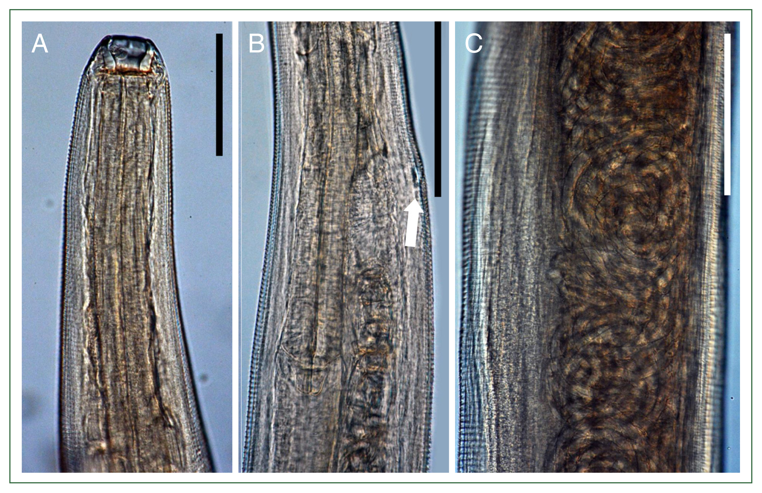
The nematodes were observed to be white in color, with elongated bodies. They had a well-developed scalariform buccal capsule and a comparatively short esophagus (4%–6% of body length) (
Fig. 2A). The body lengths of male worms ranged from 9,692 to 11,857 μm (10,598 μm on average), and width between 295 and 320 μm (315 μm on average) in width. Buccal capsule size was measured at 23–27 (24) μm by 25–27 (26) μm. Nerve ring was located 255–295 (275) μm from the anterior extremity. The esophagus was simple and directly connected to the intestine, measuring 541–586 (565) μm in length. The posterior extremity of the worms curved ventrally, with a very short tail measuring 16–23 (20) μm. Males had 2 spicules, with the right spicule significantly larger than left, at ratio of 14–16:1. The right spicule was thin and elongated, measuring 855–1,027 (944) μm, while the left spicule was short and stout, measuring 55–64 (59) μm.
The female worms measured 13,432–15,326 (14,480) μm by 320–369 (344) μm in body size. Their buccal capsules were 25–32 (30) μm by 25–32 (30) μm in size. The nerve ring was located 273–345 (315) μm from the anterior extremity. The length of esophagus was 573–664 (631) μm. The vulva was situated 482–550 (524) μm from the anterior extremity, approximately 3%–4% of the body length (
Fig. 2B). The uterus, filled with numerous ova in the posterior part of the body, developed into an embryonated form near the vulva region (
Fig. 2C). The tail was short, measuring 59–68 (64) μm in length.
The specimens collected both of raccoon dog and leopard cat were identified as
T. callipaeda by the keys in the literatures [
10,
11].
Discussion
Korea is considered to be an endemic region and 1 of the original distribution areas of this nematode. However, studies on the parasitic nematodes are limited to approximately 40 human infections, along with 5 papers on canine infections [
12–
17]. In Korea, the parasitic infection has generally been neglected unless there are unusual or novel cases, as demonstrated by previous authors [
12,
17], who suggested that infections have been underestimated. It has yet to be evaluated whether dogs are the primary reservoir hosts, as previously recorded, or if there are indeed wild animals involved in transmission in the natural environment.
In the present study, we described 2 cases of thelaziasis identified in wild animals. The infections were found in 2 different species, i.e., a raccoon dog and a leopard cat marking the first reported cases of wild animals in Korea. As noted in previous cases, the animals in this study did not exhibit significant clinical issues, with only minor symptoms such as blood congestion, epiphora and ocular discharge being observed [
17]. Although both infections were detected at the same wildlife rescue center, considering the timing of detection and the history of animals, the 2 cases are considered to have occurred independently. Notably, a case represents a nosocomial infection, while the other is presumed to have been acquired in the wild prior to rescue. The raccoon dog, whose infection was detected in August 2017, had been reared in an enclosure at the wildlife center since its rescue in 2011, making it likely that this infection was acquired within the facility. In contrast, the leopard cat was rescued from a location over 15 kilometers from the center where the raccoon dog was reared, and its infection was detected on the day of rescue, suggesting it was infected in wild.
Considering environmental factors, it is suspected that
Thelazia infections in the 2 wild animals were transmitted through arthropod vectors that had obtained larvae from animals raised by humans near the locations [
18–
20]. The raccoon dog’s enclosure was an outdoor pen with an open roof, surrounded by nearby residential areas and mountains. Similarly, the leopard cat was rescued from a plain that included rice field and farmlands, with various residential houses in the vicinity. Although we were unable to investigate the presence of the parasitic infections in animals such as dogs raised in these residential areas, previous cases of
Thelazia infections in local dogs suggest a plausible source of infection [
13,
15–
17]. Since the first record of thelaziasis in Korea, and up until recent cases, veterinarians who examined the same wildlife species had never found the parasites (personal communication), suggesting that the prevalence of
Thelazia infection in wild animals may be very low. Given that the parasite thrives in environments where arthropod vectors are more likely to access their hosts in immobile conditions, such as tethered dogs, it is likely that animals raised in human settlements serve as the primary reservoir hosts rather than wild animals. According to a study on the experimental culturing of the species under laboratory conditions, arthropod hosts mostly harbor only a larva per individual, with a maximum observed number of 5 [
20]. Considering the publication, in the present cases which 5 and 44 parasites were detected, it is likely that both the raccoon dog and the leopard cat were repeatedly exposed to these flies. Given their histories, it is evident that both animals had limited mobility, either being confined to enclosures or unable to move due to pelvis fractures.
This study might contributed to further understanding of the life cycle and ecology of
T. callipaeda in Korea, particularly by confirming, for the first time, infections in wild animals, which had not been previously documented. Although this report is limited to 2 cases, making it difficult to ascertain the actual infection status and distribution of
T. callipaeda in wild animals in the nature environment, it has at least demonstrated that infections are indeed occurring. In previous studies [
2,
4–
9], the final host range of
T. callipaeda was found to be quite broad, and considering this, it is plausible that these parasites could also be found in other Korean wildlife such as rabbits, martens, badgers, wild boars and bears. Given the susceptibility of this parasite, there is also a high likelihood that foxes, which have been the focus of recent restoration efforts, could be exposed to these parasites in the wild, just as one of the foxes have been exposed to
Sarcocystis pilosa in a previous case [
21]. Therefore, veterinarians working with wildlife should be aware of the potential for parasitic infections in animals susceptible to
T. callipaeda. Although veterinarians mentioned not having encountered this parasite before, this may be due to a lack of attention to parasitic infections and the difficulty of conducting eye examination in wild animals without anesthesia, leading to an underestimation of the infection. Therefore, we must pay special attention to the zoonosis like thelaziasis and should try to maintain good hygienic conditions for the outside facility.
In conclusion, we reported the first case of
T. callipaeda infection in Korea, found in wild animals with limited mobility. The raccoon dog and leopard cat are the first wildlife definitive hosts in Korea, and in both cases, factors that limited their immobility were described. This has filled a significant gap in the understanding of the ecology and has provided knowledge that can be applied to the prevention of
T. callipaeda infections. However, many aspects of
T. callipaeda in Korea remain unexplored. Important questions that have been studies in adjacent countries, such as the species of intermediate hosts that transmit
T. callipaeda and genetic diversity of
Thelazia are still unknown in Korea [
20,
22,
23]. Future research should address these gaps to provide more practical and effective response to
T. callipaeda infections, which remain a neglected disease in Korea.
Notes
-
Author contributions
Conceptualization: Jang J, Choe S
Data curation: Jang J
Formal analysis: Jang J
Funding acquisition: Choe S
Investigation: Jang J, Choe S
Methodology: Jang J, Eom KS, Choe S
Project administration: Park YS, Yun YM
Resources: Park YS
Software: Jang J, Choe S
Visualization: Jang J, Choe S
Validation: Yun YM, Eom KS
Supervision: Choe S
Writing – original draft: Jang J, Choe S
Writing – review and editing: Park YS, Yun YM, Eom KS, Choe S
-
Conflict of interest
The authors declare no conflict of interest related to this study.
-
Acknowledgments
This research was supported by the National Research Foundation of Korea (grant No. 2020R1C1C1013563). The authors appreciate to all the members of the Chungnam Wild Animal Rescue Center for their support during our research.
Fig. 1A close-up view of the left eye of a leopard cat, Prionailurus bengalensis infected with Thelazia callipaeda. The wild cat was found with a severe injury to its pelvis and was rescued in Asan-si (city). It was under anesthesia during the examination of parasites.

Fig. 2Microscopic morphologies of a female Thelazia callipaeda collected from a raccoon dog, Nyctereutes procyonoides, in this study. (A) Anterior extremity showing a well-developed buccal capsule. Scale bar=100 μm. (B) Vulva located at the mid-level of the esophagus (arrow). Scale bar=200 μm. (C) Coiled larvae in the uterus. Scale bar=100 μm.
References
- 1. Yamaguti S. Systema Helminthum. Vol. 3. The Nematodes of Vertebrates. Part I. Interscience Publishers. New York, USA. 1961. p 679.
- 2. Anderson RC. Nematode Parasites of Vertebrates: Their Development and Transmission. 2nd ed. CABI Publishing; Guilford, UK. 2000. p. 404-407.
- 3. Otranto D, Traversa D. Thelazia eyeworm: an original endo- and ecto-parasitic nematode. Trends Parasitol 2005;21(1):1-4.
https://doi.org/10.1016/j.pt.2004.10.008
- 4. Otranto D, Cantacessi C, Mallia E, Lia RP. First report of Thelazia callipaeda (Spirurida, Thelaziidae) in wolves in Italy. J Wildl Dis 2007;43(3):508-511.
https://doi.org/10.7589/0090-3558-43.3.508
- 5. Otranto D, Dantas-Torres F, Mallia E, DiGeronimo PM, Brianti E, et al. Thelazia callipaeda (Spirurida, Thelaziidae) in wild animals: report of new host species and ecological implications. Vet Parasitol 2009;166(3–4):262-267.
https://doi.org/10.1016/j.vetpar.2009.08.027
- 6. HodžIć A, Latrofa MS, Annoscia G, Alić A, Beck R, et al. The spread of zoonotic Thelazia callipaeda in the Balkan area. Parasit Vectors 2014;7:352.
https://doi.org/10.1186/1756-3305-7-352
- 7. Odoevskaya IM, Khrustalev AV, Shaitanov VM, Seriodkin IV, Panayotova-Pencheva MS. Occurrence of the nematode Thelazia callipaeda Railliet and Henry, 1910 (Spirurida, Thelaziidae) in wild carnivores in the Russian Far East. Acta Zool Bulg 2015;67:561-566.
- 8. Doi K, Tokiwa T, Imoto M, Chou S, Yamasaki F, et al. Molecular characterization of oriental eyeworm (Thelazia callipaeda) detected from raccoon (Procyon lotor) and Japanese raccoon dog (Nyctereutes viverrinus) in Kanto region, Japan. Parasit Vectors 2023;16:116.
https://doi.org/10.1186/s13071-023-05736-x
- 9. Kitajima A, Tokiwa T, Doi K, Kotani K, Otsubo H, et al. New host record of Thelazia callipaeda (Nematoda: Spirurida) with a notably wide host range and shared zoonotic lineage in Japan. Parasitol Int 2024;102:102913.
https://doi.org/10.1016/j.parint.2024.102913
- 10. Skrjabin KI, Sobolev AA, Ivashkin VM. Spirurata of animals and man and the disease caused by them. In: Skrjabin KI, editor. Essentials of Nematology. 16:Akademii Nauk SSSR; Moscow, Soviet Union. 1967. p. 8-58.
- 11. Otranto D, Lia RP, Traversa D, Giannetto S. Thelazia callipaeda (Spirurida, Thelaziidae) of carnivores and humans: morphological study by light and scanning electron microscopy. Parassitologia 2003;45(3–4):125-133.
- 12. Sohn WM, Na BK, Yoo JM. Two cases of human thelaziasis and a brief review of Korean cases. Korean J Parasitol 2011;49(3):265-271.
https://doi.org/10.3347/kjp.2011.49.3.265
- 13. Choi DK, Cho SY. A case of human thelaziasis concomitantly found with a reservoir host. J Korean Ophthalmol Soc 1978;19:125-129.
- 14. Ryang YS, Lee KJ, Lee DH, Cho YK, Im JA, et al. Scanning electron microscopy of Thelazia callipaeda Railliet and Henry, 1910 in the eye of a dog. Biomed Sci Lett 1999;5(1):41-49.
- 15. Seo M, Yu JR, Park HY, Huh S, Kim SK, et al. Enzoocity of the dogs, the reservoir host of Thelazia callipaeda, in Korea. Korean J Parasitol 2002;40(2):101-103.
https://doi.org/10.3347/kjp.2002.40.2.101
- 16. Kim JT, Cho SW, Kim HS, Ryu SY, Jun MH, et al. Scanning electron microscopical findings of Thelazia callipaeda from a domestic dog. J Vet Sci CNU 2005;13(1):21-25.
- 17. Choe S, Kim S, Nath TC, Kim JH. Eight cases of canine thelaziosis found in two localities in Korea. Parasites Hosts Dis 2023;61(3):325-331.
https://doi.org/10.3347/PHD.23031
- 18. Otranto D, Brianti E, Cantacessi C, Lia RP, Máca J. The zoophilic fruitfly Phortica variegata: morphology, ecology, and biological niche. Med Vet Entomol 2006;20(4):358-364.
https://doi.org/10.1111/j.1365-2915.2006.00643.x
- 19. Otranto D, Cantacessi C, Testini G, Lia RP. Phortica variegata as an intermediate host of Thelazia callipaeda under natural conditions: evidence for pathogen transmission by a male arthropod vector. Int J Parasitol 2006;36(10–11):1167-1173.
https://doi.org/10.1016/j.ijpara.2006.06.006
- 20. Wang L, Li D, Yin C, Tang H, Luo B, et al. Laboratory culture and life cycle of Thelazia callipaeda in intermediate and definitive hosts. Pathogens 2022;11(9):1066.
https://doi.org/10.3390/pathogens11091066
- 21. Jo Y, Lee SJ, Bia MM, Choe S, Jeong DH. First report of Sarcocystis pilosa from a red fox (Vulpes vulpes) released for the re-introduction project in South Korea. Animals (Basel) 2023;14(1):89.
https://doi.org/10.3390/ani14010089
- 22. Jin Y, Liu Z, Wei J, Wen Y, He N, et al. A first report of Thelazia callipaeda infection in Phortica okadai and wildlife in national nature reserves in China. Parasit Vectors 2021;14(1):13.
https://doi.org/10.1186/s13071-020-04509-0
- 23. Otranto D, Testini G, De Luca F, Hu M, Shamsi S, et al. Analysis of genetic variability within Thelazia callipaeda (Nematoda: Thelazioidea) from Europe and Asia by sequencing and mutation scanning of the mitochondrial cytochrome c oxidase subunit 1 gene. Mol Cell Probes 2005;19(5):306-313.
https://doi.org/10.1016/j.mcp.2005.05.001