Abstract
Extracellular vesicles (EVs) secreted by Trichomonas vaginalis are now recognized as important mediators of host-parasite communication and play crucial roles in the development of trichomoniasis. These membrane-enclosed vesicles, which include exosomes and microvesicles, carry a diverse array of cargo, including proteins, lipids, and nucleic acids, as well as virulence-associated components such as adhesion molecules, proteases, and tRNA-derived small RNAs. Functionally, EVs enhance parasite attachment to vaginal epithelial cells, facilitate colonization, and contribute to host tissue damage. They also influence host immune responses, exhibiting both pro-inflammatory and immunosuppressive activities. Furthermore, the composition of EVs can be modulated by endosymbionts such as Trichomonasvirus, thereby influencing host-pathogen interactions. In addition, EVs mediate parasite-parasite communication, thereby promoting parasite adaptation and survival. This review summarizes current knowledge on the composition and functional roles of T. vaginalis EVs, highlighting their involvement in adhesion, immune modulation, and infection, as well as their potential utility as diagnostic biomarkers and therapeutic targets.
-
Key words: Trichomonas vaginalis, extracellular vesicles, exosome, Trichomonasvirus, pathogenesis, adherence
Introduction
Trichomonas vaginalis is a single-celled protozoan parasite that causes trichomoniasis in humans. This parasite is an extracellular pathogen that primarily infects host tissues by adhering to urogenital epithelial cells [
1]. This cytoadherence to vaginal and prostate epithelium is enhanced by the secretion of extracellular vesicles (EVs), including exosome-like vesicles. Notably,
T. vaginalis was among the early protozoan parasites reported to release small EVs, or exosomes, capable of altering the physical and biochemical properties of host cells, thereby facilitating host-parasite interactions [
2].
The International Society for Extracellular Vesicles recommends the use of the umbrella term "EVs" to describe all membrane-derived vesicles [
3]. EVs are generally categorized into 3 main groups based on their size and biogenesis: exosomes, microvesicles, and apoptotic bodies. Exosomes are small (30–150 nm), lipid bilayer vesicles with a characteristic cup-shaped morphology, formed through the inward budding of endosomal membranes to generate intraluminal vesicles within multivesicular bodies. Upon fusion of multivesicular bodies with the plasma membrane, the intraluminal vesicles are released into the extracellular space as exosomes. Their biogenesis may occur via endosomal sorting complexes required for transport (ESCRT)-dependent or ESCRT-independent pathways, and they carry diverse molecular cargo for intercellular communication. Microvesicles (50–1,000 nm), in contrast, originate from outward budding of the plasma membrane, a process often triggered by increased intracellular Ca²⁺ levels, which activate cytoskeletal remodeling and phospholipid redistribution. In contrast, apoptotic bodies (1–5 μm) are larger vesicles formed during programmed cell death (apoptosis), encapsulating cellular components as cells undergo fragmentation.
Generally,
T. vaginalis releases 2 types of EVs: small EVs (exosomes, 30–150 nm) generated within endosomal multivesicular bodies [
2,
4-
7], and microvesicles (50–1,000 nm) or larger vesicles (>200 nm) that bud directly from the flagellar membrane of the parasite [
8-
10].
T. vaginalis EVs (TvEVs) are internalized by host cells predominantly through caveolae-mediated and lipid raft-mediated endocytosis, a process mediated by host cell cholesterol and caveolin-1. This uptake mechanism is facilitated by interactions with heparan sulfate proteoglycans on the host cell surface, leading to increased production of pro-inflammatory cytokines, enhanced parasite adherence, and modulation of host cell metabolism [
11].
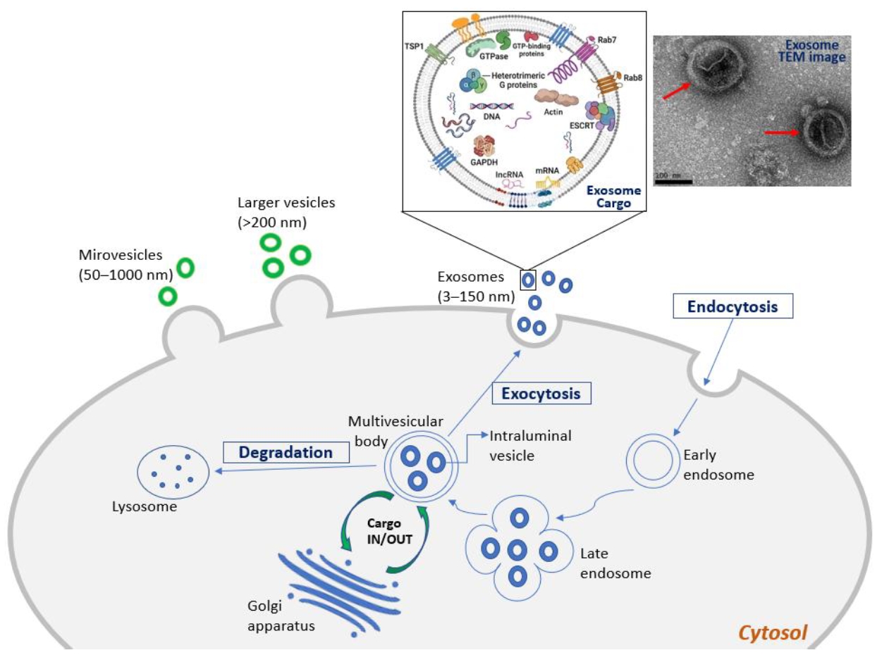
Fig. 1 illustrates the biogenesis of TvEVs and biomolecular cargo of exosome, along with the characterization of exosomes by transmission electron microscope.
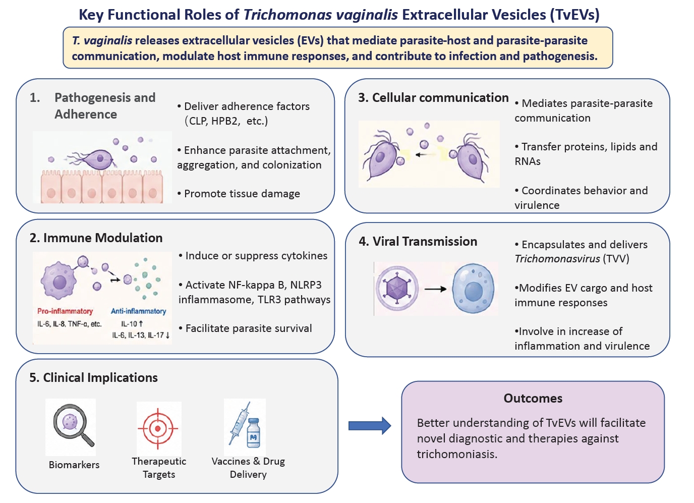
Collectively, TvEVs function as intercellular messengers, delivering a diverse cargo that consists of conserved eukaryotic EV components and parasite-specific molecules. These specific molecules include virulence-associated factors such as adhesins, proteases (e.g., GP63-like proteins), tetraspanins, and regulatory small RNAs. Subsequently, these EVs promote parasite adherence, modulate host immune responses, and influence inflammatory processes to support parasite survival and colonization. In this review, we focus on the key roles and functions of EVs in
T. vaginalis (
Fig. 2), with
Table 1 [
2,
4-
15] summarizing the major studies on TvEVs published from 2013 to the present.
Key Roles on EV of T. vaginalis
Pathogenesis and adherence
T. vaginalis releases exosomes, small EV that can fuse with host cells. Adherence of the parasite to epithelial cells represents the initial step in host infection [
1]. Studies have shown that exosome enhances the parasite's ability to attach to host cells, serving as a key mechanism during the early stages of colonization. Notably, co-inoculation of
T. vaginalis with exosomes derived from highly adherent strains significantly increases parasite attachment to vaginal and prostate epithelial cells [
2]. In addition, the parasite can lyse host epithelial cells, leading to inflammation and tissue damage, further underscoring the role of exosomes in promoting pathogenicity.
Exosomes are also thought to deliver bioactive molecules into host cells, altering the expression of adhesion-related factors and thereby facilitating parasite attachment. Adhesion is further enhanced when both parasites and host cells are pre-exposed to exosomes, suggesting that these vesicles help coordinate host-parasite interactions. Moreover, Molgora et al. [
12] demonstrated that parasite-derived EVs enhance
T. vaginalis adherence to host cell monolayers in vitro [
2] and are associated with improved parasite survival in a murine model. These findings provide the first in vivo evidence that TvEVs contribute to host-parasite interactions and support earlier in vitro findings that TvEVs facilitate parasite colonization within the host [
2,
11].
Evidence suggests that the molecules involved in parasite-parasite aggregation may also mediate parasite-host binding, with exosomes potentially depositing adhesion ligands onto host cell surfaces. Notably, exosomes derived from a highly adherent
T. vaginalis strain (B7RC2) can transfer a high-adhesion phenotype to a less adherent strain (G3), significantly enhancing their survival, aggregation, and attachment to host cells. Acting as mediators of parasite-parasite communication, EVs transfer virulence-associated proteins, such as cadherin-like protein (CLP), that improve the adhesive capacity of recipient parasites [
10]. Salas et al. [
10] revealed that
T. vaginalis coordinates behavior via cytonemes and EVs, with EVs influencing the formation of communication structures in neighboring parasites. In addition, Kochanowsky et al. [
13] reported that TvEVs upregulate heteropolysaccharide binding protein (HPB2) expression and facilitate the transfer of adherence factors between parasites, thereby increasing overall virulence. However, it remains unclear whether these effects are driven by molecules on the exosome surface or by cargo delivered into the host cells.
In addition to exosomes, the parasite also produces ectosomes, larger vesicles that bud directly from the plasma membrane, which are released in greater amounts during host-cell contact. This observation suggests that ectosomes may likewise contribute to host-parasite interactions and play a role in disease pathogenesis [
8].
The immunomodulatory roles of exosomes derived from
T. vaginalis have been extensively studied, with evidence demonstrating that they can exert both pro-inflammatory and anti-inflammatory effects in vitro and in vivo. The internalization of exosomes by ectocervical cells induces IL-6 secretion and modest IL-8 release. Conversely, exosomes that fuse with host cells may also deliver regulatory molecules that suppress IL-8 secretion from ectocervical cells, thereby limiting neutrophil recruitment to infection sites [
2]. In addition, Nievas et al. [
8] reported that
T. vaginalis releases microvesicles and larger vesicles in the presence of HeLa cells, supporting a role for parasite-derived EVs in modulating host cell interactions.
Further evidence from Olmos-Ortiz et al. [
4] showed that pretreatment with these vesicles in a murine model enhances IL-10 production and suppresses pro-inflammatory cytokines, including IL-6, IL-13, and IL-17, in RAW264.7 macrophages, indicating an anti-inflammatory effect. Furthermore, Govender et al. [
5] showed that these vesicles activate NF-κB signaling, promoting parasite adherence and persistence, modulating host cell pathways, and inducing metabolic changes in macrophages.
More recently, Huang et al. [
14] reported that TvEVs trigger pro-inflammatory cytokine production through the TLR3-dependent signaling pathway, activating NF-κB/NLRP3 in human THP-1 macrophages and PI3K/NF-κB signaling in epithelial cells. Collectively, these findings highlight the dual role of TvEVs in supporting parasite survival and pathogenesis.
Exosomes can fuse with host cells and deliver their cargo, thereby altering the host cellular environment. These vesicles act as key mediators of both parasite-parasite and parasite-host communication. Twu et al. [
2] demonstrated that exosomes carry a diverse range of molecules, including proteins, RNA, cytoskeletal components, membrane proteins, and adherence factors involved in metabolism, binding, and virulence. These vesicles also contain parasite-specific factors, such as BspA-like proteins and GP63-like metalloproteases, which are likely to be involved in the pathogenicity of trichomoniasis.
Previous studies have identified 3 tetraspanins (TvTSP1, TvTSP6, and TvTSP8) on the surface of
T. vaginalis [
15,
16], supporting its ability to secrete exosomes, as tetraspanins are widely recognized as classical markers of small EVs. Twu et al. [
2] further showed that exosomes enriched in TvTSP1 enhance parasite adherence to host cells, indicating that tetraspanins play an important role in virulence. In addition, Salas et al. [
10] reported that TvTSPs are involved in multiple aspects of cell biology and physiology by regulating both intracellular and intercellular processes. TvTSPs are critical membrane organizers that contribute to parasite survival, infection, and intercellular communication within the human urogenital tract. Research indicates that TvTSPs function as key virulence factors in the parasite's biology, facilitating adhesion to host cells, promoting cellular aggregation, and inducing inflammatory responses [
17].
Proteomic analysis of
T. vaginalis exosomes has revealed that approximately 73% of their protein content is orthologous to proteins found in mammalian exosomes. Twu et al. [
2] reported that a member of the ESCRT-III component, VPS32, plays a central role in EV biogenesis and cargo sorting, and its overexpression has been shown to enhance EV secretion, as well as host cell binding and uptake. Salas et al. [
18] demonstrated that VPS32 also promotes parasite adherence to prostate epithelial cells, which contributes to pathogenesis. Furthermore, proteomic studies of exosomes have been performed by Rada et al. [
6], Twu et al. [
2], Govender et al. [
5], which identified 1633, 215, and 171 proteins, respectively. In comparison, our study on core exosome proteome [
7] has discovered a total of 72 common exosomal-specific proteins across 6
T. vaginalis isolates. Among these, 16 core exosomal-specific proteins consistently found when comparing with 3 previous studies on
T. vaginalis exosomes. These core proteins include tetraspanins (TvTSP1), VPS32, Rab proteins, and heterotrimeric G proteins [
7].
Additionally, TvEVs contain small RNAs ranging from 25 to 200 nucleotides, accounting for approximately 55% of the total RNA content and predominantly consisting of tRNA-derived fragments. Among these, 5′ tRNA halves are the most abundant species [
9]. Given that
T. vaginalis lacks canonical microRNA genes, these RNA fragments are likely to play important roles in gene regulation and parasite communication [
19]. However, their enrichment may result from stress conditions such as serum starvation during EV isolation, highlighting the need for further studies to clarify their functional significance.
T. vaginalis is an extracellular parasite that damages host tissues through both mechanical interactions and a range of virulence factors. This parasite can harbor a double-stranded RNA virus endosymbiont,
Trichomonasvirus (TVV), which belongs to the family
Pseudototiviridae and specifically infects
T. vaginalis without causing lethal effects to its host parasite [
20]. The TVV genome is non-segmented, approximately 4.5–5.5 kilobase pairs in length, and encodes 2 overlapping open reading frames corresponding to the capsid protein (CP) and the RNA-dependent RNA polymerase (RdRp) [
21]. Infection with TVV has been shown to enhance the expression of cysteine proteases and P270, thereby promoting the ability of
T. vaginalis to degrade hemoglobin, fibronectin, collagen IV, and components of the host epithelial basement membrane [
22].
Both exosomes and microvesicles transport proteins and RNA, and may also encapsulate TVV virions [
8]. These EVs can be internalized by host cells, thereby enhancing parasite adherence and contributing to infection and pathogenesis [
2,
13,
14]. Accordingly, it has been proposed that TVV infection may alter the composition of EVs released by the parasite, or that the virus itself could be delivered to host cells via EVs to induce an antiviral immune response.
Recent studies have highlighted the clinical relevance of TVV and exosomes in
T. vaginalis infections [
5,
7,
15]. Notably, TVV can be released into the extracellular environment together with host-derived proteins and RNA via exosomes. For example, exosomes from TVV-positive parasites have been associated with decreased secretion of pro-inflammatory cytokines such as IL-8, IL-6, and TNF-α by mononuclear leukocytes, as well as reduced activation of NF-κB, IL-8, and RANTES signaling pathways in endocervical epithelial cells [
5]. Govender et al. [
5] reported that exosomes from virus-positive
T. vaginalis exert immunosuppressive effects; however, these findings contrast with those of Rada et al. [
6]. Rada et al. [
6] provided the first evidence that TVV particles can be packaged into exosomes and released by
T. vaginalis. Exosomes from TVV-positive isolates have been shown to induce stronger pro-inflammatory responses in HaCaT epithelial cells compared with those from TVV-negative isolates. In addition, exosomes released by
T. vaginalis may facilitate the transfer of TVV to host cells, although the underlying mechanisms remain unclear. The presence of TVV appears to modify the molecular cargo of these vesicles, thereby enhancing their capacity to stimulate host immune responses [
6].
Although the clinical identification and epidemiological studies have characterized different TVV types, research on how TVV infection regulates host immune responses remains limited, and the classification of TVV types remains unresolved. Further investigation is required to clarify how these viral components contribute to inflammatory pathogenesis in trichomoniasis.
Diagnostic and therapeutic potential
Proteomic analyses of TvEVs have identified numerous proteins, including the GP63 protease, BspA family proteins, and the surface immunogen P270, which may serve as potential biomarkers for trichomoniasis diagnosis and for elucidating disease pathogenesis. In addition, these vesicles carry tRNA-derived small RNAs (tsRNAs), whose expression profiles reflect the physiological and pathological states of the originating cells. As EV-associated tsRNAs change dynamically during infection and disease progression, they serve as promising molecular biomarkers for characterizing specific disease states, monitoring infection dynamics, and evaluating therapeutic responses. In
T. vaginalis, EV-derived tsRNAs may also indicate parasite physiology and TVV burden, providing a potential tool for tracking infection progression and clinical outcomes of trichomoniasis [
6].
Future studies should explore strategies to target key adherence factors, such as CLP or HPB2, delivered by EVs to disrupt parasite communication or enable targeted drug delivery to infection sites. TvEVs have also been shown to modulate host cytokine profiles, highlighting their potential as targets for therapeutic interventions to mitigate excessive inflammatory responses during infection. A deeper understanding of EV-mediated host immune modulation and infection establishment will improve target specificity and therapeutic efficacy, and may also support the development of vaccines and immunotherapies.
Conclusion
Studies on TvEVs consistently indicate that they function as central mediators of both parasite-host and parasite-parasite communication, playing complex roles in disease pathogenesis. Functionally, TvEVs enhance parasite adherence to host epithelial cells by delivering adhesion-related molecules and altering host cell surface properties. Moreover, they can either promote pro-inflammatory responses or suppress host immune defenses, thereby facilitating parasite survival and infection. The presence of the endosymbiotic TVV further modifies EV composition, enhances host inflammatory responses, and may contribute to more severe disease outcomes.
In summary, evidence from both in vivo and in vitro studies supports that TvEVs are not merely byproducts of cellular processes but active contributors to virulence and disease progression. They integrate multiple mechanisms, including virulence factor delivery, immune modulation, and intercellular communication, to promote infection establishment and persistence. A deeper understanding of their biogenesis, cargo selection, and functional roles will be crucial for the future development of novel diagnostic markers and effective therapeutic strategies for trichomoniasis.
Notes
-
Author contributions
Conceptualization: Ong SC. Data curation: Luo HW, Syu JW. Formal analysis: Luo HW, Syu JW. Supervision: Tang P. Validation: Luo HW, Syu JW, Chiu CH. Writing – original draft: Ong SC. Writing – review & editing: Ong SC, Tang P.
-
Conflict of interest
The authors have no conflicts of interest to declare.
-
Funding
This work was supported by grants from the National Science and Technology Council of Taiwan (NSTC 108-2923-B-182-001-MY3 and NSTC 113-2320-B-182-017-MY3) awarded to Tang P. Ong SC is supported by an NSTC Postdoctoral Fellowship (NSTC 114-2811-B-182-015).
Fig. 1.Biogenesis and protein cargo of
Trichomonas vaginalis extracellular vesicles. Microvesicles and larger vesicles are produced by outward budding of the plasma membrane, whereas exosomes are formed through the endosomal pathway and released following fusion of the multivesicular bodies with the plasma membrane. Exosomes carry diverse bioactive cargo, including proteins, lipids, and nucleic acids, as well as a range of common proteins, highlighting their role in mediating intercellular communication. The transmission electron microscope (TEM) image (red arrows) shows exosomes with the characteristic cup-shaped morphology, observed by TEM (JEOL JEM-2100Plus) following negative staining with 1% uranyl acetate, as described in our previous study [
7]. These vesicles have a diameter of approximately 100–150 nm, consistent with the size range of exosome. Scale bar = 100 nm.
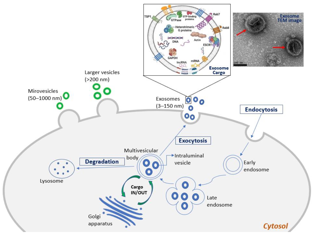
Fig. 2.Schematic representation illustrating the major physiological functions and clinical applications of extracellular vesicles (EVs) released by Trichomonas vaginalis (TvEVs).
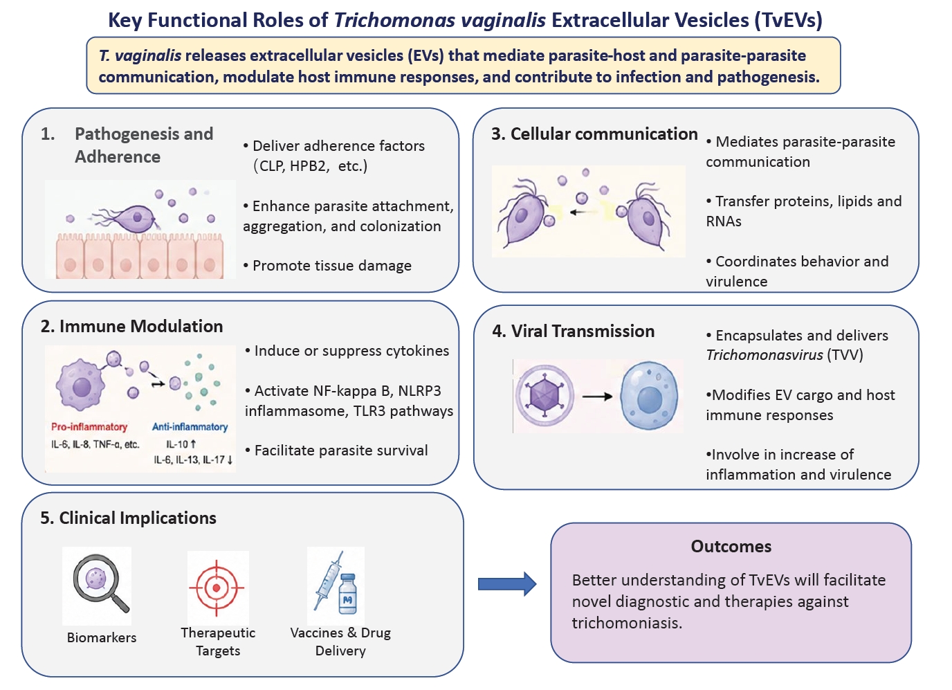
Table 1.Key literature supporting major studies on TvEVs
Table 1.
|
Reference |
TV strain |
EV type |
EV isolation method |
Protein analysis |
Biomolecular cargo content |
In vitro/in vivo (animal) experiments |
RNA analysis and viral experiments |
Research applications |
|
Twu et al. (2013) [2] |
B7RC2 (ATCC 50167) |
Exosome |
UC with sucrose gradient |
215 proteins detected by MudPIT-based mass spectrometry |
Small RNAs, TSPs, Alix, Rabs, HSP70, BspA, TcTP, GP63-like |
Immunolocalization of parasite with human cervical Ect cells |
mRNA analysis by Agilent 2000 Bioanalyzer |
Immunomodulation |
|
Annexin V-FITC in PKH-67 |
Increase parasite adhesion to host cells |
|
Olmos-Ortiz et al. (2017) [4] |
GT-21 |
Exosome-like vesicles |
UC |
Not reported |
Not reported |
Nitric oxide dosage and cytokine expression in murine macrophages |
Not reported |
Immunomodulation |
|
Cytokine expression in vivo after EVs stimulation in mice |
|
Nievas et al. (2018) [8] |
B7RC2 (PA) and Jt wild type |
MV-like structures |
UC |
LC–MS/MS |
TSP, RAB proteins, ARF, endoplasmin, GP63-like, BspA, P270, CPs, metallopeptidases |
Immunolocalization anti-HA tag antibody PKH67 |
Not reported |
Interaction of MVs and LVs with parasites and host cells |
|
Rai et al. (2019) [11] |
B7RC2 |
EV |
UC |
Not reported |
Not reported |
Cell uptake with EVs CFSE-labeled and R18 labeled |
Not reported |
Involved in host-parasite and parasite-parasite interaction |
|
Artuyants et al. (2020) [9] |
B7RC2 |
EV |
UC |
Not reported |
Not reported |
sEVs derived from TVV+ parasite stimulates a proinflammatory response in human HaCaT cells |
Analysis of small RNA in EVs cargo |
Revealed a preferential RNA cargo in EVs, which might be involved in parasite-host communication |
|
RNA sequencing |
|
Govender et al. (2020) [5] |
Isolate 347V+, 347V-, B7RC2, UR1, OC7, OC8 |
sEV |
Total exosome isolation reagent |
171 proteins detected by TMT peptide labeling |
Zinc ion binding and transferase activity proteins |
EVs stimulation NF-kB activity in endocervical cells |
TVVs modulate small RNA cargo in EVs |
Suppression of the immune response |
|
EVs stimulation of cytokines in PBMC |
|
Endocervical cells and PBMC viability |
|
Ong et al. (2022) [15] |
ATCC 30236, 30238, 50148, 50143, PRA-98 and T1 |
sEV |
UC with 30% sucrose |
LC–MS/MS |
TSP1, TSP6 & TSP8, TVV capsid proteins |
Not reported |
Identification of TVV subspecies by RT-PCR, RNA sequencing and NGS |
Encapsulation of TVV particles |
|
Rada et al. (2022) [6] |
TV79-49c1+ and TV79-49c1–
|
sEV |
UC with 5%–50% OptiPrep |
1633 proteins detected by label-free quantitative mass spectrometry |
TSP1, Rab small GTPases, HSP70, TSG101, BspA, calpains, GP63-like metallopeptidases, small regulatory tsRNA |
sEVs derived from TVV+ parasite stimulates a proinflammatory response in human HaCaT cells |
TVVs modulate small RNA cargo in EVs |
Encapsulation of TVV particles |
|
RNA sequencing |
|
Salas et al. (2023) [10] |
G3, B7RC2, and CDC1132 |
MVs (cytoneme structure) and exosomes |
UC |
LC–MS/MS |
VPS32, Rab proteins, BspA, cathepsin L-like cysteine peptidase, GP63-like metallopeptidases, peptidase T-like |
Parasites were seeded onto human prostate BPH-1 cells to quantify binding to the host cells |
Not reported |
Increase parasite adhesion to host cells |
|
Molgora et al. (2023) [12] |
B7RC2 |
EV |
UC |
Not reported |
Not reported |
Delivering parasites into the murine urogenital tract via transurethral catheterization |
Not reported |
Increase the adherence of TV to host cell monolayers in vitro |
|
Increased survival of the parasite in murine |
|
Ong et al. (2024) [7] |
ATCC 30236, 30238, 50148, 50143, PRA-98, and T1 |
Exosome |
UC with 30% sucrose |
Total of 1207 exosomal-specific proteins detected by LC–MS/MS |
TSP1, ribosomal proteins, Rabs, heterotrimeric G proteins, actin, VPS32, GP63-like |
Not reported |
Not reported |
Established a reference core exosome proteome of TV |
|
Kochanowsky et al. (2024) [13] |
B7RC2 and G3 |
EV |
Total exosome isolation reagent |
303 proteins detected by MS/MS |
TSPs, small Rab & Ras GTPases, VSPs, HPB, CLP |
Co-infection with EVs in mouse |
RNA sequencing |
Identify TvEVs as a mediator of parasite-parasite communication |
|
Huang et al. (2025) [14] |
ATCC 30236 and ATCC 50143 |
EV |
Total exosome isolation reagent |
LC–MS/MS |
Adhesins, cysteine proteases |
Mice were inoculated intravaginally with TvEVs |
RNA sequencing |
Investigate the molecular mechanisms driving inflammation induced by TvEVs |
|
Co-incubation of labeled EV with THP-1 macrophages and human Ect cells |
Involved in host-parasite interaction |
References
- 1. Ryan CM, de Miguel N, Johnson PJ. Trichomonas vaginalis: current understanding of host-parasite interactions. Essays Biochem 2011;51:161-75. https://doi.org/10.1042/bse0510161
- 2. Twu O, de Miguel N, Lustig G, et al. Trichomonas vaginalis exosomes deliver cargo to host cells and mediate host: parasite interactions. PLoS Pathog 2013;9:e1003482. https://doi.org/10.1371/journal.ppat.1003482
- 3. Théry C, Witwer KW, Aikawa E, et al. Minimal information for studies of extracellular vesicles 2018 (MISEV2018): a position statement of the International Society for Extracellular Vesicles and update of the MISEV2014 guidelines. J Extracell Vesicles 2018;7:1535750. https://doi.org/10.1080/20013078.2018.1535750
- 4. Olmos-Ortiz LM, Barajas-Mendiola MA, Barrios-Rodiles M, et al. Trichomonas vaginalis exosome-like vesicles modify the cytokine profile and reduce inflammation in parasite-infected mice. Parasite Immunol 2017;39. https://doi.org/10.1111/pim.12426
- 5. Govender Y, Chan T, Yamamoto HS, Budnik B, Fichorova RN. The role of small extracellular vesicles in viral-protozoan symbiosis: lessons from Trichomonasvirus in an isogenic host parasite model. Front Cell Infect Microbiol 2020;10:591172. https://doi.org/10.3389/fcimb.2020.591172
- 6. Rada P, Hrdý I, Zdrha A, et al. Double-stranded RNA viruses are released from Trichomonas vaginalis inside small extracellular vesicles and modulate the exosomal cargo. Front Microbiol 2022;13:893692. https://doi.org/10.3389/fmicb.2022.893692
- 7. Ong SC, Luo HW, Cheng WH, et al. The core exosome proteome of Trichomonas vaginalis. J Microbiol Immunol Infect 2024;57:246-56. https://doi.org/10.1016/j.jmii.2024.02.003
- 8. Nievas YR, Coceres VM, Midlej V, et al. Membrane-shed vesicles from the parasite Trichomonas vaginalis: characterization and their association with cell interaction. Cell Mol Life Sci 2018;75:2211-26. https://doi.org/10.1007/s00018-017-2726-3
- 9. Artuyants A, Campos TL, Rai AK, et al. Extracellular vesicles produced by the protozoan parasite Trichomonas vaginalis contain a preferential cargo of tRNA-derived small RNAs. Int J Parasitol 2020;50:1145-55. https://doi.org/10.1016/j.ijpara.2020.07.003
- 10. Salas N, Blasco Pedreros M, Dos Santos Melo T, et al. Role of cytoneme structures and extracellular vesicles in Trichomonas vaginalis parasite-parasite communication. Elife 2023;12:e86067. https://doi.org/10.7554/eLife.86067
- 11. Rai AK, Johnson PJ. Trichomonas vaginalis extracellular vesicles are internalized by host cells using proteoglycans and caveolin-dependent endocytosis. Proc Natl Acad Sci USA 2019;116:21354-60. https://doi.org/10.1073/pnas.1912356116
- 12. Molgora BM, Mukherjee SK, Baumel-Alterzon S, et al. Trichomonas vaginalis adherence phenotypes and extracellular vesicles impact parasite survival in a novel in vivo model of pathogenesis. PLoS Negl Trop Dis 2023;17:e0011693. https://doi.org/10.1371/journal.pntd.0011693
- 13. Kochanowsky JA, Mira PM, Elikaee S, et al. Trichomonas vaginalis extracellular vesicles upregulate and directly transfer adherence factors promoting host cell colonization. Proc Natl Acad Sci USA 2024;121:e2401159121. https://doi.org/10.1073/pnas.2401159121
- 14. Huang CY, Chiu SF, Chu LJ, et al. Trichomonas vaginalis extracellular vesicles activate the NLRP3 inflammasome and TLR3-mediated inflammatory cascades in host cells. PLoS Pathog 2025;21:e1013216. https://doi.org/10.1371/journal.ppat.1013216
- 15. Ong SC, Cheng WH, Ku FM, et al. Identification of endosymbiotic virus in small extracellular vesicles derived from Trichomonas vaginalis. Genes (Basel) 2022;13:531. https://doi.org/10.3390/genes13030531
- 16. de Miguel N, Lustig G, Twu O, et al. Proteome analysis of the surface of Trichomonas vaginalis reveals novel proteins and strain-dependent differential expression. Mol Cell Proteomics 2010;9:1554-66. https://doi.org/10.1074/mcp.M000022-MCP201
- 17. Coceres VM, Alonso AM, Nievas YR, et al. The C-terminal tail of tetraspanin proteins regulates their intracellular distribution in the parasite Trichomonas vaginalis. Cell Microbiol 2015;17:1217-29. https://doi.org/10.1111/cmi.12431
- 18. Salas N, Coceres VM, Melo TDS, et al. VPS32, a member of the ESCRT complex, modulates adherence to host cells in the parasite Trichomonas vaginalis by affecting biogenesis and cargo sorting of released extracellular vesicles. Cell Mol Life Sci 2021;79:11. https://doi.org/10.1007/s00018-021-04083-3
- 19. Wang ZS, Zhou HC, Wei CY, et al. Global survey of miRNAs and tRNA-derived small RNAs from the human parasitic protist Trichomonas vaginalis. Parasit Vectors 2021;14:87. https://doi.org/10.1186/s13071-020-04570-9
- 20. Goodman RP, Ghabrial SA, Fichorova RN, Nibert ML. Trichomonasvirus: a new genus of protozoan viruses in the family Totiviridae. Arch Virol 2011;156:171-9. https://doi.org/10.1007/s00705-010-0832-8
- 21. Wang AL, Wang CC. The double-stranded RNA in Trichomonas vaginalis may originate from virus-like particles. Proc Natl Acad Sci USA 1986;83:7956-60. https://doi.org/10.1073/pnas.83.20.7956
- 22. Luo HW, Ong SC, Syu JW, et al. Viral load dependent cellular heterogeneity in Trichomonas vaginalis at the single cell level. iScience 2025;29:114260. https://doi.org/10.1016/j.isci.2025.114260