Abstract
Human echinococcosis, caused by Echinococcus spp., remains a significant public health concern in endemic regions and manifests primarily as cystic echinococcosis (CE) and alveolar echinococcosis (AE), both characterized by chronic infection and heterogeneous clinical outcomes. This review synthesizes current evidence on host genetic determinants that influence immune responses, susceptibility, and disease persistence in CE and AE. Available studies indicate that polymorphisms in genes encoding key immunoregulatory cytokines, particularly IL10 and TGFB1, are associated with an anti-inflammatory immune profile that favors parasite persistence through enhanced regulatory activity and suppression of protective effector responses. Cytokine pathways involving IL-4, IL-13, and IFN-γ further modulate the balance between Th2-mediated and Th1-mediated immune responses, thereby influencing infection outcomes. In addition, innate immune pathways, including Toll-like receptor signaling, affect early parasite recognition and downstream immune responses, although direct human genetic evidence for Toll-like receptor polymorphisms in CE and AE remains limited. Human leukocyte antigen class II polymorphisms may also contribute to inter-individual differences in antigen presentation and CE susceptibility, although reported associations remain population-specific and require further validation. Collectively, these findings support a mechanistic framework in which host immunogenetic variation shapes the balance between immune activation and tolerance and may influence disease trajectory. Integration of genetic markers with clinical and imaging data may improve risk stratification and support the development of more targeted therapeutic strategies. Future research should prioritize large-scale, multicenter studies, standardized phenotyping, and functional validation to clarify the clinical relevance of identified variants.
-
Key words: Echinococcosis, host genetics, polymorphism, immune tolerance, human leukocyte antigen, Toll-like receptors
Introduction
Echinococcus infection and its life cycle
Echinococcosis is a zoonotic parasitic disease caused by cestodes of the genus
Echinococcus, primarily
Echinococcus granulosus and
Echinococcus multilocularis, which are responsible for cystic echinococcosis (CE) and alveolar echinococcosis (AE), respectively [
1,
2]. The parasite life cycle involves 2 hosts: a definitive host, typically canids such as dogs and foxes, and an intermediate host, including livestock and humans. Infection occurs when intermediate hosts ingest parasite eggs shed in the feces of definitive hosts. Following ingestion, larvae penetrate the intestinal wall and develop into hydatid cysts, predominantly in the liver and lungs, leading to progressive tissue damage and clinical disease [
2].
Echinococcosis remains a significant global health concern, particularly in pastoral and resource-limited regions. CE is widely distributed across South America, the Mediterranean basin, Eastern Europe, Africa, and Asia, whereas AE is more prevalent in the Northern Hemisphere, including parts of China, Central Europe, and Russia [
3]. Despite the implementation of control strategies such as dog deworming, livestock inspection, and public health education, the disease persists in endemic regions and continues to pose major challenges [
4]. The global burden of echinococcosis is substantial, with estimates indicating approximately one million disability-adjusted life years lost annually, alongside considerable economic losses in both human health and livestock production [
5]. These impacts are exacerbated by delayed diagnosis, limited access to healthcare, and the complexity of treatment in advanced cases.
In humans, echinococcosis manifests primarily as CE or AE, which differ markedly in their biological behavior and clinical progression. CE is characterized by the formation of fluid-filled hydatid cysts, most commonly in the liver and lungs, although other organs may also be affected [
6]. These cysts typically grow slowly and may remain asymptomatic for years, but can cause clinical symptoms such as abdominal pain, nausea, or organ dysfunction due to mass effect. Cyst rupture may result in severe allergic reactions or secondary dissemination [
7]. In contrast, AE exhibits a more aggressive and infiltrative growth pattern, resembling a malignant tumor. The parasite forms irregular, invasive lesions in the liver that can spread to adjacent tissues and distant organs. Clinical manifestations include jaundice, weight loss, and hepatic dysfunction, and the disease is often fatal if left untreated [
3,
8].
Diagnosis of echinococcosis relies primarily on imaging techniques, including ultrasound, computed tomography, and magnetic resonance imaging, supported by serological assays such as ELISA and indirect hemagglutination tests [
9,
10]. Molecular methods, including PCR-based detection, provide additional specificity in complex or ambiguous cases [
11]. Management strategies depend on disease stage and include surgical intervention, antiparasitic chemotherapy such as albendazole, active surveillance in selected cases, and percutaneous techniques such as PAIR. PAIR refers to puncture, aspiration, injection of a scolicidal agent, and re-aspiration, and is mainly used for selected uncomplicated CE cysts [
12-
15]. However, treatment outcomes remain variable, particularly in AE, where complete cure is often difficult to achieve.
The Importance of Studying Genetic Determinants of Susceptibility to Echinococcus
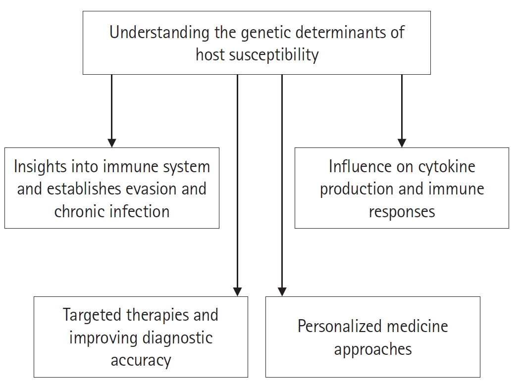
Understanding the genetic determinants of host susceptibility to
Echinococcus infection is critical for elucidating the mechanisms underlying parasite persistence and disease progression. Host genetic variation, particularly in genes regulating cytokine production and immune responses, plays a pivotal role in shaping infection outcomes by influencing the balance between protective immunity and immune tolerance. Identifying these genetic factors provides a foundation for the development of targeted therapeutic strategies and improved diagnostic approaches. Furthermore, integrating genetic information into clinical practice may enable personalized medicine, allowing treatment strategies to be tailored according to individual immune and genetic profiles. As summarized in
Fig. 1, investigating host genetic susceptibility not only advances our understanding of host–parasite interactions but also supports the development of more precise and effective interventions.
This review is based on a targeted literature survey of published studies relevant to host genetic and immunological factors in echinococcosis. Relevant articles were identified through major scientific databases and selected based on their relevance to CE and AE, host immune responses, and genetic susceptibility. Both clinical and experimental studies were included to provide a comprehensive overview of the topic. No formal systematic review protocol was applied.
Host Immune Response to Echinococcus Infection
Mechanisms of immune response against Echinococcus
The host immune response to Echinococcus infection involves coordinated interactions between innate and adaptive immune systems. Following infection, parasite-derived antigens are recognized by host immune cells, initiating a cascade of immunological responses that determine infection outcome.
Innate immune response to Echinococcus
The innate immune system constitutes the first line of defense against
Echinococcus. Innate immune cells, including macrophages, dendritic cells, and neutrophils, express pattern-recognition receptors such as Toll-like receptors (TLRs), which recognize pathogen-associated molecular patterns. In cystic and alveolar echinococcosis, TLR2 and TLR4 have been implicated in immune regulation, although their precise roles remain incompletely characterized. TLR signaling can activate MyD88-dependent pathways involving NF-κB and mitogen-activated protein kinases, thereby promoting pro-inflammatory cytokine production. Activated macrophages may also produce nitric oxide, which contributes to preventing the dissemination of hydatid cyst layers [
16]. Evidence from helminth infection models further suggests that parasite-derived excretory–secretory products can modulate innate immune responses, including alterations in co-stimulatory molecule expression and cytokine production, thereby contributing to immune evasion strategies [
17].
Adaptive immune response to Echinococcus
Adaptive immune responses play an important role in controlling Echinococcus multilocularis infection. In a chronic murine model of alveolar echinococcosis, PD-L1 blockade reduced parasite burden, increased CD4
+ and CD8
+ effector T cells, decreased regulatory T cells, and restored dendritic cells and Kupffer cells/macrophages. These findings indicate that PD-L1 signaling regulates coordinated innate and adaptive immune responses and influences parasite control and tissue inflammatio [
18]. In addition, both cellular and humoral immune components are involved in responses to parasite-derived antigens. Studies have demonstrated that exposure to hydatid cyst antigens induces the production of pro-inflammatory cytokines, such as IFN-γ and TNF-α, as well as parasite-specific IgG antibodies, highlighting the role of antigen-specific immune responses in host defense [
19].
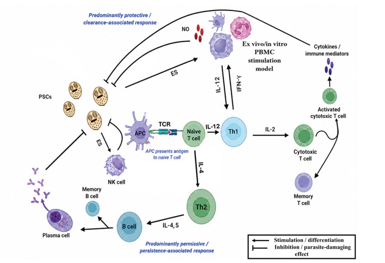
Fig. 2 summarizes the principal immune mechanisms involved in host responses to
Echinococcus infection.
Echinococcus species have evolved multiple sophisticated strategies to evade host immune defenses, enabling long-term survival and chronic infection.
Modulation of host immune response
Echinococcus infection is associated with the activation of immunosuppressive pathways that promote immune tolerance. In patients with AE, increased expression of the Tim-3/Galectin-9 pathway was associated with elevated IL-10 and TGF-β expression, altered CD8
+ T-cell activity, and reduced cytotoxic T-lymphocyte responses. These immunoregulatory changes may impair parasite clearance and contribute to persistent infection [
20]. In addition, parasite-derived antigens and ES products can modulate immune-cell responses in ex vivo and in vitro settings. These effects may involve peripheral blood mononuclear cells, dendritic cells, and CD4
⁺ T-cell subsets, and may be reflected by reduced protoscolex-killing activity and nitric oxide production, impaired dendritic-cell function, suppression of Th1 development, and induction of CD4
⁺CD25
⁺Foxp3
⁺ regulatory T cells, thereby contributing to immune evasion and chronic infection [
21,
22].
Antigenic diversity and immune interference
Rather than classical antigenic switching,
Echinococcus exhibits antigenic diversity across different cyst compartments and host tissues. In
E. granulosus, antigens from the laminated layer (LL), protoscoleces, and hydatid cyst fluid have been shown to display immunogenically distinct protein profiles, with the LL containing prominent immunogenic proteins [
23].
Induction of regulatory T cells
A key immune evasion mechanism involves the expansion of Tregs, which suppress protective immune responses. Studies demonstrate increased Foxp3
⁺ Treg activity and associated cytokines (IL-10 and TGF-β), contributing to a tolerogenic environment that favors parasite survival [
24-
26].
Macrophage polarization and immune regulation
Parasite-driven immune responses are often associated with a shift toward Th2-type immunity, characterized by cytokines such as IL-4 and IL-13. These cytokines promote the polarization of macrophages toward an alternatively activated phenotype, which exhibits reduced microbicidal activity, including decreased nitric oxide production, thereby facilitating parasite persistence [
27].
Physical and molecular barriers
The hydatid cyst contains parasite-derived molecules that play key roles in host–parasite interactions. These components exhibit antigenic diversity and bioactive properties that contribute to immune modulation and parasite survival within the host [
28].
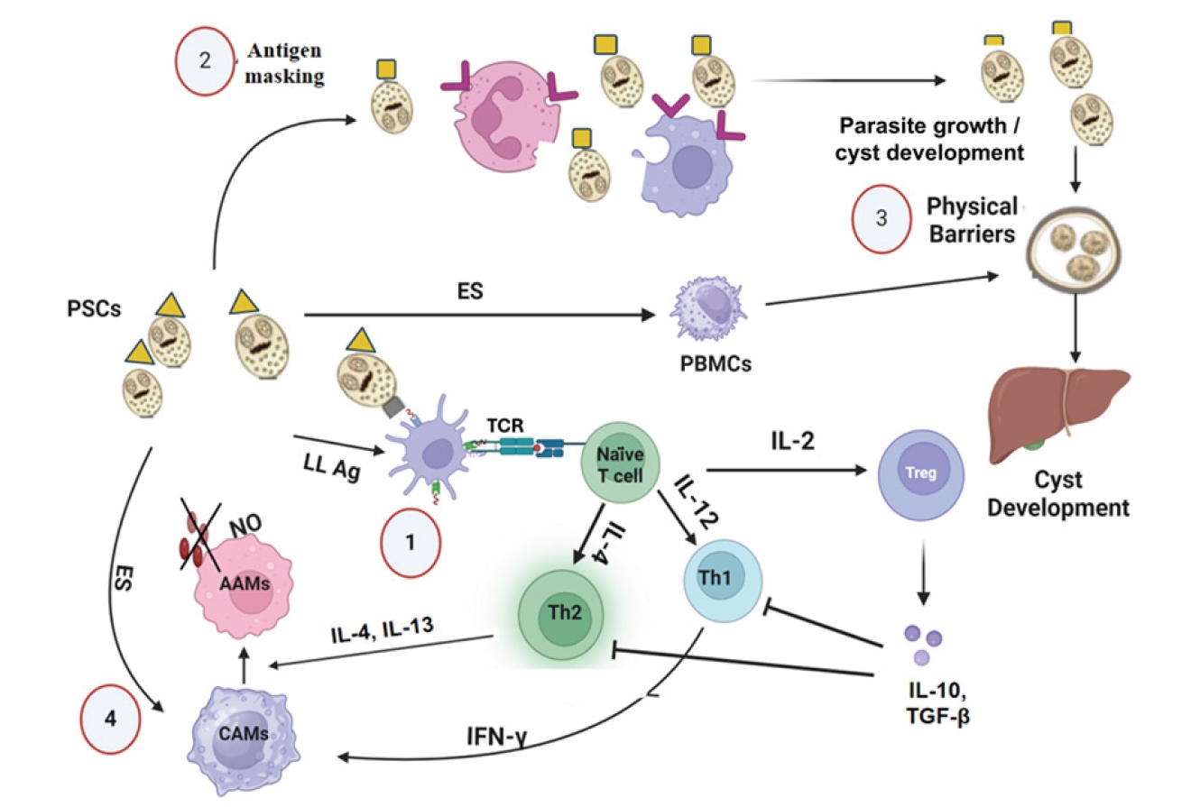
Fig. 3 provides a schematic overview of the principal immune evasion mechanisms of
Echinococcus.
Genetic Determinants of Host Susceptibility
Overview of host genetic factors influencing susceptibility
Host genetic factors play an important role in determining susceptibility to parasitic infections, including
E. granulosus and
E. multilocularis. Variations in genes involved in immune regulation can influence how effectively the host recognizes, responds to, and controls infection [
29].
In particular, genetic variations such as cytokine gene polymorphisms and other immune-related factors have been implicated in shaping host–parasite interactions and infection susceptibility [
29]. These variations may influence immune regulation and contribute to differences in infection outcomes [
29].
Beyond host genetic influences, helminth infections are also characterized by strong parasite-driven immunomodulation. Parasites such as
E. granulosus can manipulate host immune pathways, promoting regulatory and anti-inflammatory responses that support chronic infection, while excessive inflammatory responses may contribute to host tissue damage despite limiting parasite survival [
30-
32].
IL-10: immunoregulation and disease persistence
IL-10 is a key anti-inflammatory cytokine that suppresses pro-inflammatory responses and promotes immune tolerance. In CE, elevated IL-10 levels have been associated with chronic infection and parasite persistence, particularly in patients with active cystic disease [
33,
34]. Functional studies indicate that IL-10, together with IL-4, impairs parasite killing by reducing nitric oxide production and suppressing Th1-mediated immunity [
22], while parasite-derived factors from
E. granulosus may further enhance regulatory immune responses [
21]. At the genetic level, the IL10 rs1800896 variant has been identified in an exploratory genomic screening study related to
E. granulosus susceptibility in an endemic population [
35]. However, this evidence is based on a small cohort and lacks case-control validation.
TGFB1: immune tolerance and parasite survival
TGF-β1 is a multifunctional cytokine involved in immune suppression, immune tolerance, and tissue remodeling. In CE and AE, increased TGF-β expression has been associated with parasite persistence, immune tolerance, and tissue remodeling or fibrosis, supporting its potential role in chronic infection [
36-
38]. TGFB1 rs1800469 was also reported as a high-frequency candidate variant in the same exploratory
E. granulosus–related study [
35]. However, this evidence remains preliminary and lacks validation in independent cohorts. Overall, TGFB1 should be regarded as a biologically plausible candidate gene involved in immune tolerance and tissue remodeling in CE and AE, although its genetic association with susceptibility remains preliminary and requires validation in larger, independent cohorts.
IL-4 and IL-13: Th2 polarization and immune deviation
IL-4 and IL-13 are key mediators of Th2 immune responses, promoting humoral immunity and IgE production. These cytokines play a central role in helminth infections by driving Th2 polarization and modulating host immune responses [
39,
40]. In
E. multilocularis infections, parasite-derived antigens have been shown to induce IL-4 and IL-13 production, contributing to a Th2-skewed immune environment. This Th2 deviation may counterbalance Th1-associated protective responses and contribute to conditions that favor parasite persistence [
40].
IFNG: Th1-mediated protective immunity
IFN-γ is a key cytokine in Th1-mediated immune responses and plays a central role in macrophage activation and parasite clearance. In CE, parasite-derived antigens have been shown to induce IFN-γ production by CD4
⁺ and CD8
⁺ T cells, supporting Th1-mediated immunity [
41]. Genetic polymorphisms in IFNG are known to influence cytokine production and host resistance in infectious diseases. IFNG rs2779249 was reported as a high-frequency candidate variant in an exploratory hydatidosis study related to
E. granulosus; however, no direct validated association with disease susceptibility has yet been established [
35]. Overall, IFNG represents a biologically plausible candidate gene in CE, although current genetic evidence remains limited and requires further validation.
TLR2 and TLR4: innate immune recognition and regulation
TLRs, particularly TLR2 and TLR4, play important roles in innate immune recognition and early host responses to
Echinococcus infection. In CE, hydatid cyst-derived antigens from
E. granulosus have been shown to modulate TLR2 and TLR4 expression in peripheral blood mononuclear cells, suggesting that the parasite may interfere with innate immune activation and downstream cytokine responses [
42]. In AE, experimental murine studies have also implicated TLR signaling in immune regulation; for example, TLR2 expression has been associated with IL-10 production during
E. multilocularis infection, indicating a possible role in promoting regulatory or tolerogenic immune responses [
43]. Overall, current evidence supports the involvement of TLR2 and TLR4 signaling in immune modulation during CE and AE. However, direct human genetic evidence linking specific TLR polymorphisms to susceptibility or disease progression in CE or AE remains limited. Therefore, TLR2 and TLR4 should currently be interpreted as biologically plausible candidate pathways rather than validated genetic risk markers in echinococcosis.
HLA genes: antigen presentation and genetic susceptibility
Human leukocyte antigen (HLA) class II molecules are central to antigen presentation and the activation of adaptive immune responses by presenting peptide antigens to T cells. The high polymorphism of HLA genes results in substantial inter-individual variability in the repertoire of peptides that can be presented, thereby influencing immune recognition and response to pathogens [
44].
Several case-control studies have investigated the association between HLA class II polymorphisms and susceptibility to CE. Specific alleles, including HLA-DRB1*07, HLA-DQB1*09, and HLA-DQB1*02, have been associated with an increased risk of CE in children, while HLA-DQB1*02 and HLA-DRB1*03 have been reported more frequently in patients with suppuration-complicated cysts [
45]. In contrast, a reduced frequency of HLA-DRB1*03 has been observed in patients compared to healthy controls, suggesting a potential protective role against infection [
46].
These findings collectively indicate that HLA genetic variation may influence host susceptibility to CE by modulating antigen presentation and subsequent T-cell–mediated immune responses. However, the reported associations remain heterogeneous and may vary across populations, highlighting the need for larger, multicenter studies to better clarify the role of HLA class II polymorphisms in the natural history of E. granulosus infection.
Integrated perspective on host genetic regulation
Recent studies highlight the critical role of host–parasite cross-talk mechanisms involving TLR signaling, apoptosis, and inflammasome activation in both CE and AE, with these pathways contributing to immune modulation and disease progression [
16]. In addition, exploratory genetic studies have identified associations between polymorphisms in immune-related genes—such as IL10, IL17A, IFNG, FOXP3, TGFB1, and VDR—and susceptibility to
E. granulosus infection. These findings suggest that genetic variability in immune regulatory pathways may modulate host responses; however, most evidence is derived from small-scale studies and should be considered preliminary [
35]. Furthermore, the immunological landscape of hepatic AE has been increasingly characterized in recent years, with bibliometric analyses identifying major research trends, hotspots, and emerging themes related to host–parasite immune interactions, diagnosis, and treatment strategies. This growing body of research reflects increasing interest in AE immunology and may help guide future investigations in the field [
47]. Because many reported genetic associations are derived from small-scale, exploratory, or population-specific studies, the variants summarized in
Table 1 should be interpreted as candidate susceptibility markers rather than validated clinical predictors [
35,
44-
46].
Table 1 summarizes specific gene functions, proposed immunological roles, disease context where available, and evidence summaries for reported candidate associations [
35,
44-
46].
Table 2 summarizes immune-regulatory pathways and host–parasite interaction mechanisms in echinococcosis, with disease context and evidence type specified where available [
21,
33,
34,
37,
38,
42,
43].
Accumulating evidence suggests that host immunogenetic variation may contribute to inter-individual differences in immune responses and susceptibility in echinococcosis, although current genetic evidence remains limited and is derived mainly from exploratory
E. granulosus/CE-related studies. Polymorphisms in immunoregulatory genes, including IL10 rs1800896 and TGFB1 rs1800469, have been reported as candidate variants in an exploratory genomic analysis, but their functional relevance requires further validation [
35]. Functional and tissue-based evidence indicates that IL-4- and IL-10-associated responses can reduce Th1-related parasite-killing mechanisms, whereas IL-10- and TGF-β-associated immune regulation may contribute to parasite persistence in CE [
22,
36]. In AE, longitudinal immune profiling has shown that antigen-specific IFN-γ responses increase after cure, supporting the association between stronger Th1-type responses and improved disease control [
48]. Innate immune pathways further contribute to this balance. In AE, an experimental murine study showed that
E. multilocularis infection is associated with altered TLR2 and TLR4 expression and changes in cytokines such as IL-10, IFN-γ, and IL-5, with TLR2 expression positively correlated with IL-10 production, suggesting a possible role in immune tolerance [
43]. In parallel, host genetic background modulates adaptive immune responses. The HLA-B8, DR3, DQ2 haplotype has been associated with altered cytokine production profiles, including increased IL-10 secretion and modulation of Th1/Th2 responses, which may contribute to disease severity and clinical course in AE [
49]. Despite these advances, some available evidence is derived from small-scale or population-specific studies, including recent exploratory genomic analyses [
35]. Consequently, the functional significance and clinical applicability of many candidate variants remain to be fully established. Future studies integrating large-scale genomics with functional immunological approaches and standardized clinical phenotyping will be essential to clarify the mechanistic links between host genetics, immune modulation, and disease outcomes.
Understanding the genetic determinants of host susceptibility to CE and AE remains a critical research priority. To date, most available evidence is derived from candidate gene studies and small-scale association analyses, which provide limited insight into the complex genetic architecture underlying disease susceptibility. Recent exploratory genomic investigations in endemic populations have identified polymorphisms in immune-related genes, including IL10, IL17A, IFNG, FOXP3, and TGFB1, suggesting potential genetic contributions to host susceptibility. However, these findings are based on relatively small cohorts and require validation in larger, well-designed studies [
35]. In this context, genome-wide approaches, particularly genome-wide association studies, represent a promising strategy for identifying novel susceptibility loci in an unbiased manner. Although the genome-wide association study approach has been widely applied to other infectious and complex diseases, its application to CE and AE remains limited. In addition to host genetic variation, gene–environment interactions are likely to play a significant role in shaping susceptibility to echinococcosis. Environmental and epidemiological factors, such as geographic distribution, parasite genotype diversity, host nutritional status, and co-infections, may interact with genetic predisposition to influence disease progression and severity. Integrative approaches combining genomic, environmental, and epidemiological data are therefore essential for a more comprehensive understanding of disease heterogeneity. Furthermore, advances in molecular and genomic technologies offer new opportunities for improving diagnosis and disease monitoring. The detection of
E. granulosus cell-free DNA and the characterization of parasite genotypes in clinical samples highlight the potential of molecular tools to complement conventional diagnostic methods. Such approaches may contribute to improved diagnostic sensitivity and enable better tracking of transmission dynamics, although further refinement and validation are required before routine clinical application [
50]. Finally, future research should prioritize the integration of large-scale genomics, functional immunology, and standardized clinical phenotyping. Linking genetic variants to specific immune pathways, such as cytokine regulation, innate immune signaling, and antigen presentation, will be essential to bridge the gap between genetic susceptibility and disease pathogenesis. These efforts may ultimately support the development of personalized diagnostic and therapeutic strategies for echinococcosis.
Conclusion
This review highlights the emerging role of host genetic factors in shaping susceptibility and immune responses in CE and AE. Available evidence suggests that variants in immunoregulatory and immune-response genes, including IL10, TGFB1, IFNG, FOXP3, IL17A, and HLA loci, may contribute to inter-individual differences in host–parasite interactions and disease progression. However, most reported associations are derived from small-scale, exploratory, or population-specific studies, particularly in E. granulosus–related settings, and require validation in larger, well-characterized cohorts. In addition, although cytokine regulation, TLR signaling, and Th1/Th2/Treg balance are biologically relevant to CE and AE pathogenesis, direct human genetic evidence for several candidate pathways remains limited. Advancing this field will require integrative approaches combining large-scale genomics, functional immunology, standardized clinical phenotyping, and disease-specific analysis of CE and AE. Such efforts may clarify the mechanistic links between genetic variation and immune regulation and support the development of improved diagnostic, prognostic, and targeted disease-management strategies in endemic regions.
Notes
-
Author contributions
Conceptualization: Shamsan E, Chuanchuan L, Haining F. Data curation: Shamsan E. Formal analysis: Shamsan E, Chuanchuan L. Funding acquisition: Haining F. Investigation: Shamsan E. Methodology: Shamsan E, Chuanchuan L, Haining F. Project administration: Haining F, Chuanchuan L. Resources: Haining F. Software: Shamsan E, Chuanchuan L. Supervision: Chuanchuan L, Haining F. Validation: Shamsan E, Chuanchuan L. Visualization: Shamsan E. Writing – original draft: Shamsan E. Writing – review & editing: Chuanchuan L, Haining F.
-
Conflict of interest
The authors have no conflicts of interest to declare.
-
Funding
This work was supported by the National Clinical Key Specialty Construction Project of Hepatobiliary Surgery (Hydatidosis) at Qinghai University Affiliated Hospital (Qinghai [2023]-125) and the Qinghai Scholar Program (2025-qhxz-fhn).
Fig. 1.Schematic summary of the importance of studying genetic determinants of susceptibility to Echinococcus infection.
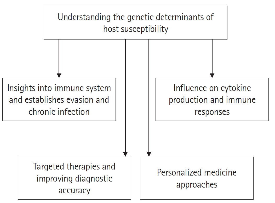
Fig. 2.Immune responses to Echinococcus protoscoleces (PSCs). PSC-derived excretory-secretory (ES) products are recognized by antigen-presenting cells (APCs), which present parasite antigens to naïve T cells and promote Th1/Th2 differentiation. IL-12 and IL-4 indicate cytokine signals that promote Th1 and Th2 differentiation, respectively. Th1-associated responses, including IFN-γ production, immune-cell activation, and nitric oxide (NO) production, are generally considered protective and may contribute to parasite clearance. Th2-associated humoral responses involving IL-4, IL-5, B-cell activation, plasma-cell differentiation, and antibody production may contribute to parasite recognition or containment, whereas Th1-associated IFN-γ/NO responses are more directly associated with parasite clearance. Peripheral blood mononuclear cells (PBMCs)-related interactions shown in the figure represent ex vivo/in vitro PBMC stimulation models and do not imply in vivo localization of PSCs in peripheral blood. TCR, T-cell receptor; NK, natural killer.
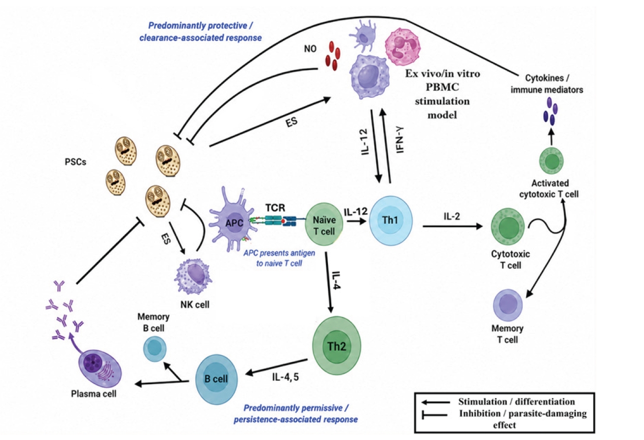
Fig. 3.Mechanisms of immune evasion by Echinococcus spp. The figure summarizes 4 major immune-evasion mechanisms: (1) laminated layer antigens (LL Ag) may impair dendritic-cell maturation and promote Th2/Treg-skewed responses associated with IL-10 and TGF-β production; (2) antigen masking reduces immune recognition; (3) cyst barriers restrict immune-cell access and support cyst development; and (4) parasite-derived excretory-secretory (ES) products promote alternative macrophage activation, reduce nitric oxide (NO) production, and impair parasite clearance. ES products may also modulate peripheral blood mononuclear cell responses in ex vivo/in vitro stimulation models; this does not imply that protoscoleces (PSCs) are normally present in peripheral blood. Treg, regulatory T cell; AAMs, alternatively activated macrophages; CAMs, classically activated macrophages.
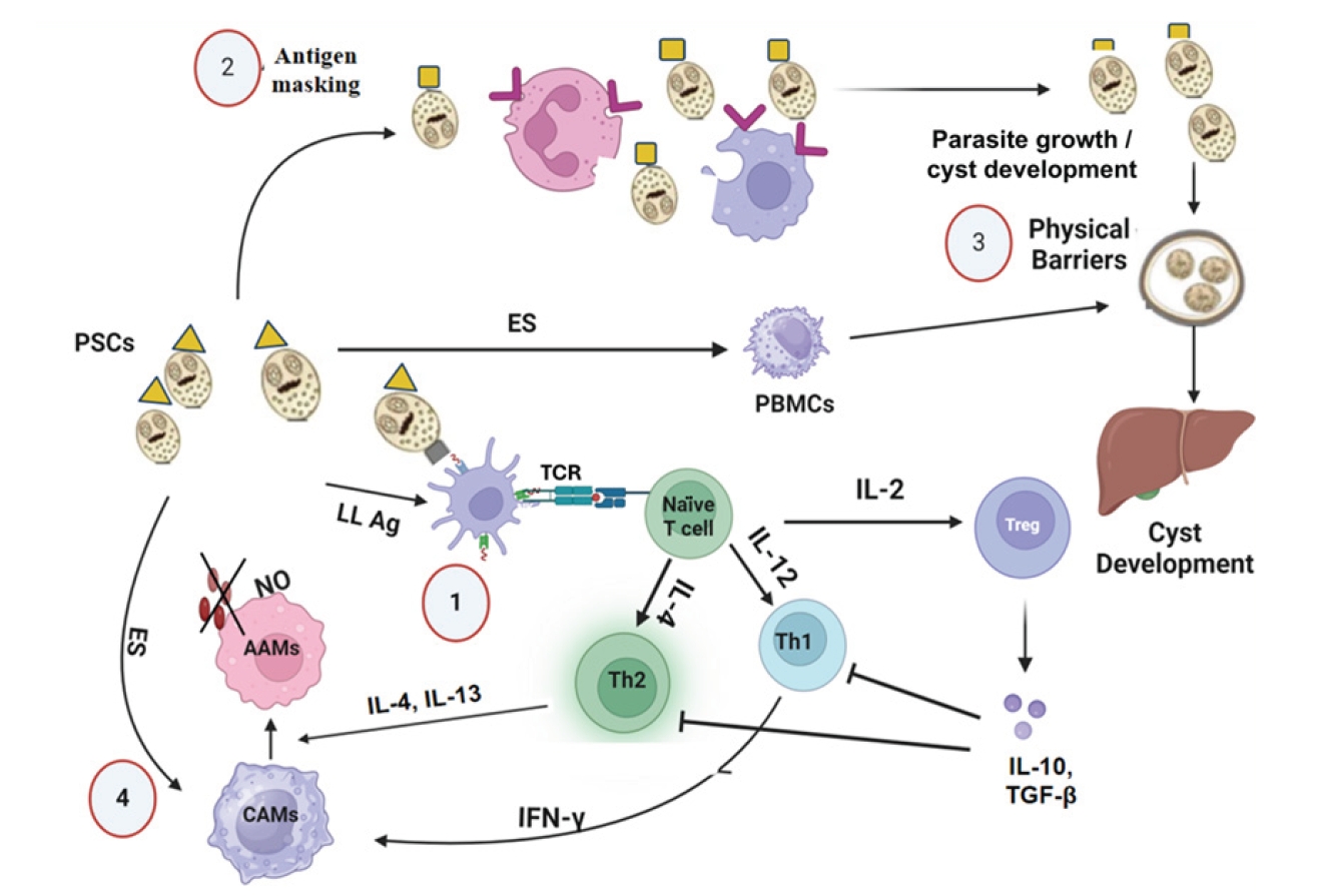
Table 1.Candidate host genetic susceptibility factors in echinococcosis with disease context where available
Table 1.
|
Gene/locus |
Variant/allele |
Disease context |
Functional role |
Evidence summary |
References |
|
IL10 |
rs1800896 |
CE-related exploratory evidence |
Immune regulatory cytokine |
Candidate variant; requires validation |
[35] |
|
TGFB1 |
rs1800469 |
CE-related exploratory evidence |
Immune regulation and fibrosis |
Candidate variant; preliminary evidence |
[35] |
|
IL17A |
rs2275913 |
CE-related exploratory evidence |
Th17 immune response |
Candidate variant; requires validation |
[35] |
|
IFNG |
rs2779249 |
CE-related exploratory evidence |
Th1 cytokine response |
Candidate variant; association unconfirmed |
[35] |
|
FOXP3 |
rs11568821 |
CE-related exploratory evidence |
Regulatory T-cell function |
Candidate variant; limited validation |
[35] |
|
HLA-DRB1/HLA-DQB1 |
HLA-DRB1*07, HLA-DQB1*02, HLA-DQB1*09 |
CE |
Antigen presentation |
Reported increased-risk alleles in pediatric CE |
[45] |
|
HLA-DRB1 |
HLA-DRB1*03 |
CE |
Antigen presentation |
Possible protective association |
[46] |
|
HLA class II (general) |
HLA variants and haplotypes |
General HLA biology; not echinococcosis-specific |
Peptide-presentation diversity |
Mechanistic rationale only |
[44] |
Table 2.Immune regulation and host–parasite interaction mechanisms in echinococcosis with disease context where available
Table 2.
|
Pathway/molecule |
Disease context |
Observed effect |
Immunological role |
Evidence type |
References |
|
TLR2/TLR4 |
CE |
Downregulation or modulation by parasite antigens |
Innate immune recognition and immune evasion |
Functional in vitro study |
[42] |
|
TLR2/TLR4 + cytokines |
AE |
Altered expression linked with IL-10, IFN-γ, IL-5 changes |
Regulation of innate–adaptive immune signaling |
Murine infection model |
[43] |
|
IL-10 |
CE |
Elevated or infection-associated expression in CE patients |
Anti-inflammatory immune regulation |
Clinical cytokine profiling |
[33] |
|
IFN-γ |
CE |
Increased in hepatic CE patients; Th1-type cytokines more evident in inactive cysts |
Th1-associated immune response and possible parasite control |
Clinical cytokine profiling |
[33,34] |
|
TGF-β–Smad pathway |
CE and AE |
Activated/upregulated in experimental CE and AE models |
Immune tolerance, altered T/NK-cell responses, and AE-associated fibrosis |
Mouse studies and human AE tissue analysis |
[37,38] |
|
FOXP3⁺ T cells |
CE |
Induction of CD4⁺CD25⁺Foxp3⁺ Tregs |
DC impairment, Treg induction, and immune evasion |
In vitro BMDC–T-cell co-culture study |
[21] |
|
Th1/Th2 balance |
CE |
Th2 dominance in active cysts; Th1/Th17 in inactive stages |
Regulation of infection progression and parasite persistence |
Human clinical studies |
[34] |
References
- 1. Grezzi L, González C, Díaz Á, Casaravilla C. The acute inflammatory potential of particles from the Echinococcus granulosus laminated layer is moderated by its calcium inositol hexakisphosphate component. Parasite Immunol 2024;46:e13040. https://doi.org/10.1111/pim.13040
- 2. Craig PS, Rogan MT, Campos-Ponce M. Echinococcosis: disease, detection and transmission. Parasitology 2003;127 Suppl:S5-20.
- 3. Moro P, Schantz PM. Echinococcosis: a review. Int J Infect Dis 2009;13:125-33. https://doi.org/10.1016/j.ijid.2008.03.037
- 4. Budke CM, Deplazes P, Torgerson PR. Global socioeconomic impact of cystic echinococcosis. Emerg Infect Dis 2006;12:296-303. https://doi.org/10.3201/eid1202.050499
- 5. Nedjim SA, Bencherki Y, Daghdagh Y, et al. Diagnosis and multidisciplinary management of a large primary retroperitoneal hydatid cyst: report of a sporadic case. Afr J Urol 2024;30:29. https://doi.org/10.1186/s12301-024-00431-4
- 6. Siyadatpanah A, Brunetti E, Emami Zeydi A, Moghadam YD, Agudelo Higuita NI. Cerebral cystic echinococcosis. Case Rep Infect Dis 2020;2020:1754231. https://doi.org/10.1155/2020/1754231
- 7. Hogea MO, Ciomaga BF, Muntean MM, et al. Cystic echinococcosis in the early 2020s: a review. Trop Med Infect Dis 2024;9:36. https://doi.org/10.3390/tropicalmed9020036
- 8. Craig PS, Hegglin D, Lightowlers MW, Torgerson PR, Wang Q. Echinococcosis: control and prevention. Adv Parasitol 2017;96:55-158. https://doi.org/10.1016/bs.apar.2016.09.002
- 9. Erganis S, Sarzhanov F, Al FD, Cağlar K. Comparison of methods in the serologic diagnosis of cystic echinococcosis. Acta Parasitol 2024;69:1122-31. https://doi.org/10.1007/s11686-024-00840-z
- 10. Kronenberg PA, Deibel A, Gottstein B, et al. Serological assays for alveolar and cystic echinococcosis: a comparative multi-test study in Switzerland and Kyrgyzstan. Pathogens 2022;11:518. https://doi.org/10.3390/pathogens11050518
- 11. Grimm J, Krickl J, Beck A, et al. Establishing and evaluation of a polymerase chain reaction for the detection of Echinococcus multilocularis in human tissue. PLoS Negl Trop Dis 2021;15:e0009155. https://doi.org/10.1371/journal.pntd.0009155
- 12. Mönnink GLE, Stijnis C, van Delden OM, Spijker R, Grobusch MP. Percutaneous versus surgical interventions for hepatic cystic echinococcosis: a systematic review and meta-analysis. Cardiovasc Intervent Radiol 2021;44:1689-96. https://doi.org/10.1007/s00270-021-02911-4
- 13. Akhan O, Erdoğan E, Ciftci TT, et al. Comparison of the long-term results of puncture, aspiration, injection and re-aspiration (PAIR) and catheterization techniques for the percutaneous treatment of CE1 and CE3a liver hydatid cysts: a prospective randomized trial. Cardiovasc Intervent Radiol 2020;43:1034-40. https://doi.org/10.1007/s00270-020-02477-7
- 14. Hemphill A, Stadelmann B, Rufener R, et al. Treatment of echinococcosis: albendazole and mebendazole: what else? Parasite 2014;21:70. https://doi.org/10.1051/parasite/2014073
- 15. Stojkovic M, Rosenberger KD, Steudle F, Junghanss T. Watch and wait management of inactive cystic echinococcosis: does the path to inactivity matter: analysis of a prospective patient cohort. PLoS Negl Trop Dis 2016;10:e0005243. https://doi.org/10.1371/journal.pntd.0005243
- 16. Bakhtiar NM, Spotin A, Mahami-Oskouei M, Ahmadpour E, Rostami A. Recent advances on innate immune pathways related to host-parasite cross-talk in cystic and alveolar echinococcosis. Parasit Vectors 2020;13:232. https://doi.org/10.1186/s13071-020-04103-4
- 17. Bakshi M, Hebert D, Gulbronson C, et al. Ostertagia ostertagi mediates early host immune responses via macrophage and toll-like receptor pathways. Infect Immun 2021;89:e00017-21. https://doi.org/10.1128/IAI.00017-21
- 18. Jebbawi F, Bellanger AP, Lunström-Stadelmann B, et al. Innate and adaptive immune responses following PD-L1 blockade in treating chronic murine alveolar echinococcosis. Parasite Immunol 2021;43:e12834. https://doi.org/10.1111/pim.12834
- 19. Vatankhah A, Ghaffari SMR, Vatankhah RG, et al. Characterization of cellular and humoral immune responses to alkaline phosphatase from fertile hydatid cysts in the human peripheral blood. J Cell Physiol 2019;234:2765-77. https://doi.org/10.1002/jcp.27092
- 20. Zhao H, Ma Y, Tian F, et al. Expression of Tim-3/Galectin-9 pathway and CD8+T cells and related factors in patients with cystic echinococcosis. Exp Parasitol 2023;254:108623. https://doi.org/10.1016/j.exppara.2023.108623
- 21. Wang Y, Zhou H, Shen Y, et al. Impairment of dendritic cell function and induction of CD4(+)CD25(+)Foxp3(+) T cells by excretory-secretory products: a potential mechanism of immune evasion adopted by Echinococcus granulosus. BMC Immunol 2015;16:44. https://doi.org/10.1186/s12865-015-0110-3
- 22. Amri M, Mezioug D, Touil-Boukoffa C. Involvement of IL-10 and IL-4 in evasion strategies of Echinococcus granulosus to host immune response. Eur Cytokine Netw 2009;20:63-8. https://doi.org/10.1684/ecn.2009.0154
- 23. Amni F, Hajizadeh M, Elmi T, et al. Different manifestation of Echinococcus granulosus immunogenic antigens in the liver and lungs of intermediate host. Comp Immunol Microbiol Infect Dis 2021;74:101573. https://doi.org/10.1016/j.cimid.2020.101573
- 24. Gottstein B, Soboslay P, Ortona E, et al. Immunology of alveolar and cystic echinococcosis (AE and CE). Adv Parasitol 2017;96:1-54. https://doi.org/10.1016/bs.apar.2016.09.005
- 25. Wang J, Cardoso R, Marreros N, et al. Foxp3(+) T regulatory cells as a potential target for immunotherapy against primary infection with Echinococcus multilocularis eggs. Infect Immun 2018;86:e00542-18. https://doi.org/10.1128/IAI.00542-18
- 26. Yarahmadov T, Wang J, Sanchez-Taltavull D, et al. Primary infection by E. multilocularis induces distinct patterns of cross talk between hepatic natural killer T cells and regulatory T cells in mice. Infect Immun 2022;90:e0017422. https://doi.org/10.1128/iai.00174-22
- 27. Peón AN, Ledesma-Soto Y, Terrazas LI. Regulation of immunity by Taeniids: lessons from animal models and in vitro studies. Parasite Immunol 2016;38:124-35. https://doi.org/10.1111/pim.12289
- 28. Ahn CS, Kim JG, Han X, Kang I, Kong Y. Comparison of Echinococcus multilocularis and Echinococcus granulosus hydatid fluid proteome provides molecular strategies for specialized host-parasite interactions. Oncotarget 2017;8:97009-24. https://doi.org/10.18632/oncotarget.20761
- 29. Ye C, Zhang L, Tang L, et al. Host genetic backgrounds: the key to determining parasite-host adaptation. Front Cell Infect Microbiol 2023;13:1228206. https://doi.org/10.3389/fcimb.2023.1228206
- 30. Siracusano A, Delunardo F, Teggi A, Ortona E. Host-parasite relationship in cystic echinococcosis: an evolving story. Clin Dev Immunol 2012;2012:639362. https://doi.org/10.1155/2012/639362
- 31. Zakeri A, Hansen EP, Andersen SD, Williams AR, Nejsum P. Immunomodulation by helminths: intracellular pathways and extracellular vesicles. Front Immunol 2018;9:2349. https://doi.org/10.3389/fimmu.2018.02349
- 32. Legorreta-Herrera M, Rodriguez-Sosa M, Satoskar AR. Editorial: regulation of the host's immune system by parasitic infections and its implications. Front Cell Infect Microbiol 2022;12:1118692. https://doi.org/10.3389/fcimb.2022.1118692
- 33. Beyhan YE, Albayrak C, Guven I. IL-4, IL-10, TNF-α and IFN-γ cytokine levels in patients with cystic echinococcosis and fascioliasis. Iran J Public Health 2022;51:1339-47. https://doi.org/10.18502/ijph.v51i6.9679
- 34. Yasen A, Li W, Aini A, et al. Th1/Th2/Th17 cytokine profile in hepatic cystic echinococcosis patients with different cyst stages. Parasite Immunol 2021;43:e12839. https://doi.org/10.1111/pim.12839
- 35. Jaramillo-Valverde L, Garcia-Paitan MY, Landeo D, Santivañez SJ, Cacacabelos R. Exploratory study on genetic variants related to hydatidosis susceptibility and albendazole pharmacogenetics in the Cusco region in Peru. Front Biosci (Schol Ed) 2025;17:40566. https://doi.org/10.31083/FBS40566
- 36. De Biase D, Prisco F, Pepe P, et al. Evaluation of the local immune response to hydatid cysts in sheep liver. Vet Sci 2023;10:315. https://doi.org/10.3390/vetsci10050315
- 37. Yin S, Chen X, Zhang J, et al. The effect of Echinococcus granulosus on spleen cells and TGF-β expression in the peripheral blood of BALB/c mice. Parasite Immunol 2017;39. https://doi.org/10.1111/pim.12415
- 38. Wang J, Zhang C, Wei X, et al. TGF-β and TGF-β/Smad signaling in the interactions between Echinococcus multilocularis and its hosts. PLoS One 2013;8:e55379. https://doi.org/10.1371/journal.pone.0055379
- 39. Wang M, Qiao F, Li Z, et al. Impact of Echinococcus granulosus antigens on monocyte development and dendritic cell differentiation. Iran J Immunol 2023;20:348-58. https://doi.org/10.22034/iji.2023.98163.2557
- 40. Aumüller E, Schramm G, Gronow A, et al. Echinococcus multilocularis metacestode extract triggers human basophils to release interleukin-4. Parasite Immunol 2004;26:387-95. https://doi.org/10.1111/j.0141-9838.2004.00724.x
- 41. Yang J, Zhao Y, Fu Y, et al. Recombinant antigen P29 of Echinococcus granulosus induces Th1, Tc1, and Th17 cell immune responses in sheep. Front Immunol 2023;14:1243204. https://doi.org/10.3389/fimmu.2023.1243204
- 42. Soleymani N, Taran F, Nazemshirazi M, et al. Dysregulation of ovine toll-like receptors 2 and 4 expression by hydatid cyst-derived antigens. Iran J Parasitol 2021;16:219-28. https://doi.org/10.18502/ijpa.v16i2.6271
- 43. Apaer S, Tuxun T, Ma HZ, et al. Expression of toll-like receptor 2, 4 and related cytokines in intraperitoneally inoculated Balb/C mice with Echinococcus multilocularis. Int J Clin Exp Pathol 2017;10:7947-55.
- 44. Karnaukhov V, Paes W, Woodhouse IB, et al. HLA variants have different preferences to present proteins with specific molecular functions which are complemented in frequent haplotypes. Front Immunol 2022;13:1067463. https://doi.org/10.3389/fimmu.2022.1067463
- 45. Lukmanova GI, Gumerov AA, Elicheva ZM, Lukmanova LI. Distribution of HLA specificity frequencies in patients with cystic echinococcosis. Med Parazitol (Mosk) 2011;14-6.
- 46. Madih AN, Ravari MS, Yousefi M, et al. HLA class II profile in patients with different stages of cystic echinococcosis according to the WHO ultrasound imaging classification. Parasitol Res 2024;123:330. https://doi.org/10.1007/s00436-024-08353-4
- 47. Tulading A, Wu J, Zhang YF, et al. Immunological landscape of hepatic alveolar echinococcosis: a bibliometric analysis. World J Hepatol 2025;17:102001. https://doi.org/10.4254/wjh.v17.i2.102001
- 48. Grüner B, Peters L, Hillenbrand A, et al. Echinococcus multilocularis specific antibody, systemic cytokine, and chemokine levels, as well as antigen-specific cellular responses in patients with progressive, stable, and cured alveolar echinococcosis: a 10-year follow-up. PLoS Negl Trop Dis 2022;16:e0010099. https://doi.org/10.1371/journal.pntd.0010099
- 49. Godot V, Harraga S, Beurton I, et al. Resistance/susceptibility to Echinococcus multilocularis infection and cytokine profile in humans. II. Influence of the HLA B8, DR3, DQ2 haplotype. Clin Exp Immunol 2000;121:491-8. https://doi.org/10.1046/j.1365-2249.2000.01309.x
- 50. Moradi M, Meamar AR, Akhlaghi L, Roozbehani M, Razmjou E. Detection and genetic characterization of Echinococcus granulosus mitochondrial DNA in serum and formalin-fixed paraffin embedded cyst tissue samples of cystic echinococcosis patients. PLoS One 2019;14:e0224501. https://doi.org/10.1371/journal.pone.0224501